. 2005 Jan 4;102(1):227-31.
doi: 10.1073/pnas.0406361102. Epub 2004 Dec 22.

Microtubule-binding drugs offset tau sequestration by stabilizing microtubules and reversing fast axonal transport deficits in a tauopathy model

Affiliations

Microtubule-binding drugs offset tau sequestration by stabilizing microtubules and reversing fast axonal transport deficits in a tauopathy model

Bin Zhang et al. Proc Natl Acad Sci U S A. .

Abstract

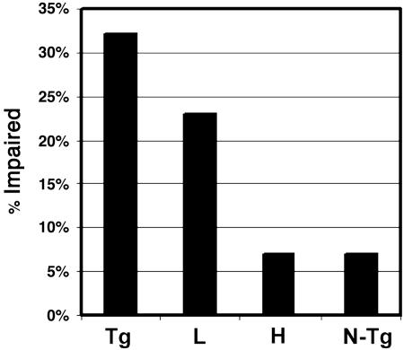
We tested the hypothesis that microtubule (MT)-binding drugs could be therapeutically beneficial in tauopathies by functionally substituting for the MT-binding protein tau, which is sequestered into inclusions of human tauopathies and transgenic mouse models thereof. Transgenic mice were treated for 12 weeks with weekly i.p. injections of 10 or 25 mg/m(2) paclitaxel (Paxceed). Both doses restored fast axonal transport in spinal axons, wherein MT numbers and stable (detyrosinated) tubulins were increased, compared with sham treatment, and only Paxceed ameliorated motor impairments in tau transgenic mice. Thus, MT-stabilizing drugs could have therapeutic potential for treating neurodegenerative tauopathies by offsetting losses of tau function that result from the sequestration of this MT-stabilizing protein into filamentous inclusions.


Figures

Fig. 1.
Fig. 1.
Dose-dependent improvement of FAT in ventral roots of T44 tau Tg mice treated with Paxceed. SDS/PAGE shows improved FAT in spinal L5 ventral roots of T44 line 7 tau Tg mice at 13 months of age treated with low-dose (A–C) or high-dose (D–F) Paxceed compared with age-matched sham-treated T44 tau Tg mice, respectively. Fluorographs (A and D) show an increase in FAT of a variety of [35S]methionine-labeled proteins (indicated by symbols) in the 13-month-old T44 tau Tg mice treated with low and high doses of Paxceed. Graphs (B, C, E, and F) illustrate quantification of proteins conveyed by FAT in pairs of age-matched T44 tau Tg mice treated with or without Paxceed. The symbols in A and D correspond to proteins analyzed in the graphs in B and E, respectively. n = 3. *, P < 0.05; **, P < 0.01.
Fig. 2.
Fig. 2.
Axonal degeneration and axonal MTs in spinal ventral roots of Paxceed-treated T44 tau Tg mice. (A) A representative semithin section micrograph shows the L5 ventral root of a 12-month-old non-Tg mouse with regular large and small myelinated axons evenly distributed within the nerve. However, the ventral root axons from T44 tau Tg mice and sham- and nontreated Tg mice are irregularly shaped (B and C) and a prominent endoneurial space is found in tau Tg mice (C). The ventral roots in tau Tg mice treated with low- and high-dose Paxceed contain more regularly shaped axons with less endoneurial space than sham-treated and untreated tau Tg mice (D and E). MTs (large arrows) and neurofilaments (small arrows) as indicated in the Inset (A′) in the ventral root axons viewed by EM in micrographs of T44 tau Tg mice treated with different doses of Paxceed compared with sham- and nontreated tau Tg mice at 12 months of age (A′–E′). M, mitochondria. [Scale bars: A, 10 μm; A′, 100 nm.] (F) Graph summarizing the MT density in the five treatment groups. n = 3. *, P < 0.05; **, P < 0.01.
Fig. 3.
Fig. 3.
An increase in stable detyrosinated tubulin (Glu-Tub) in the ventral roots (VR) of tau Tg mice treated with Paxceed. Shown are quantitative Western blot analyses of proteins in the ventral roots, spinal cords, and brains of T44 tau Tg mice treated with different doses of Paxceed compared with sham-treated tau Tg mice at 12 months of age. Mid-sized neurofilament (NFM) immunobands in the same blot show equal levels in the treated (Right) and sham-treated (Left) tau Tg mice (A). SC, spinal cord. The levels of α- and β-tubulin in the ventral roots of T44 tau Tg mice treated with low- and high-dose Paxceed are comparable to those in sham-treated tau Tg mice (A and B). However, T44 tau Tg mice treated with low- and high-dose Paxceed demonstrate a significant increase in stable detyrosinated tubulin (Glu-Tub) levels in most ventral root segments compared with sham-treated tau Tg mice (A–C; only data on high-dose treatment are shown). (D) T44 tau Tg mice treated with different doses of Paxceed reveal no detectable change in the spinal cords (Right) and brains (Left) compared with age-matched sham-treated control Tg mice. AC-Tub, acetylated tubulin; Tyr-Tub, tyrosinated tubulin. *, P < 0.05; **, P < 0.01.
Fig. 4.
Fig. 4.
No detectable change in numbers of tau spheroids in the spinal cord of tau Tg mice treated with Paxceed. Immunohistochemistry with the polyclonal anti-tau antibody 17026 in the spinal cord of T44 tau Tg mice with and without Paxceed treatment demonstrates tau spheroids in the spinal cords at 12 months of age as indicated in boxes (A). High-power photomicrographs in Insets demonstrate intense tau immunoreactivity in tau spheroids in both Paxceed-treated and sham-treated Tg mice. [Scale bars: A, 100 μm; Inset, 10 μm]. Quantitation (average number of tau spheroids per section of the spinal cord) revealed no detectable change in tau spheroids in the spinal cords of T44 tau Tg mice treated with Paxceed (B). ANOVA, P = 0.5016.
Fig. 5.
Fig. 5.
Improved motor behavior in tau Tg mice treated with low (L) and high (H) doses of Paxceed. Non-Tg (N-Tg), and T44 tau Tg mice with or without Paxceed from 9–12 months of age (n = 20 per group) were assessed by three blinded observers for motor impairments. A χ2 test for linear trend showed that Paxceed improved motor behavior (P = 0.0289).

References

    1. Lee, V. M.-Y., Goedert, M. & Trojanowski, J. Q. (2001) Annu. Rev. Neurosci. 24, 1121-1159. - PubMed
    1. Drechsel, D. N., Hyman, A. A., Cobb, M. H. & Kirschner, M. W. (1992) Mol. Biol. Cell 3, 1141-1154. - PMC - PubMed
    1. Morfini, G., Pigino, G., Beffert, U., Busciglio, J. & Brady, S. T. (2002) Neuromol. Med. 2, 89-99. - PubMed
    1. Bramblett, G. T., Goedert, M., Jakes, R., Merrick, S. E., Trojanowski, J. Q. & Lee, V. M.-Y. (1993) Neuron 10, 1089-1099. - PubMed
    1. Lee, V. M.-Y., Balin, B. J., Otvos, L., Jr., & Trojanowski, J. Q. (1991) Science 251, 675-678. - PubMed

Publication types