. 2005 Jan 4;102(1):204-9.
doi: 10.1073/pnas.0406092102. Epub 2004 Dec 22.

Endothelial-specific expression of caveolin-1 impairs microvascular permeability and angiogenesis

Affiliations

Endothelial-specific expression of caveolin-1 impairs microvascular permeability and angiogenesis

Philip M Bauer et al. Proc Natl Acad Sci U S A. .

Abstract

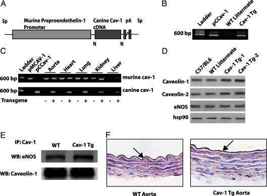
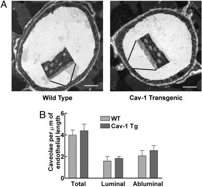
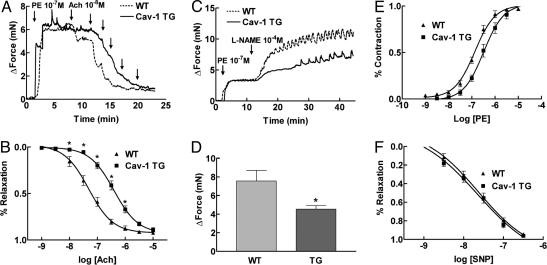
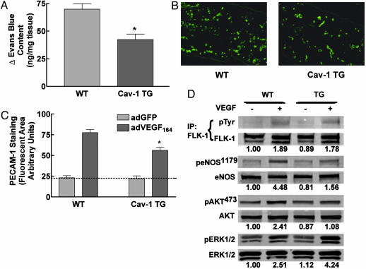
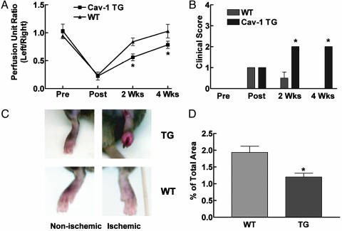
The functions of caveolae and/or caveolins in intact animals are beginning to be explored. Here, by using endothelial cell-specific transgenesis of the caveolin-1 (Cav-1) gene in mice, we show the critical role of Cav-1 in several postnatal vascular paradigms. First, increasing levels of Cav-1 do not increase caveolae number in the endothelium in vivo. Second, despite a lack of quantitative changes in organelle number, endothelial-specific expression of Cav-1 impairs endothelial nitric oxide synthase activation, endothelial barrier function, and angiogenic responses to exogenous VEGF and tissue ischemia. In addition, VEGF-mediated phosphorylation of Akt and its substrate, endothelial nitric oxide synthase, were significantly reduced in VEGF-treated Cav-1 transgenic mice, compared with WT littermates. The inhibitory effect of Cav-1 expression on the Akt-endothelial nitric oxide synthase pathway was specific because VEGF-stimulated phosphorylation of mitogen-activated protein kinase (ERK1/2) was elevated in the Cav-1 transgenics, compared with littermates. These data strongly support the idea that, in vivo, Cav-1 may modulate signaling pathways independent of its essential role in caveolae biogenesis.


Figures

Fig. 1.
Fig. 1.
Generation of endothelium-specific TG mice. (A) Construct used for generating the Cav-1 TG mice. The canine Cav-1 cDNA was driven by the preproendothelin promoter. (B) Expression of the transgene vs. endogenous Cav-1. Primers specific for canine Cav-1 recognize the plasmid containing canine Cav-1 (pCCav-1) and TG Cav-1 (Cav-1 TG) but not endogenous Cav-1 (WT littermate). (C) The presence of both WT and transgene Cav-1 by RT-PCR in various tissues. pMCav-1 is the murine Cav-1 cDNA (D) The increased expression of Cav-1 and Cav-2 in aortic extracts from two TG lines (TG-1 and TG-2), compared with C57Bl6 or WT littermates. (E) Immunoprecipitation of Cav-1 from TG lung extracts increases the amount of eNOS co-associated (Lower, total Cav-1 immunoprecipitated; Upper, coassociated eNOS). (F) Immunohistochemical evidence for expression of Cav-1 transgene in the endothelium. WT and Cav-1 TG aortic sections were stained with a Cav-1 mAb. Note the presence of intense labeling of Cav-1 in the EC, with little difference in the intensity of Cav-1 in underlying smooth muscle.
Fig. 2.
Fig. 2.
TG expression of Cav-1 in endothelium does not change caveolae number. (A) Representative transmission electron micrographs of coronary capillaries from WT or TG mice. (Insets) Magnifications (×4) to demonstrate the caveolae anatomy. (Scale bar: 1 μm.) (B) Quantification of luminal and abluminal caveolae structures. Data (mean ± SEM) are from 50 sections per mouse with n = 3 mice per group.
Fig. 3.
Fig. 3.
Endothelial expression of Cav-1 impairs eNOS-dependent vasodilation. (A) Representative bioassay trace from WT littermate (intermittent line) and Cav-1 TG (solid line) mice. Aortic rings were precontracted with PE, and ACh-induced dilations were examined. (B) Statistical analysis of the full dose–response curves to ACh demonstrates a 10-fold change in the EC50 value for ACch in Cav-1 TG mice. (C) Basal eNOS activation is reduced in Cav-1 TG. Vessels were contracted with PE and then incubated with a NOS inhibitor L-NAME to remove basal NO synthesis. (D) Cav-1 TG mice show a deficiency in basal NO synthesis. (E and F) The deficit in eNOS activation does not influence the ability of the vessels to contract to PE or to relax to the NO donor drug, SNP. Data are mean ± SEM, with n = 5 animals and two aortic rings per animal. *, P < 0.05.
Fig. 4.
Fig. 4.
VEGF-mediated functions are reduced in Cav-1 TG mice. (A) Cav-1 TG mice exhibit reduced (30 min) VEGF-stimulated vascular leakage. Data are mean ± SEM (n = 6). (B) PECAM-1-positive vascular structures in the ears of WT (Left) or Cav-1 TG (Right) mice injected with AdVEGF (after 6 days). (C) AdVEGF-mediated angiogenesis was reduced in Cav-1 TG (mean ± SEM, with n = 6 animals per group, and 10 fields were quantified for each ear). (D) VEGF-mediated signal transduction to Akt and eNOS are reduced in Cav-1 TG mice. Mice were injected with saline or VEGF, and lung tissue was processed for Western blot analysis. The numbers below represent densitometric evaluation of the data, with a value of 1.0 reflecting the basal level of phosphorylation of each signaling protein (FLK, eNOS, Akt, and ERK, respectively). Similar results were obtained in three additional experiments. *, P < 0.05.
Fig. 5.
Fig. 5.
Ischemia-induced arteriogenesis and angiogenesis are impaired in Cav-1 TG mice. (A and B) Arteriectomy was performed in Cav-1 TG and WT mice, and blood flow (A) and clinical index (B) were quantified over time. Data are mean ± SEM (n = 6). (C) The inflamed, necrotic foot seen in nonischemic (Upper Left) and ischemic (Upper Right) Cav-1 TG mice, compared with littermate controls (Lower). (D) The reduction in blood-flow recovery and clinical outcome were attributable to a decrease in lower-limb angiogenesis. Data are mean ± SEM, with n = 6 animals per group. Four fields per section and five sections per animal were quantified. *, P < 0.05.

Similar articles

Cited by

References

    1. Bruns, R. R. & Palade, G. E. (1968) J. Cell Biol. 37, 244-276. - PMC - PubMed
    1. Fielding, P. E. & Fielding, C. J. (1995) Biochemistry 34, 14288-14292. - PubMed
    1. Smart, E. J., Ying, Y., Donzell, W. C. & Anderson, R. G. (1996) J. Biol. Chem. 271, 29427-29435. - PubMed
    1. Schnitzer, J. E., Oh, P. & McIntosh, D. P. (1996) Science 274, 239-242. - PubMed
    1. Anderson, R. G., Kamen, B. A., Rothberg, K. G. & Lacey, S. W. (1992) Science 255, 410-411. - PubMed

Publication types

LinkOut - more resources