. 2005 Mar;16(3):1213-22.
doi: 10.1091/mbc.e04-07-0599. Epub 2004 Dec 22.

Dynamics of GBF1, a Brefeldin A-sensitive Arf1 exchange factor at the Golgi

Affiliations

Dynamics of GBF1, a Brefeldin A-sensitive Arf1 exchange factor at the Golgi

Ting-Kuang Niu et al. Mol Biol Cell. 2005 Mar.

Abstract

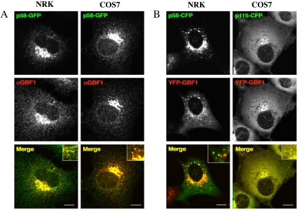
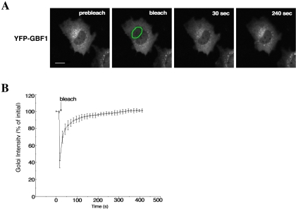
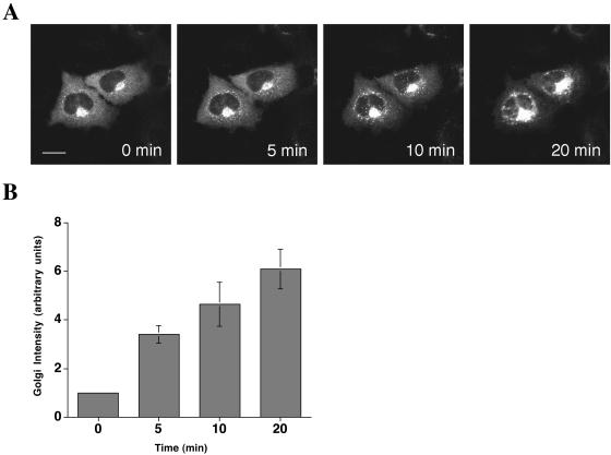
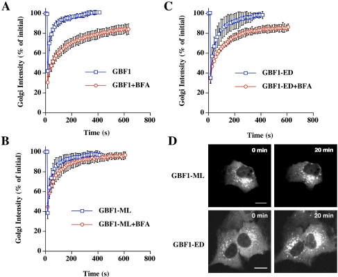
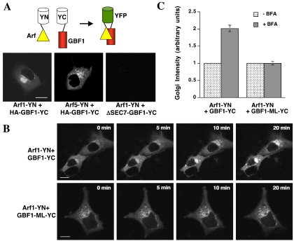
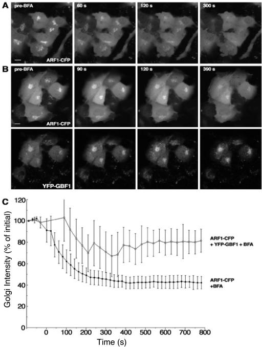
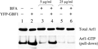
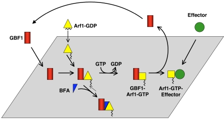
Trafficking through the Golgi apparatus requires members of the Arf family of GTPases, whose activation is regulated by guanine nucleotide exchange factors (GEFs). Once activated, Arf-GTP recruits effectors such as coat complexes and lipid-modifying enzymes to specific membrane sites, creating a domain competent for cargo concentration and transport. GBF1 is a peripherally associated Arf GEF involved in both endoplasmic reticulum-Golgi and intra-Golgi transport. The mechanism of GBF1 binding to membranes is unknown. As a first step to understanding the mechanism of membrane association, we constructed a yellow fluorescent protein-tagged version of GBF1 and performed fluorescence recovery after photobleaching analysis to determine its residence time on Golgi membranes. We find that GBF1 molecules are not stably associated with the Golgi but rather cycle rapidly on and off membranes. The drug brefeldin A (BFA), an uncompetitive inhibitor of the exchange reaction that binds to an Arf-GDP-Arf GEF complex, stabilizes GBF1 on Golgi membranes. Using an in vivo assay to monitor Arf1-GTP levels, we show that GBF1 exchange activity on Arf1 is inhibited by BFA in mammalian cells. These results suggest that an Arf1-GBF1-BFA complex is formed and has a longer residence time on Golgi membranes than GBF1 or Arf1 alone.


Figures

Figure 1.
Figure 1.
Colocalization of GBF1 with p58 and p115 in NRK and COS7 cells. (A) NRK and COS7 cells were transfected with p58-GFP (green). Twenty-four hours after transfection, cells were fixed and stained with antibodies to GBF1 (red). Insets in merge images show colocalization of p58-GFP and endogenous GBF1. (B) NRK cells were cotransfected with p58-CFP (green) and YFP-GBF1 (red) and COS7 cells with p115-CFP (green) and YFP-GBF1 (red). Cells were visualized by 24 h after transfection. Insets in merge images show colocalization of p58-CFP or p115-CFP with overexpressed YFP-GBF1. Bar, 10 μm.
Figure 2.
Figure 2.
Kinetics of GBF1 binding to and dissociation from Golgi membranes. (A) NRK cells expressing YFP-GBF1 were photographed before photobleaching to obtain a prebleach image. Photobleaching of the Golgi region (outlined in green) was carried out by illumination with high-intensity laser light. After bleaching, images were taken at 13-s intervals to monitor the recovery of fluorescence to the bleached area. Images at representative time points of postbleach are shown. Bar, 10 μm. (B) Quantification of Golgi FRAP in NRK cells expressing YFP-GBF1 (n = 5). Error bars indicate the SD.
Figure 3.
Figure 3.
GBF1 is stabilized on Golgi membranes by BFA. (A) NRK cells expressing YFP-GBF1 were treated with 1 μg/ml BFA for 20 min. Images were taken every 30 s after the addition of BFA (see movie in supplemental data). (B) Relative Golgi intensity was measured at representative time points (n = 4). Error bars indicate the SD. Bar, 10 μm.
Figure 4.
Figure 4.
Golgi membrane binding and dissociation kinetics of GBF1 and Sec7 domain mutants in the presence and absence of BFA. Quantification of Golgi FRAP in NRK cells expressing YFP-GBF1 (n = 7) (A), YFP-GBF1-ML (n = 6) (B), or YFP-GBF1-ED (n = 7) (C) with or without addition of 1 μg/ml BFA. In the presence of BFA, FRAP was performed after 20 min of treatment. Error bars indicate the SD. (D) Images of cells expressing YFP-GBF1-ED and YFP-GBF1-ML with and without 1 μg/ml BFA. Bar, 10 μm.
Figure 5.
Figure 5.
Bimolecular fluorescence complementation analysis to monitor GBF1-Arf1 interaction in cells. (A) Schematic diagram showing principle of bimolecular fluorescence complementation assay (top). COS7 cells were transfected with Arf1-YN and either HA-GBF1-YC or ΔSEC7-GBF1-YC and were imaged 24 h after transfection (bottom). Bar, 10 μm. (B) COS7 cells were transfected with Arf1-YN and either HA-GBF1-YC or HA-GBF1-M794L-YC and were treated for the indicated times with 5 μg/ml BFA. Cells were imaged 24 h after transfection. Bar, 10 μm. (C) Quantitation of the results shown in B (n = 5). Error bars indicate the SD.
Figure 6.
Figure 6.
More Arf remains associated with Golgi membranes after BFA treatment in cells expressing GBF1. (A) NRK cells expressing Arf1-CFP were treated with 1 μg/ml BFA. Images at representative time points are shown. (B) NRK cells expressing Arf1-CFP and YFP-GBF1 were treated with 1 μg/ml BFA. Images of Arf1 (top) and GBF1 (bottom) taken at the same time point are shown. (C) Quantification of Arf1 release from membranes with (n = 4) or without (n = 6) YFP-GBF1 after BFA treatment. Error bars indicate the SD. Bar, 10 μm.
Figure 7.
Figure 7.
Arf-GTP pull-down assay. COS7 cells were cotransfected with Arf1-HA and empty vector (lanes 1, 3, and 5) or Arf1-HA and YFP-GBF1 (lanes 2, 4, and 6). Sixteen hours after transfection, cells were either lysed directly (lanes 1 and 2) or treated with 5 μg/ml BFA (lanes 3 and 4) or 25 μg/ml BFA (lanes 5 and 6) for 20 min before lysis. Arf-GTP pull-downs by using GST-GGA3-GAT bound to glutathione-Sepharose was performed as described in Materials and Methods.
Figure 8.
Figure 8.
Model for Arf activation at the Golgi. The Arf GEF GBF1 (red) and Arf1-GDP (yellow triangle) are recruited independently to Golgi membranes. The exchange reaction proceeds through several short-lived intermediates, and GBF1 is released from membranes upon GTP binding to Arf1. Arf1-GTP (yellow square) remains associated with membranes where it recruits effectors. BFA (blue triangle) binds to an early reaction intermediate, a GBF1–Arf–GDP complex, which is stabilized on Golgi membranes.

References

    1. Antonny, B., and Schekman, R. (2001). ER export: public transportation by the COPII coach. Curr. Opin. Cell Biol. 13, 438-443. - PubMed
    1. Beraud-Dufour, S., Robineau, S., Chardin, P., Paris, S., Chabre, M., Cherfils, J., and Antonny, B. (1998). A glutamic finger in the guanine nucleotide exchange factor ARNO displaces Mg2+ and the beta-phosphate to destabilize GDP on ARF1. EMBO J. 17, 3651-3659. - PMC - PubMed
    1. Boman, A. L. (2001). GGA proteins: new players in the sorting game. J. Cell. Sci. 114, 3413-3418. - PubMed
    1. Chantalat, S., Courbeyrette, R., Senic-Matuglia, F., Jackson, C. L., Goud, B., and Peyroche, A. (2003). A novel Golgi membrane protein is a partner of the ARF exchange factors Gea1p and Gea2p. Mol. Biol. Cell 14, 2357-2371. - PMC - PubMed
    1. Claude, A., Zhao, B. P., Kuziemsky, C. E., Dahan, S., Berger, S. J., Yan, J. P., Armold, A. D., Sullivan, E. M., and Melancon, P. (1999). GBF 1, A novel Golgi-associated BFA-resistant guanine nucleotide exchange factor that displays specificity for ADP-ribosylation factor 5. J. Cell Biol. 146, 71-84. - PMC - PubMed

Publication types

MeSH terms

LinkOut - more resources