. 2005 Mar;16(3):1082-94.
doi: 10.1091/mbc.e04-05-0427. Epub 2004 Dec 22.

Novel role for Na,K-ATPase in phosphatidylinositol 3-kinase signaling and suppression of cell motility

Affiliations

Novel role for Na,K-ATPase in phosphatidylinositol 3-kinase signaling and suppression of cell motility

Sonali P Barwe et al. Mol Biol Cell. 2005 Mar.

Abstract

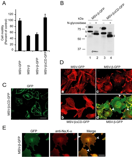
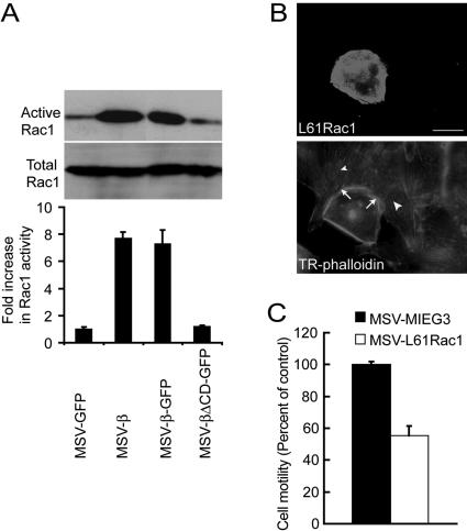
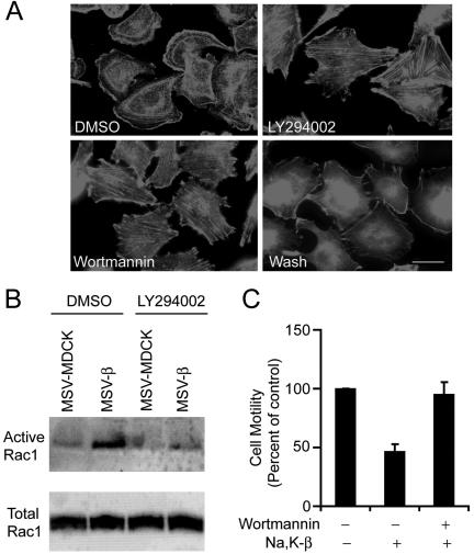
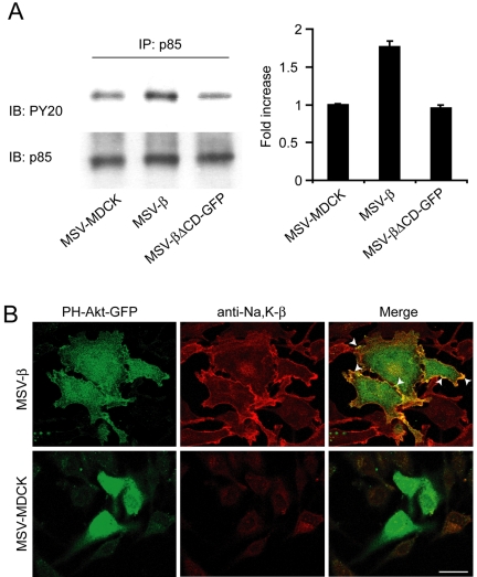
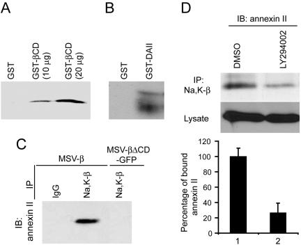
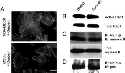
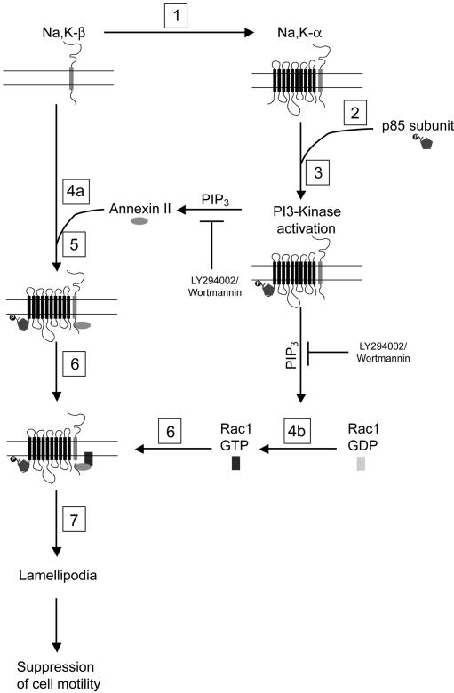
The Na,K-ATPase, consisting of alpha- and beta-subunits, regulates intracellular ion homeostasis. Recent studies have demonstrated that Na,K-ATPase also regulates epithelial cell tight junction structure and functions. Consistent with an important role in the regulation of epithelial cell structure, both Na,K-ATPase enzyme activity and subunit levels are altered in carcinoma. Previously, we have shown that repletion of Na,K-ATPase beta1-subunit (Na,K-beta) in highly motile Moloney sarcoma virus-transformed Madin-Darby canine kidney (MSV-MDCK) cells suppressed their motility. However, until now, the mechanism by which Na,K-beta reduces cell motility remained elusive. Here, we demonstrate that Na,K-beta localizes to lamellipodia and suppresses cell motility by a novel signaling mechanism involving a cross-talk between Na,K-ATPase alpha1-subunit (Na,K-alpha) and Na,K-beta with proteins involved in phosphatidylinositol 3-kinase (PI3-kinase) signaling pathway. We show that Na,K-alpha associates with the regulatory subunit of PI3-kinase and Na,K-beta binds to annexin II. These molecular interactions locally activate PI3-kinase at the lamellipodia and suppress cell motility in MSV-MDCK cells, independent of Na,K-ATPase ion transport activity. Thus, these results demonstrate a new role for Na,K-ATPase in regulating carcinoma cell motility.


Figures

Figure 1.
Figure 1.
Na,K-β expression suppresses cell motility by reorganization of the actin cytoskeleton. (A) Migration of MSV-MDCK cells expressing either GFP, Na,K-β, GFP-tagged Na,K-β, or GFP-tagged Na,K-βΔCD across Transwell filters. The error bars represent the SD of the mean of three independent measurements done in triplicates. (B) MSV-β-GFP and MSV-βΔCD-GFP cell lysates treated without or with N-glycosidase were separated on SDS-PAGE and blotted with anti-Na,K-β antibody. In lane 1, the faint 54-kDa band (a) indicates the endogenous fully glycosylated Na,K-β. The 80-kDa band (c) represents the fully glycosylated Na,K-β-GFP chimera, whereas the 65-kDa band (b) represents a high mannose form of the Na,K-β-GFP. In lane 2, the 33-kDa band (d) is the core Na,K-β protein, the band at 59 kDa (e) is the fully deglycosylated Na,K-β-GFP chimera, and the faint band above is incompletely digested form of the Na,K-β-GFP chimera. In lane 3, the 76-kDa band (f) represents the Na,K-βΔCD-GFP chimera. In lane 4, the 55-kDa band (g) represents the full deglycosylated Na,K-βΔCD-GFP chimera. (C) GFP fluorescence in MSV-βΔCD-GFP cells showing plasma membrane localization of Na,K-βΔCD-GFP. (D) Confocal microscope projections obtained from optical slices show actin organization (red) of MSV-GFP (a), MSV-βΔCD-GFP (b), and MSV-β-GFP (c), and merged image showing colocalization of Na,K-β-GFP with filamentous actin (yellow) at the lamellipodia (arrowhead) (d). The GFP fluorescence in a and b is not shown to reveal stress fibers clearly. (E) Immunofluorescence of MSV-β-GFP cells, stained with anti-Na,K-α antibody followed by anti-Cy3 antibody, Na,K-β-GFP fluorescence, and merged image showing colocalization of the two (yellow) at the lamellipodia (arrowhead). Bar, 25 μm.
Figure 2.
Figure 2.
Na,K-β–mediated reorganization of the actin cytoskeleton and suppression of cell motility is Rac1 dependent. (A) Immunoblot showing active and total endogenous Rac1 in MSV-GFP, MSV-β, MSV-β-GFP, and MSV-βΔCD-GFP cells. The graph represents mean ± SD of three independent experiments. (B) Fluorescence of MSV-MDCK cells transiently transfected with L61 Rac1 showing GFP-L61 Rac1 and Texas Red-labeled phalloidin. Arrows show the lamellipodia in a L61 Rac1-transfected cell. Arrowheads show the stress fibers in untransfected cells. Bar, 25 μm. (C) Migration of MSV-MDCK cells stably transfected with MIEG3 vector or L61 Rac1. The error bars show SD of the mean of three independent experiments done in triplicates.
Figure 3.
Figure 3.
Requirement of PI3-kinase for Na,K-β–mediated suppression of cell motility and enhancement of Rac1 activity. (A) Phalloidin staining showing actin organization of MSV-β cells treated with DMSO, LY294002, wortmannin, or wortmannin followed by washout for 1 h (Wash). Bar, 25 μm. (B) Immunoblot showing active and total levels of endogenous Rac1 in DMSO- or LY294002-treated MSV-MDCK and MSV-β cells. (C) Migration of MSV-β cells treated with DMSO or wortmannin across Transwell filters. The error bars show SD of the mean of two independent experiments done in triplicates.
Figure 4.
Figure 4.
Analysis of the activation of PI3-kinase in MSV-β cells. (A) MSV-MDCK, MSV-β, and MSV-βΔCD-GFP cells were lysed, and p85 was immunoprecipitated from 1 mg of total cell lysates. The amount of tyrosine phosphorylated p85 in the immunoprecipitates was normalized to total p85 detected by immunoblotting with anti-phosphotyrosine and anti-p85 antibodies, respectively. The graph represents mean ± SD from three independent experiments. (B) Colocalization (arrowheads) of PH-Akt-GFP and Na,K-β (stained with anti-Na,K-β antibody followed by anti-Cy3 antibody) in MSV-MDCK and MSV-β cells. Bar, 25 μm.
Figure 5.
Figure 5.
Na,K-β cytoplasmic tail interacts with annexin II in a PI3-kinase dependent manner. (A) Ten or 20 μg of GST-βCD was incubated with 1 mg of MSV-β cell lysate and the annexin II pulled down by GST-βCD was determined by immunoblotting. (B) GST-annexin II (GST-DAII) was incubated with MSV-β cell lysate as described in Materials and Methods. The blots were then probed with anti-Na,K-β antibody to detect Na,K-β interaction with annexin II. (C) Na,K-β from MSV-β and MSV-βΔCD-GFP cell lysates was immunoprecipitated (IP) with anti-Na,K-β antibody and analyzed by immunoblotting (IB) for the presence of annexin II. (D) MSV-β cells were treated with DMSO or LY294002 and lysed. One milligram of total protein was used for immunoprecipitating Na,K-β. The amount of annexin II bound to Na,K-β was detected by immunoblotting. The graph represents the mean ± SD of three independent experiments.
Figure 6.
Figure 6.
Analysis of the role of Na,K-α in PI3-kinase signaling. (A) Immunoblot showing levels of Na,K-α from the lysates of indicated cells. (B) p85 was immunoprecipitated (IP) from 1 mg of MSV-MDCK, MSV-β and MSV-βΔCD-GFP cell lysates, and coprecipitating Na,K-α was detected by immunoblotting (IB). The graph represents mean ± SD of three independent experiments. (C) MSV-MDCK, MSV-β, and MSV-βΔCD-GFP cells were lysed and Na,K-α was immunoprecipitated. The precipitate was analyzed by immunoblotting for tyrosine phosphorylated p85 by using anti-phosphotyrosine antibody. The blots were stripped and reprobed for p85 to confirm the presence of p85 (our unpublished data). The graph represents mean ± SD of three independent experiments. (D) GST pull-down assay using GST, GST-αCD, or GST-βCD–coupled agarose beads with MSV-β cell lysate. The immunoblot shows the amount of p85 bound to GST-αCD. (E) The amount of tyrosine phosphorylated p85 in MSV-βΔCD-GFP cells and MSV-βΔCD-GFP cells transfected with Na,K-α (MSV-βΔCD-GFP-α) was determined by immunoprecipitating p85 from 1 mg of cell lysates, followed by blotting with anti-phosphotyrosine antibody. The graph represents mean ± SD of three independent experiments. (F) Phalloidin staining showing the actin organization of MSV-βΔCD-GFP-α cells. Bar, 25 μm. Note abundant stress fibers and lack of lamellipodia (G) Motility of MSV-βΔCD-GFP (1) and MSV-βΔCD-GFP-α (2) cells in a Transwell motility assay. The error bars show the SD of the mean of triplicate measurements done twice, expressed as a percentage of the control (MSV-βΔCD-GFP).
Figure 7.
Figure 7.
Analysis of the role of Na,K-ATPase pump activity in PI3-kinase signaling and Rac1 activation. (A) Phalloidin staining showing actin organization in MSV-MDCK and MSV-β cells treated with ouabain. Bar, 25 μm. For B–D, MSV-β cells treated with DMSO or ouabain were used. Immunoblot showing the active and total levels of endogenous Rac1 (B), annexin II bound to Na,K-β (C), and p85 bound to Na,K-α (D). The blots are representative of three independent experiments.
Figure 8.
Figure 8.
Model showing the mechanism of Na,K-β–mediated suppression of cell motility. 1) Repletion of Na,K-β in MSV-MDCK cells increases Na,K-α levels. 2) High levels of Na,K-α lead to the increased tyrosine phosphorylation of p85 and its recruitment to the plasma membrane. 3) This causes activation of PI3-kinase and the generation of PIP3. Increased levels of PIP3 induce 4a) binding of annexin II to Na,K-β cytoplasmic tail and 4b) activation of Rac1. 5) A complex containing both Na,K-ATPase subunits, annexin II, and PI3 kinase is assembled at the plasma membrane. 6) Annexin II sequesters active Rac1 into this complex. 7) Formation of lamellipodia is accomplished, leading to suppression of cell motility.

References

    1. Abbott, A., and Ball, W. J., Jr. (1993). The epitope for the inhibitory antibody M7-PB-E9 contains Ser-646 and Asp-652 of the sheep Na+,K(+)-ATPase alpha-subunit. Biochemistry 32, 3511-3518. - PubMed
    1. Anilkumar, G., Rajasekaran, S. A., Wang, S., Hankinson, O., Bander, N. H., and Rajasekaran, A. K. (2003). Prostate-specific membrane antigen association with filamin A modulates its internalization and NAALADase activity. Cancer Res. 63, 2645-2648. - PubMed
    1. Balch, C., and Dedman, J. R. (1997). Annexins II and V inhibit cell migration. Exp. Cell Res. 237, 259-263. - PubMed
    1. Birchmeier, C., Birchmeier, W., and Brand-Saberi, B. (1996). Epithelial-mesenchymal transitions in cancer progression. Acta Anat. 156, 217-226. - PubMed
    1. Blok, L. J., Chang, G. T., Steenbeek-Slotboom, M., van Weerden, W. M., Swarts, H. G., De Pont, J. J., van Steenbrugge, G. J., and Brinkmann, A. O. (1999). Regulation of expression of Na+,K+-ATPase in androgen-dependent and androgen-independent prostate cancer. Br. J. Cancer 81, 28-36. - PMC - PubMed

Publication types

MeSH terms

LinkOut - more resources