. 2005 Jan 26;24(2):382-94.
doi: 10.1038/sj.emboj.7600533. Epub 2004 Dec 23.

The ancient origin of the complement system

Affiliations

The ancient origin of the complement system

Yong Zhu et al. EMBO J. .

Abstract

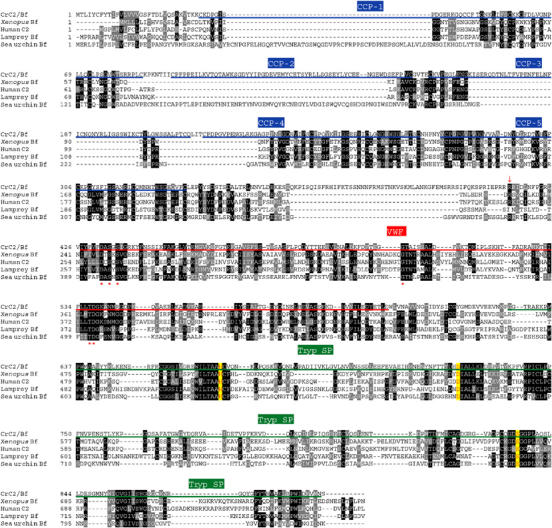
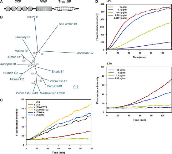
The complement system has been thought to originate exclusively in the deuterostomes. Here, we show that the central complement components already existed in the primitive protostome lineage. A functional homolog of vertebrate complement 3, CrC3, has been isolated from a 'living fossil', the horseshoe crab (Carcinoscorpius rotundicauda). CrC3 resembles human C3 and shows closest homology to C3 sequences of lower deuterostomes. CrC3 and plasma lectins bind a wide range of microbes, forming the frontline innate immune defense system. Additionally, we identified CrC2/Bf, a homolog of vertebrate C2 and Bf that participates in C3 activation, and a C3 receptor-like sequence. Furthermore, complement-mediated phagocytosis of bacteria by the hemocytes of horseshoe crab was also observed. Thus, a primitive yet complex opsonic complement defense system is revealed in the horseshoe crab, a protostome species. Our findings demonstrate an ancient origin of the critical complement components and the opsonic defense mechanism in the Precambrian ancestor of bilateral animals.


Figures

Figure 1
Figure 1
Isolation and characterization of CrC3. Bacteria-binding proteins from horseshoe crab plasma were recovered by elution from S. aureus cells incubated with the horseshoe crab cell-free plasma (‘−', control S. aureus treated in saline; ‘+', S. aureus incubated with plasma), under different buffer conditions as indicated. (A) Profile of plasma proteins extracted from the bacteria at indicated conditions. Protein sample derived from ∼0.25 ml each of bacteria/plasma was loaded per lane (see Materials and methods). (B) Protein profiles of bacteria after extraction (∼0.25 ml of bacteria per lane). (C) Comparison of total protein profiles of S. aureus cells (‘−', untreated bacterial cells; ‘+', bacterial cells incubated with plasma), treated with triethanolamine (pH 11.5 (B)), that were solubilized in nonreducing condition (−2-ME) and reducing condition (+2-ME). Protein extract derived from ∼0.25 ml each of bacterial cells was loaded per lane. (D) 2D SDS–PAGE (nonreducing 1st D and reducing 2nd D) analysis of protein profile of S. aureus cells (treated with plasma) that were solubilized in nonreducing condition. CrC3 of ∼250 kDa (p250) observed in the 1st D was resolved into p75, p50, p36(CrC3), and p34 after reduction.
Figure 2
Figure 2
CrC3 is a homolog of vertebrate C3. (A) Schematic modular organization of CrC3, a standard modular structure of vertebrate C3/C4/C5. The scheme is drawn as revealed by conserved domain search at http://www.ncbi.nlm.nih.gov/Structure/cdd/wrpsb.cgi. Conserved domain database references: A2M_N (pfam01835.11), N-terminal region of the TEP super family; ANATO (pfam01821.11; smart00104.10), anaphylatoxin-like domain; A2M (pfam00207.11), the C-terminal region of the α2M/TEP/C3/C4/C5 family; and C345C, an additional module at the extreme C-terminus of C3/C4/C5 (pfam01759.11, smart00643.10). (B) Alignment of C3/C4/C5-characteristic ANATO region of CrC3 with the most homologous sequences in frog C4 (BAA11188.1), hagfish C3 (P98094), human C4 (P01028), lamprey C3 (Q00685), mouse C4 (P01029), mouse C5 (P06684), pig C5 (1C5A), and snake C3 (Q01833). The six most-conserved cysteine residues critical for maintaining the structure of C345a peptides through three pairs of disulfide bonds are double-underlined. (C) Phylogeny of CrC3 and related TEPs. The scale bar corresponds to 0.1 estimated amino-acid substitutions per site. The confidence scores (in %) of a bootstrap test of 1000 replicates are indicated in red for major branching nodes. Selected proteins are: aTEP, Anopheles TEP-I; dTEPI, Drosophila TEP1; dTEPII, Drosophila TEP2; dTEPIII, Drosophila TEP3; dTEPIV, Drosophila TEP4; cTEP, Caenorhabditis TEP2; SeC3, cnidaria C3-like protein; BbC3, Amphioxus C3; SpC3, sea urchin C3; HrC3, Halocynthia C3; CiC3, Ciona C3; HaC3, hagfish C3; LaC3, lamprey C3; TrC3, trout C3; CaC3, carp C3-H1; XeC3, Xenopus C3; ChC3, chicken C3; CoC3, cobra C3; CVF, cobra venom factor; HuC3, human C3; GpC3, guinea pig C3; RaC3, rat C3; MoC3, mouse C3; CaC4, carp C4; ShC4, shark C4; ChC4, chicken C4; XeC4, Xenopus C4; MoC4, mouse C4; MoSPL, mouse sex limited protein (M21576); HuC4A, human C4A (K02403); HuC4B, human C4B (U24578); CaC5, carp C5; MoC5, mouse C5; HuC5, human C5; LiA2M, horseshoe crab α2M; LaA2M, lamprey α2M; ChOvo, chicken ovostatin; RaA1I, rat alpha1-inhibitor; HuPZP, human pregnancy zone protein; GpA2M, guinea pig α2M; HuA2M, human α2M; RaA2M, rat α2M; RaA1M, rat alpha1-macroglobulin; MoA2M, mouse α2M; CiTEP, Ciona intestinalis TEP; OmA2M, Ornithodoros (soft tick) α2M. For further information including database accession numbers, see Supplementary Table 1. (D) HA inhibits the attachment of CrC3 to bacteria. HA was added into the plasma to a final concentration of 0.2 or 1.0 M. S. aureus cells were incubated with naïve plasma or plasma supplemented with HA (see Materials and methods) at room temperature for ∼15 min. Washed bacterial cells were lysed in SDS–PAGE loading buffer and analyzed by nonreducing SDS–PAGE. ‘−', untreated bacterial cells; ‘+', bacterial cells incubated with horseshoe crab plasma with/without HA supplemented; protein samples derived from ∼50 μl bacteria (OD600 nm ∼0.5) or bacteria/plasma were loaded per lane.
Figure 3
Figure 3
Sequence comparison of CrC3 and the human C3, C4, and C5. The multiple sequence alignment of CrC3 with human C3 (P01024)/C4B (U24578)/C5 (P01031) was produced with ClustalX. All the cysteine residues of human C3 (except that in the thioester site GCGEQ) and conserved cysteines in the other sequences are highlighted. Other characteristic sites, including proteolytic cleavage sites, thioester sites, catalytic His sites, are also annotated accordingly. The inferred regions of p75, p50, and p34 in the CrC3 sequence are marked.
Figure 4
Figure 4
Similar profiles of plasma proteins bound to representative microbes. The major proteins that bind Gram-positive bacteria S. aureus, Gram-negative bacteria E. coli, and fungus K. marxianus are CL5a, CL5b, and CrC3. CrC3 bound to all microbes is fragmented into p75, p50, p36(CrC3), and p34. (A) Comparison of protein profiles of three microbes (untreated or treated with horseshoe crab plasma) under reducing and nonreducing conditions of solubilization (±2-ME). Protein samples derived from ∼0.2 ml each of microbial cells/plasma were loaded per lane. (B) 2D SDS–PAGE (nonreducing 1st D and reducing 2nd D) analysis of protein extracts from E. coli and K. marxianus that were incubated with horseshoe crab plasma.
Figure 5
Figure 5
Multiple alignment of CrC2/Bf and related proteins selected. The deduced amino-acid sequence of CrC2/Bf was aligned with C2 and/or Bf from Xenopus, human, lamprey, and sea urchin. For the database accession numbers of the selected sequences and information on various domains (CCP, VWF, and Tryp_SP), refer to the legend of Figure 6. The predicted active sites for serine protease activity are shown in yellow boxes, and factor D cleavage site is shown by a downward arrow. Consensus Mg2+-binding sites are marked (*).
Figure 6
Figure 6
Identification of CrC2/Bf and PAMP molecule-triggered Tryp_SP activity. (A) Conserved domain architecture of CrC2/Bf revealed by database search at http://www.ncbi.nlm.nih.gov/Structure/cdd/wrpsb.cgi. Database references: complement control protein module (CCP), smart00033, pfam00084; von Willebrand factor type A domain (VWF), smart00327, pfam00092; trypsin-like serine protease (Tryp_SP), smart00020, pfam00089. (B) Unrooted phylogenetic tree of CrC2/Bf with other C2s and Bfs. The scale bar corresponds to 0.1 estimated amino-acid substitutions per site. The bootstrap test scores (in %) of 1000 replicates are indicated at the major branch nodes. Database accession numbers of the compared sequences are: human C2, AAB97607; human Bf, CAA51389; mouse C2, AAA37381; mouse Bf, P04186; Xenopus Bf, BAA06179; zebra fish Bf, AAC05096; carp C2/Bf, BAA34707; medaka fish, BAA12207; shark Bf, BAB63203; puffer fish C2/Bf, CAD21938; sea urchin Bf, AAC79682; ascidian C2, AAK00631; lamprey Bf, BAA027630. (C) PAMP molecules (LPS and LTA) trigger a Tryp_SP activity in the horseshoe crab plasma. The enzymatic assay was carried out as follows: 5 μl of plasma was added in replacement of the recombinant factor C into a 100 μl reaction volume of PyroGene™ Endotoxin Detection System (BioWhittaker™). LPS or LTA resuspended in pyrogen-free water was added into the reaction at different concentrations. The fluorescence intensity of the reaction was monitored for 2 h at excitation/emission wavelength of 380/440 nm (both at 2.5 nm slit) with plate reader using Luminescence Spectrometer LS50B (Perkin Elmer). The fluorescence intensity is corelational to the amount of substrate cleaved by the activated Tryp_SP activity. (D) LTA-triggered Tryp_SP activity requires Ca2+ and Mg2+. Enzymatic assays were carried out as above, with the addition of EDTA, CaCl2, or MgCl2 at 10 mM.
Figure 7
Figure 7
Smaller fragment (p25) of CrC3 appeared in the horseshoe crab plasma after incubation of plasma with representative microbes. (A) Profiles of polypeptides extracted through C18 minicolumn, from naïve and pathogen-treated plasma samples. Extracted polypeptides were resolved by tricine–SDS–PAGE. The samples derived from ∼0.5 ml each of plasma were loaded per lane. The gel was stained with Coomassie blue. The apparent pathogen-induced polypeptides (p25 and p16) were excised, in-gel digested with trypsin, and identified by MS. p25 was found to be a fragment of CrC3, while p16 was found to be a coagulin homolog. The implication of induction of p16 is not clear yet. (B) Position of p25 (C3c counterpart) in the predicted p34 region (underlined) of CrC3 (showing only the 666–959 position of the amino-acid sequence; see Figure 3). The peptide sequences from p25, which were identified by MS-MS, are highlighted in yellow. (C) An inferred organization of four CrC3 fragments constituting the complete CrC3 molecule. The predicted anaphylactic peptide region on p34 is marked red; the thioester site on p50 is marked with a star. The interchain disulfide bridging pattern is predicted based on the alignment of CrC3 with human C3.

References

    1. Armstrong PB, Melchior R, Swarnakar S, Quigley JP (1998) Alpha2-macroglobulin does not function as a C3 homologue in the plasma hemolytic system of the American horseshoe crab, Limulus. Mol Immunol 35: 47–53 - PubMed
    1. Azumi K, De Santis R, De Tomaso A, Rigoutsos I, Yoshizaki F, Pinto MR, Marino R, Shida K, Ikeda M, Ikeda M, Arai M, Inoue Y, Shimizu T, Satoh N, Rokhsar DS, Du Pasquier L, Kasahara M, Satake M, Nonaka M (2003) Genomic analysis of immunity in a Urochordate and the emergence of the vertebrate immune system: ‘waiting for Godot'. Immunogenetics 55: 570–581 - PubMed
    1. Beutler B (2004) Innate immunity: an overview. Mol Immune 40: 845–859 - PubMed
    1. Chen SC, Yen CH, Yeh MS, Huang CJ, Liu TY (2001) Biochemical properties and cDNA cloning of two new lectins from the plasma of Tachypleus tridentatus: Tachypleus plasma lectin 1 and 2+. J Biol Chem 276: 9631–9639 - PubMed
    1. Davis AE III, Harrison RA, Lachmann PJ (1984) Physiologic inactivation of fluid phase C3b: isolation and structural analysis of C3c, C3d,g (alpha 2D), and C3g. J Immunol 132: 1960–1966 - PubMed

Publication types