. 2005 Jan 26;24(2):428-37.
doi: 10.1038/sj.emboj.7600524. Epub 2004 Dec 23.

The VirE3 protein of Agrobacterium mimics a host cell function required for plant genetic transformation

Affiliations

The VirE3 protein of Agrobacterium mimics a host cell function required for plant genetic transformation

Benoît Lacroix et al. EMBO J. .

Abstract

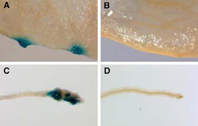
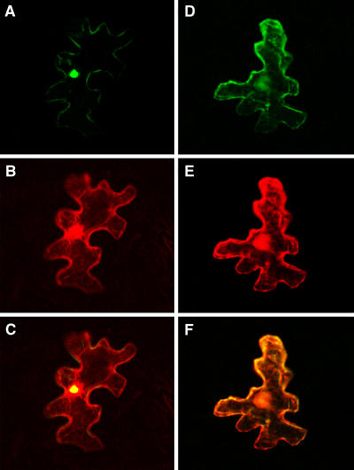
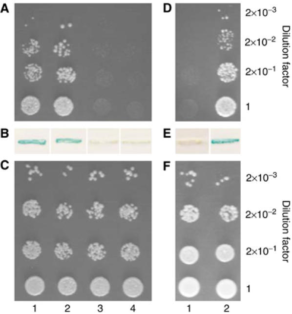
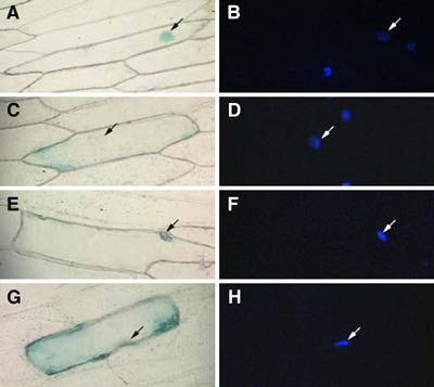
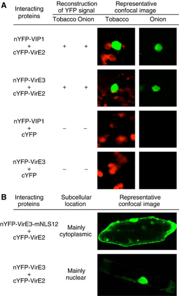
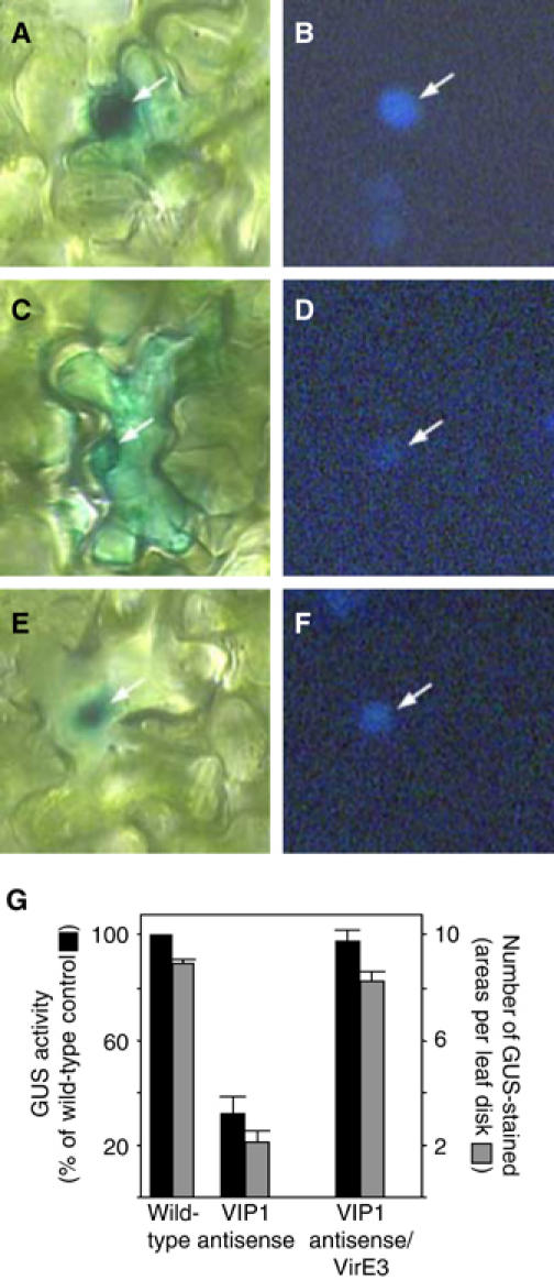
To genetically transform plants, Agrobacterium exports its transferred DNA (T-DNA) and several virulence (Vir) proteins into the host cell. Among these proteins, VirE3 is the only one whose biological function is completely unknown. Here, we demonstrate that VirE3 is transferred from Agrobacterium to the plant cell and then imported into its nucleus via the karyopherin alpha-dependent pathway. In addition to binding plant karyopherin alpha, VirE3 interacts with VirE2, a major bacterial protein that directly associates with the T-DNA and facilitates its nuclear import. The VirE2 nuclear import in turn is mediated by a plant protein, VIP1. Our data indicate that VirE3 can mimic this VIP1 function, acting as an 'adapter' molecule between VirE2 and karyopherin alpha and 'piggy-backing' VirE2 into the host cell nucleus. As VIP1 is not an abundant protein, representing one of the limiting factors for transformation, Agrobacterium may have evolved to produce and export to the host cells its own virulence protein that at least partially complements the cellular VIP1 function necessary for the T-DNA nuclear import and subsequent expression within the infected cell.


Figures

Figure 1
Figure 1
Export of VirE3 from Agrobacterium to Arabidopsis cells. Leaf segments were inoculated with Agrobacterium-expressing mGAL4-VP16-VirE3 (A) or mGAL4-VP16-GFP (B). Root segments were inoculated with Agrobacterium-expressing mGAL4-VP16-VirE3 (C) or mGAL4-VP16-GFP (D).
Figure 2
Figure 2
Nuclear import of GFP-VirE3 in tobacco plants requires the presence of the VirE3 NLS sequences: (A) GFP-VirE3; (B) free DsRed2 produced from the GFP-VirE3-expressing construct; (C) merged image; (D) GFP-VirE3-mNLS12; (E) free DsRed2 produced from the GFP-VirE3-mNLS12-expressing construct; and (F) merged image. GFP is in green, DsRed2 is in red, and overlapping GFP and DsRed2 are in yellow. All images are single confocal sections.
Figure 3
Figure 3
VirE3 interacts with VirE2 and AtKAPα in Y2H assay. (A) Cell growth in the absence of histidine. (B) β-galactosidase assay. (C) Cell growth in the presence of histidine. Lane 1, VirE3+AtKAPα; lane 2, VirE3+VirE2; lane 3, VirE3+VirD2; lane 4, VirE3+lamin C. VirE3-mNLS12 interacts with VirE2, but not with AtKAPα in the Y2H assay. (D) Cell growth in the absence of histidine. (E) β-Galactosidase assay. (F) Cell growth in the presence of histidine. Lane 1, VirE3-mNLS12+AtKAPα; lane 2, VirE3-mNLS12+VirE2.
Figure 4
Figure 4
VirE3 NLS1 and NLS2 promote nuclear import of GUS reporter in plant cells: (A, B) GUS-NLS1; (C, D) GUS-mNLS1; (E, F) GUS-NLS2; and (G, H) GUS-mNLS2. Panels A, C, E, and G show GUS staining, and panels B, D, F, and H show DAPI staining. Arrows indicate cell nuclei.
Figure 5
Figure 5
BiFC assay for the VirE3–VirE2 interaction in planta. Positive reconstruction of YFP fluorescence indicates that the signal was observed in numerous (30–100) cells per each bombardment. Negative reconstruction of YFP fluorescence indicates that the signal was never found in any bombarded cells. Representative confocal images of bombarded cells observed in each experimental system are shown. YFP is in green, and plastid autofluorescence is in red. Note that onion cells do not contain chloroplasts. (A) Images focus on tobacco and onion cell areas with reconstructed YFP signal. (B) Images show entire onion cells. All images are single confocal sections.
Figure 6
Figure 6
VirE3 facilitates nuclear import of VirE2 in COS-1 cells: (A) GFP-VirE2; (B) GFP-VirE3; (C) GFP-VirE2+VirE3; (D) GFP-VIP1; (E) GFP-VirE2+VIP1; (F) GFP-VirE2+VirE3-mNLS12; (G, H) YFP-VirE2+CFP-VIP1; (I, J) YFP-VirE2+CFP-VirE3; and (K, L) YFP-VirE2+CFP-VirE3-mNLS12. YFP is in green (G, I, and K), and CFP is in blue (H, J, and L). Asterisks indicate cell nuclei that contain VirE2. All images are single confocal sections.
Figure 7
Figure 7
Quantitative RT–PCR analysis of VIP1 antisense/VirE3 double-transgenic plants. (A) Detection of sense VIP1 RNA-specific product (290 bp). Lanes 1–3, wild-type plants, VIP1 antisense plants, and VIP1 antisense/VirE3 double-transgenic plants, respectively. (B) Detection of antisense VIP1 RNA-specific product (290 bp) in the samples shown in panel A. (C) Detection of sense VirE3 RNA-specific product (210 bp) in the same samples shown in panel A. (D) Detection of sense actin RNA-specific product (500 bp) in the samples shown in panel A.
Figure 8
Figure 8
Restoration of GUS-VirE2 nuclear import and T-DNA gene expression in VIP1 antisense/VirE3 double-transgenic plants. (A, B) GUS-VirE2 expressed in wild-type plants. (C, D) GUS-VirE2 expressed in VIP1 antisense plants. (E, F) GUS-VirE2 expressed in VIP1 antisense/VirE3 double-transgenic plants. Panels A, C, and E represent GUS staining, and panels B, D, and F represent DAPI staining. Arrows indicate cell nuclei. (G) Expression of GUS activity contained within Agrobacterium T-DNA. Black bars indicate total GUS activity, with that in control, wild-type plants defined as 100%, and gray bars indicate the number of GUS-stained areas per inoculated leaf disk. All data represent average values of three independent experiments (20 disks each) with indicated standard deviation values.

References

    1. Atmakuri K, Ding Z, Christie PJ (2003) VirE2, a type IV secretion substrate, interacts with the VirD4 transfer protein at cell poles of Agrobacterium tumefaciens. Mol Microbiol 49: 1699–1713 - PMC - PubMed
    1. Ballas N, Citovsky V (1997) Nuclear localization signal binding protein from Arabidopsis mediates nuclear import of Agrobacterium VirD2 protein. Proc Natl Acad Sci USA 94: 10723–10728 - PMC - PubMed
    1. Bartel P, Chien C, Sternglanz R, Fields S (1993) Elimination of false positives that arise in using the two-hybrid system. BioTechniques 14: 920–924 - PubMed
    1. Braem CV, Kas K, Meyen E, Debiec-Rychter M, Van De Ven WJ, Voz ML (2002) Identification of a karyopherin alpha 2 recognition site in PLAG1, which functions as a nuclear localization signal. J Biol Chem 277: 19673–19678 - PubMed
    1. Cascales E, Christie PJ (2004) Definition of a bacterial type IV secretion pathway for a DNA substrate. Science 304: 1170–1173 - PMC - PubMed

Publication types

Associated data