. 2005 Jan 26;24(2):315-24.
doi: 10.1038/sj.emboj.7600515. Epub 2004 Dec 16.

Silencing of the Cav3.2 T-type calcium channel gene in sensory neurons demonstrates its major role in nociception

Affiliations

Silencing of the Cav3.2 T-type calcium channel gene in sensory neurons demonstrates its major role in nociception

Emmanuel Bourinet et al. EMBO J. .

Abstract

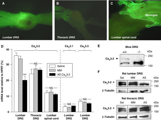
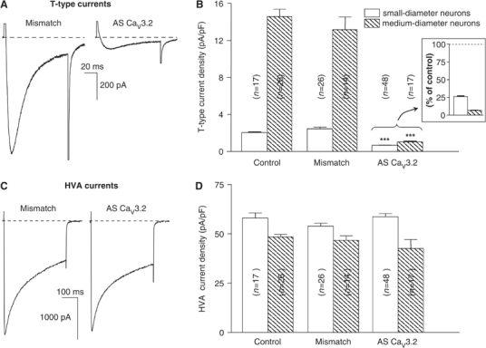
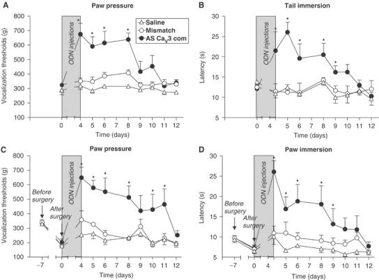
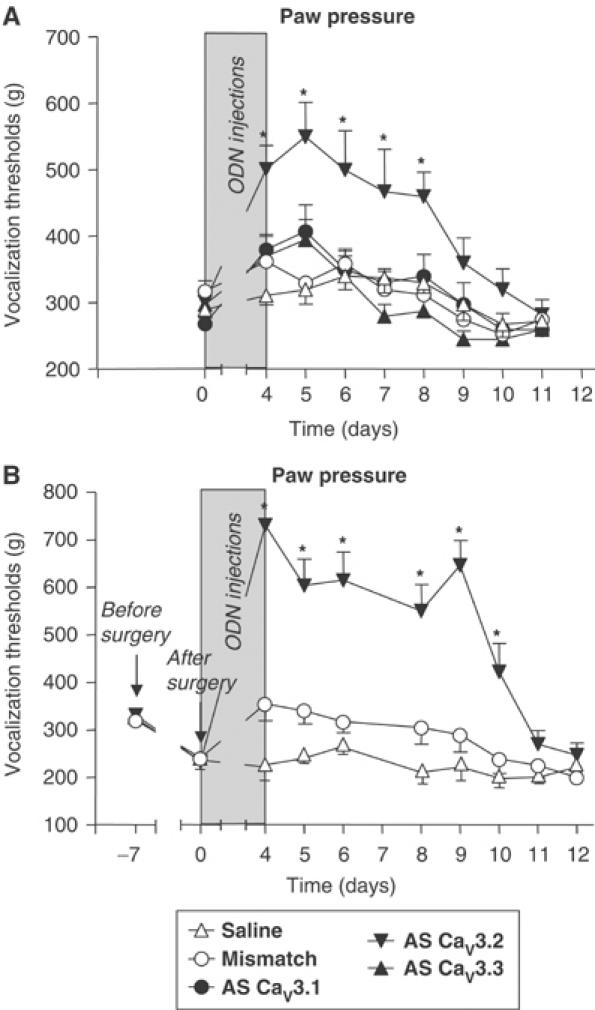
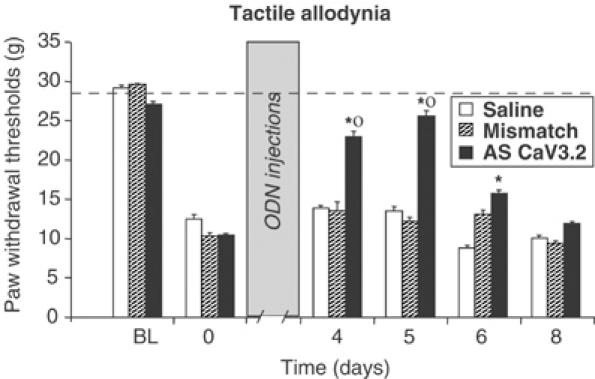
Analgesic therapies are still limited and sometimes poorly effective, therefore finding new targets for the development of innovative drugs is urgently needed. In order to validate the potential utility of blocking T-type calcium channels to reduce nociception, we explored the effects of intrathecally administered oligodeoxynucleotide antisenses, specific to the recently identified T-type calcium channel family (CaV3.1, CaV3.2, and CaV3.3), on reactions to noxious stimuli in healthy and mononeuropathic rats. Our results demonstrate that the antisense targeting CaV3.2 induced a knockdown of the CaV3.2 mRNA and protein expression as well as a large reduction of 'CaV3.2-like' T-type currents in nociceptive dorsal root ganglion neurons. Concomitantly, the antisense treatment resulted in major antinociceptive, anti-hyperalgesic, and anti-allodynic effects, suggesting that CaV3.2 plays a major pronociceptive role in acute and chronic pain states. Taken together, the results provide direct evidence linking CaV3.2 T-type channels to pain perception and suggest that CaV3.2 may offer a specific molecular target for the treatment of pain.


Figures

Figure 1
Figure 1
I.t. administration of CaV3.2-AS leads to a preferential uptake of AS in lumbar DRGs leading to a selective knockdown of CaV3.2 protein levels. After the last injection of AS-CaV3.2-FITC, bright fluorescence can be detected in freshly dissected lumbar DRG (A). In comparison, the thoracic DRGs did not show FITC staining (B). (C) In the lumbar enlargement near the ODN injection site, most of the FITC signal was retained in the meninges. The picture shows a dorsal view of the spinal cord with the meninges partially unfolded on the left side and still attached to the spinal cord on the right side. Note that a dotted line has been added to the border of the floating meninges to delineate the edge (C). Note that compared to the lumbar DRG, weaker levels of FITC fluorescence were seen on the spinal cord. (D) QRT–PCR analysis of CaV3.2 transcripts in lumbar and thoracic DRGs and in lumbar spinal cord of saline-, mismatch-, and AS-CaV3.2-treated rats (n=8 –11). Comparative analysis of CaV3.1 and CaV3.3 in lumbar DRGs of the same animal groups (n=3). (E) A rabbit Ab raised against a rat CaV3.2 fusion protein recognizes a 230 kDa band in DRG membranes (40 μg/lane) from WT but not from CaV3.2 KO mice (representative blot of three independent experiments). (F) Western blots of rat DRG membrane proteins with the anti-CaV3.2 Ab. Analysis of CaV3.2 in DRG extracts after saline, mismatch, or AS AS-CaV3.2 treatment. In each case, equal amounts of membrane proteins were loaded on 4–20% gradient polyacrylamide gels, subjected to SDS–PAGE, and transferred to nitrocellulose as described in Materials and methods. The apparent molecular size markers are indicated on the right. Blots are representative of four independent experiments. Note that the AS treatment induces a strong reduction of the CaV3.2 immunoreactivity in the lumbar DRGs (see arrowhead in (F)). Loading controls were provided by immunoblotting β-tubulin protein.
Figure 2
Figure 2
The AS treatment against the CaV3.2 subunit induces a reduction of T-type calcium currents in isolated lumbar DRG neurons. (A) Representative T-type calcium currents evoked by 100 ms long depolarizations from −100 to −40 mV in small nociceptive DRG neurons isolated from mismatch- or AS-CaV3.2-injected rats. Note the reduced size of the current in the AS-treated cell. (B) Averaged peak T-type current density (mean±s.e.m.) recorded from small- and medium-sized isolated neuron cell bodies from mismatch-, AS-, or saline-treated animals. Note that the AS treatment results in a highly significant (***P<0.001) knockdown of the T-type channel activity in both small- and medium-diameter neurons as illustrated in the inset. (C) Representative traces of ensemble HVA calcium currents evoked by 400 ms long depolarizations from −100 to +10 mV in nociceptive DRG neurons isolated from mismatch- or AS-CaV3.2-injected rats. (D) Averaged peak HVA current density (mean±s.e.m.) recorded from the same cells showed on the histogram of part B. Note that the current density levels were not altered by the treatment. Cells were prepared from four animals in each group.
Figure 3
Figure 3
Time course of the effect of i.t. injection of a generic AS to CaV3 subunits on mechanical and thermal nociception in healthy and mononeuropathic rats (CCI model). (A, B) Three groups (n=5–7) of animals were injected (i.t.) twice daily during 4 days with 10 μl containing either 12.5 μg of AS-CaV3-com (filled circles), 12.5 μg of the mismatch ODN (open circles), or the saline vehicle alone (open triangles). The rats were tested for nociceptive responses to a noxious pressure (A, paw pressure test) and to noxious thermal stimulus (B, tail immersion test). The response scale was measured in grams applied to the paw for the pressure test (vocalization thresholds) and in seconds for the latency of tail withdrawal from the water bath. Note that in both tests, the AS injection produced a 4-day significant antinociceptive effect. (C, D) For mononeuropathic rats, similarly, three groups of 5–7 animals were treated with AS-CaV3-com (filled circles), the mismatch ODN (open circles), and the saline vehicle (open triangles). Evoked mechanical (C) and thermal (D) hyperalgesia was scored using the paw pressure and the paw immersion tests. Thresholds were measured before (day −7) and after (day 0) the induction of neuropathy, then daily (days 4–12) after the end of the AS injection protocol. Note that the anti-hyperalgesic effect of AS-CaV3-com was prolonged in mononeuropathic animals compared to healthy rats. Each point represents the mean±s.e.m. from 5–7 animals per treatment group. Statistical differences between measures P<0.05 (*: AS-CaV3-com versus saline; •: AS-CaV3-com versus mismatch) were calculated by ANOVA followed by a protected least significant difference (PLSD) Fischer t-test.
Figure 4
Figure 4
Selective effects of a specific AS to CaV3.2 on mechanical nociception. (A) Five groups of animals (n=6) were injected (i.t.) twice daily during 4 days with 10 μl containing either 12.5 μg of AS-CaV3.1 (filled circles), 12.5 μg of AS-CaV3.2 (filled down-triangles), 12.5 μg of AS-CaV3.3 (filled up-triangles), 12.5 μg of the mismatch ODN (open circles), or the saline vehicle alone (open up-triangles). The rats were tested for nociceptive responses to pressure (paw pressure test). The response scale was measured in grams applied to the paw (vocalization thresholds). Note that only the AS-CaV3.2 AS injection produced a significant antinociceptive effect. (B) For mononeuropathic animals, three groups of seven animals were treated either with AS-CaV3.2 (filled down-triangles), the mismatch ODN (open circles), or the saline vehicle alone (open up-triangles). Evoked mechanical hyperalgesia was scored as in panel A. Vocalization thresholds were measured before (day −7) and after (day 0) the induction of neuropathy, and then daily (days 4–12) after the end of the AS injection protocol. Each point represents the mean±s.e.m. from 6–7 animals per treatment group. Statistical differences between measures P<0.05 (*: AS-CaV3.2 versus saline; ○: AS-CaV3.2 versus mismatch) were calculated by ANOVA followed by a PLSD Fischer t-test.
Figure 5
Figure 5
Effects of AS-CaV3.2 on tactile allodynia in mononeuropathic rats. Three groups of seven animals were injected (i.t.) twice daily during 4 days with 10 μl containing either 12.5 μg of AS-CaV3.2 (filled bars), 12.5 μg of the mismatch ODN (hatched bars), or the saline vehicle (open bars). Paw withdrawal thresholds were scored using the electronic von Frey Hair test. Thresholds were measured before injury (baseline values: BL) 14 days after the induction of neuropathy, before starting the injections (0), and then daily (days 4–8) after the end of the AS injection protocol. Each point represents the mean±s.e.m. from seven animals per treatment group. Statistical differences between measures P<0.05 (*: AS-CaV3.2 versus saline; o: AS-CaV3.2 versus mismatch) were calculated by ANOVA followed by a PLSD Fischer t-test.

References

    1. Akopian AN, Souslova V, England S, Okuse K, Ogata N, Ure J, Smith A, Kerr BJ, McMahon SB, Boyce S, Hill R, Stanfa LC, Dickenson AH, Wood JN (1999) The tetrodotoxin-resistant sodium channel SNS has a specialized function in pain pathways. Nat Neurosci 2: 541–548 - PubMed
    1. Andre S, Puech-Mallie S, Desmadryl G, Valmier J, Scamps F (2003) Axotomy differentially regulates voltage-gated calcium currents in mice sensory neurones. Neuroreport 14: 147–150 - PubMed
    1. Baccei ML, Kocsis JD (2000) Voltage-gated calcium currents in axotomized adult rat cutaneous afferent neurons. J Neurophysiol 83: 2227–2238 - PubMed
    1. Bao J, Li JJ, Perl ER (1998) Differences in Ca2+ channels governing generation of miniature and evoked excitatory synaptic currents in spinal laminae I and II. J Neurosci 18: 8740–8750 - PMC - PubMed
    1. Beedle AM, McRory JE, Poirot O, Doering CJ, Altier C, Barrere C, Hamid J, Nargeot J, Bourinet E, Zamponi GW (2004) Agonist-independent modulation of N-type calcium channels by ORL1 receptors. Nat Neurosci 7: 118–125 - PubMed

Publication types