. 2005 Jan 26;24(2):294-304.
doi: 10.1038/sj.emboj.7600510. Epub 2004 Dec 16.

Bidirectional signals transduced by DAPK-ERK interaction promote the apoptotic effect of DAPK

Affiliations

Bidirectional signals transduced by DAPK-ERK interaction promote the apoptotic effect of DAPK

Chun-Hau Chen et al. EMBO J. .

Abstract

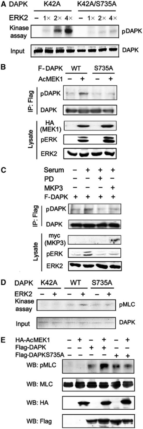
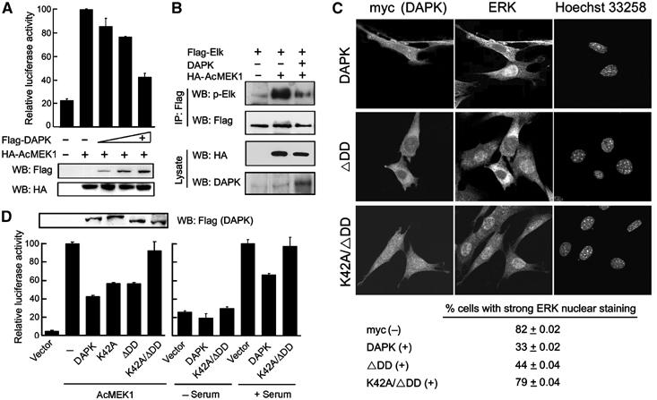
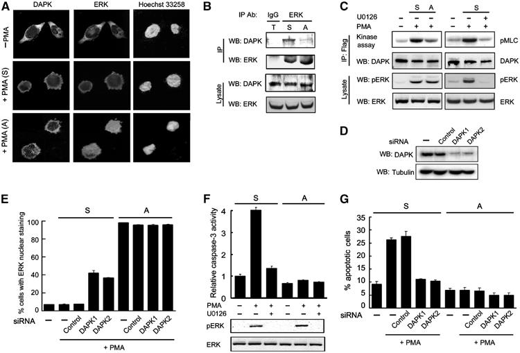
Death-associated protein kinase (DAPK) is a death domain-containing serine/threonine kinase, and participates in various apoptotic paradigms. Here, we identify the extracellular signal-regulated kinase (ERK) as a DAPK-interacting protein. DAPK interacts with ERK through a docking sequence within its death domain and is a substrate of ERK. Phosphorylation of DAPK at Ser 735 by ERK increases the catalytic activity of DAPK both in vitro and in vivo. Conversely, DAPK promotes the cytoplasmic retention of ERK, thereby inhibiting ERK signaling in the nucleus. This reciprocal regulation between DAPK and ERK constitutes a positive feedback loop that ultimately promotes the apoptotic activity of DAPK. In a physiological apoptosis system where ERK-DAPK interplay is reinforced, downregulation of either ERK or DAPK suppresses such apoptosis. These results indicate that bidirectional signalings between DAPK and ERK may contribute to the apoptosis-promoting function of the death domain of DAPK.


Figures

Figure 1
Figure 1
Identification of ERK1/2 as DAPK-binding proteins. (A) Yeast strain L40 cotransformed with Gal- and LexA-based fusion constructs was assayed for β-gal activity or His3 phenotype (−His). (B) In vitro-translated, 35S-labeled ERK1 or ERK2 was incubated with either GST or GST-DD. The pull-down products and 5% amount of input ERK1/2 were analyzed by autoradiography (upper panel). The lower panel indicates the equal input of GST and GST-DD in pull-down reactions. (C) 293T cells were transfected with ERK1 or ERK2, together with Flag-DAPK (F-DAPK) or a control plasmid (−). Cell lysates were subjected to immunoprecipitations followed by Western blot with antibodies as indicated. The amounts of ERK1/2 in cell lysates are shown on the bottom. As the ERK antibody used in this study detects ERK2 more efficiently than ERK1, a strong ERK2 band, corresponding to the endogenous ERK2, was seen in the ERK1 transfectants. (D) DAPK and ERK interact endogenously. Lysates of 293T cells were used for immunoprecipitations with control IgG or anti-ERK and followed by immunoblot with anti-DAPK or anti-ERK.
Figure 2
Figure 2
Mapping the ERK binding region in DAPK. (A) Lysates of 293T cells were incubated with GST-DAPK deletion mutants as illustrated in the upper panel. The pull-down products were assayed by Western blot with anti-ERK. The equal inputs of GST fusion proteins are shown on the bottom (input). (B) 293T cells transfected with various Flag-tagged DAPK constructs were subjected to immunoprecipitations and Western blot with antibodies as indicated. The amounts of ERK and Flag-DAPK proteins in lysates are shown on the bottom. (C) Alignment of the ERK docking sequences found in DAPK DD and several other ERK substrates. In the sequence of DAPK, the first amino acid listed is numbered. Conserved residues are highlighted. The feature of consensus D-domain sequence is indicated on the bottom; ϕ and X represent a hydrophobic amino acid and any amino acid, respectively. (D) GST fusion proteins immobilized on beads were incubated with equal amounts of 293 lysates. The pull-down products were analyzed by Western blot with anti-ERK2 antibody. The lower panel shows an equal input of the GST and GST fusion proteins in pull-down reactions.
Figure 3
Figure 3
ERK upregulates DAPK catalytic activity. (A) Serum-starved 293T cells transfected with Flag-DAPK and/or MKP3 were pretreated with 50 μM PD98059 and/or stimulated with or without serum for 15 min. Lysates with equal amounts of proteins were subjected to immunoprecipitations followed by in vitro kinase assays with MLC as a substrate or by Western blot with anti-Flag. Cell lysates were also used for Western blot analysis to detect the expression of various proteins (bottom panels). (B) Double reciprocal (Lineweaver–Burk) plots for analyzing the kinetic parameters of MLC phosphorylation catalyzed by DAPK or DAPKS735D isolated from transfected 293T cells at various conditions. The initial velocities (V0) were measured using 1 μg DAPK at increasing substrate concentrations, and kinetic analysis was performed as described in Materials and methods. (C) 293T cells transfected with various constructs were serum-starved and then lysed. DAPK kinase activity was assayed as in (A). The expression of various proteins in cell lysates is shown in the bottom panels.
Figure 4
Figure 4
ERK activates DAPK kinase activity through a direct phosphorylation at Ser 735. (A) DAPK proteins as indicated were purified from baculovirus and used as substrates in the in vitro kinase reactions with increasing amounts of purified and active ERK2. The reactions were analyzed by autoradiography (upper) or Coomassie blue staining (lower). (B, C) 293T cells transfected with plasmids as indicated were serum-starved (B), or pretreated with or without PD98059 (PD) and stimulated with or without serum for 15 min (C). Cell lysates were subjected to immunoprecipitations with anti-Flag followed by immunoblot with anti-DAPK or anti-phospho-DAPK(S735). The expression levels of various proteins in cell lysates are shown on the bottom. (D) In vitro phosphorylation of DAPK by ERK enhances the kinase activity of DAPK. Purified DAPK or its mutants immobilized on beads were subjected to in vitro kinase reactions with or without active ERK2 as described in Materials and methods. The phosphorylated and unphosphorylated DAPK or its mutants were assayed for their kinase activities using MLC as a substrate. [γ-32P]ATP was included only in the second kinase reaction. The final reaction products were analyzed by autoradiography (upper) or Coomassie blue staining (lower). (E) 293 cells transfected with plasmids as indicated were serum-starved and lysed. Cell lysates with equal amounts of proteins were used for Western blot with antibodies as indicated.
Figure 5
Figure 5
DAPK blocks ERK nuclear translocation and nuclear signaling. (A) DAPK blocks ERK signaling to Elk-1 activation. NIH3T3 cells were transfected with Gal4-Elk-1, Gal4-luciferase, pRK5β-Gal, and AcMEK1 (+) or a control vector (−), together with increasing amounts of Flag-DAPK or a control vector (−). Cells were serum-starved and lysed for luciferase and β-gal assays and Western blot analysis. (B) NIH3T3 cells transfected with plasmids as indicated were serum-starved. Cell lysates were subjected to immunoprecipitations and Western blot with antibodies as indicated. The levels of AcMEK1 or DAPK in cell lysates are shown on the bottom. (C) NIH3T3 cells transfected with myc-tagged DAPK or its mutants as indicated were serum-starved and then stimulated with serum for 2 h. Cells were triple stained with anti-myc, anti-ERK and Hoechst 33258, and visualized by confocal microscopy. The percent of cells showing stronger ERK nuclear staining than the cytoplasmic staining was quantitated and is listed on the bottom. The values shown are means±s.d. from three independent experiments, and at least 300 cells were counted for each population in each experiment. (D) NIH3T3 cells were cotransfected with Gal4-Elk-1, Gal4-luciferase, pRK5β-Gal, together with various DAPK constructs and/or AcMEK1. Transfectants were serum-starved or stimulated and then lysed for Elk-1 reporter assays as described in (A) and Western blot analysis (upper panel).
Figure 6
Figure 6
ERK activation stimulates the anoikis inducibility of DAPK. (A) NIH3T3 cells transfected with GFP together with various DAPK and AcMEK1 constructs were assayed by Western blot with antibodies as indicated. (B) Cells as in (A) were serum-starved and then assayed for their adhesion on fibronectin as described in Materials and methods. (C) NIH3T3 cells were cotransfected with p53-TA-luc and pRK5-βgal in the presence or absence of various DAPK constructs and AcMEK1, and then subjected to luciferase assays. (D, E) NIH3T3 cells transfected with various constructs were serum-starved and assayed for apoptosis using Cell Death-Detection ELISA. In (E), the expression levels of endogenous and exogenous DAPK are shown on the bottom. (F) DAPK or its mutants was introduced into MCF10A cells via retroviral-mediated gene transfer (bottom panels). Cells were seeded on plates coated with 25 ng/μl fibronectin and cultured in EGF-deprived medium for 16 h. Apoptosis was assayed as in (D).
Figure 7
Figure 7
DAPK and ERK are both required for apoptotic death of cells with elevated ERK–DAPK interaction. (A) D2 cells were seeded on culture dishes treated with (+PMA (A)) or without (−PMA) PMA in serum-free condition for 3 h, or seeded on hydrogel-coated dishes followed by PMA treatment (+PMA (S)). Cells were triple stained by anti-DAPK, anti-ERK and Hoechst 33258, and visualized by confocal microscopy. (B) PMA-treated attached (A) and suspended (S) D2 cells as described in (A) and 1:1 mixture of both populations (T) were used for immunoprecipitation followed by Western blot analyses with antibodies as indicated. ERK and DAPK expressions in each population are shown on the bottom. (C) PMA induces a higher DAPK activity in suspended cells. Flag-DAPK-transfected D2 cells plated as in (A) were pretreated with or without 20 μM U0126, and then exposed to PMA for 3 h. DAPK catalytic activity was assayed as in Figure 3. The levels of ERK and pERK in lysates are shown on the bottom. (D) D2 cells were transfected with or without (−) DAPK siRNA or control siRNA, together with a CD2 marker plasmid. Transfected cells were isolated as described in Materials and methods and lysed for Western blot analysis. (E) D2 cells transfected with or without various siRNAs together with GFP were cultured and treated as in (A). The percentage of GFP-positive cells with ERK nuclear staining was quantitated. (F) Suspended or attached D2 cells were pretreated with 20 μM U0126 and then exposed to PMA or DMSO (−) for 6 h. Cells were harvested for analyzing the activity of caspase-3 (upper panel) or the status of ERK activation (bottom panels). (G) Knockdown of endogenous DAPK protects against PMA-induced apoptosis in suspended cells. Suspended or attached D2 cells transfected as in (E) were treated with or without PMA for 6 h. Cells were assayed for apoptosis with Annexin V staining as described in Materials and methods.
Figure 8
Figure 8
Scheme depicting the promotion of apoptosis through a feedback regulatory circuit formed by DAPK–ERK interplay (see text).

References

    1. Adachi M, Fukuda M, Nishida E (2000) Nuclear export of MAP kinase (ERK) involves a MAP kinase kinase (MEK)-dependent active transport mechanism. J Cell Biol 148: 849–856 - PMC - PubMed
    1. Almeida EAC, Ilic D, Han Q, Hauck CR, Jin F, Kawakatsu H, Schlaepfer DD, Damsky CH (2000) Matrix survival signaling: from fibronectin via focal adhesion kinase to c-Jun NH2-terminal kinase. J Cell Biol 149: 741–754 - PMC - PubMed
    1. Aplin AE, Stewart SA, Assoian RK, Juliano RL (2001) Integrin-mediated adhesion regulates ERK nuclear translocation and phosphorylation of Elk-1. J Cell Biol 153: 273–282 - PMC - PubMed
    1. Ballif BA, Blenis J (2001) Molecular mechanisms mediating mammalian mitogen-activated protein kinase (MAPK) kinase (MEK)-MAPK cell survival signals. Cell Growth Differ 12: 397–408 - PubMed
    1. Bialik S, Brenick AR, Kimchi A (2004) DAP-kinase-mediated morphological changes are localization dependent and involve myosin-II phosphorylation. Cell Death Differ 11: 631–644 - PubMed

Publication types

Substances

LinkOut - more resources