. 2005 Jan 12;24(1):34-43.
doi: 10.1038/sj.emboj.7600490. Epub 2004 Dec 16.

Solution structure of choline binding protein A, the major adhesin of Streptococcus pneumoniae

Affiliations

Solution structure of choline binding protein A, the major adhesin of Streptococcus pneumoniae

Rensheng Luo et al. EMBO J. .

Abstract

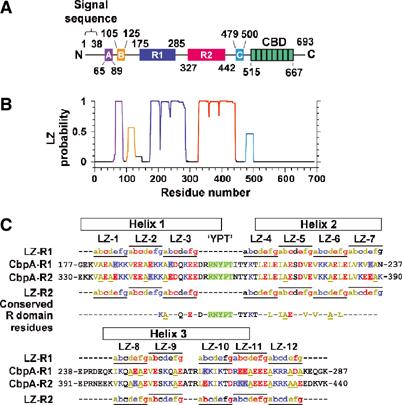
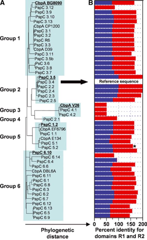
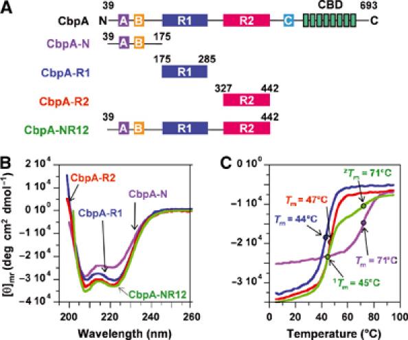
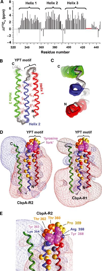
Streptococcus pneumoniae (pneumococcus) remains a significant health threat worldwide, especially to the young and old. While some of the biomolecules involved in pneumococcal pathogenesis are known and understood in mechanistic terms, little is known about the molecular details of bacterium/host interactions. We report here the solution structure of the 'repeated' adhesion domains (domains R1 and R2) of the principal pneumococcal adhesin, choline binding protein A (CbpA). Further, we provide insights into the mechanism by which CbpA binds its human receptor, polymeric immunoglobulin receptor (pIgR). The R domains, comprised of 12 imperfect copies of the leucine zipper heptad motif, adopt a unique 3-alpha-helix, raft-like structure. Each pair of alpha-helices is antiparallel and conserved residues in the loop between Helices 1 and 2 exhibit a novel 'tyrosine fork' structure that is involved in binding pIgR. This and other structural features that we show are conserved in most pneumococcal strains appear to generally play an important role in bacterial adhesion to pIgR. Interestingly, pneumococcus is the only bacterium known to adhere to and invade human cells by binding to pIgR.


Figures

Figure 1
Figure 1
Domain structure of CbpA (from the TIGR4 strain of S. pneumoniae). (A) Domains labeled A, B, R1, R2 and C exhibit multiple repeats of the LZ motif, and the domain labeled CBD (choline binding domain) contains eight repeats of the choline binding motif. (B) LZ probability for CbpA determined using COILS (Lupas et al, 1991). (C) Sequences of domains R1 and R2. The letters ag and horizontal lines indicate the locations of the 12 LZ heptad motifs. Within the LZ motifs, hydrophobic residues are colored brown (L, V, I and A), acidic residues red (D and E) and basic residues blue (K and R). Sites in the R1 and R2 sequences where an acidic residue is swapped for a basic residue, or vice versa, are indicated by red and blue shading, respectively. Residues of the conserved RNYPT motif are colored green and the three α-helices of CbpA-R2 are indicated by black rectangles. Residues that are conserved in 87 R domain sequences from 47 pneumococcal strains are also given.
Figure 2
Figure 2
R domain structure is conserved in CbpA sequences from most pneumococcal strains. (A) Phylogenetic tree showing the relationships between 47 CbpA sequences, which cluster into six groups. The TIGR4 sequence is also termed PspC 3.4 (marked by arrow). (B) The histogram illustrates the percentage of amino-acid identity (relative to TIGR4 CbpA) for the R1 and/or R2 domains (R1, blue bars; R2, red bars). Details of this analysis are found in Supplementary data.
Figure 3
Figure 3
Secondary structure of CbpA domains. (A) CbpA constructs used in this study. (B) CD spectra for CbpA-N (violet trace), -R1 (blue trace), -R2 (red trace) and -NR12 (green trace). (C) Thermal denaturation traces obtained by measuring CD ellipticity at 222 nm at different temperatures. The coloring scheme is as in (B).
Figure 4
Figure 4
Solution structure of CbpA R domains. (A) Secondary 13Cα chemical shift values for CbpA-R2 showing the three α-helices. Resonances for residues marked by red asterisks are unassigned. (B) Superposed ensemble of 20 lowest-energy structures of CbpA-R2 obtained from solution NMR data; the backbone and select hydrophobic residues (in brown color) at the two α-helix/α-helix interfaces are illustrated. Helix 1 is colored red, Helix 2 blue and Helix 3 green. The location of the YPT motif is noted. (C) End-on view of the three α-helices of domain R2, colored as in (B). (D) Contour maps of electrostatic potentials (±1kt; red, negative potential; blue, positive potential) for CbpA-R2 (left) and homology model of CbpA-R1 (right) generated using GRASP (Nicholls et al, 1991). The α-helices are colored as in (B). Tyr 358 and 363 (magenta; labeled ‘tyrosine fork'), Pro 359 (yellow) and Thr 360 and 362 (orange) are also illustrated. The Cα atoms of other conserved residues are illustrated as colored spheres (Lys 346, Arg 356 and Lys 364 (blue); Glu 352, Asp 354 and Glu 372 (red); Gln 350 and Thr 365 (gray); Ala 347, Ile 368, Ile 370, Ala 371, Val 375, Val 377, Ala 80 and Leu 382 (yellow)). (E) Close-up view of conserved residues in loop between Helix 1 and Helix 2 of CbpA-R2 that protrude into a region of neutral electrostatic potential. Key residues are illustrated as in (D).
Figure 5
Figure 5
The 800 MHz 2D 1H-15N TROSY spectra of 2H/13C/15N-labeled CbpA-R2 (A) and -NR12 (B). The individual spectra are illustrated in red (A) and green (B) ink. The blue overlays in panels A and B identify resonances that appear at identical positions in the two spectra.
Figure 6
Figure 6
Insights into the molecular mechanism of CbpA/pIgR interactions. (A) Histogram illustrating the association (ka) and dissociation (kd) rate constants derived from SPR data for different CbpA constructs interacting with immobilized sIgA. The rate constants were obtained by fitting equations for a 1:1 binding model to raw data like those illustrated in Supplementary Figure 2B. (B) Raw ITC data (top) and binding curves (bottom) for CbpA-R1 (blue), -R2 (red), -R12 (black) and -NR12 (green) binding to SC-D15. In the bottom panels, the colored circles show raw data points and the black lines show the fit of equations for a single binding site model to the raw data. Only every second data point is illustrated although all points were included in the analysis. The CbpA:SC-D15 mole ratios at the reaction end points are noted in each bottom panel. (C) Adhesion of pneumococci to NE (Detroit) cells. ΔCbpA pneumococci were transformed with pNE1 plasmids that encoded full-length CbpA or CbpA with one or two mutations within the YPT motif. ΔCbpA (control) corresponds to results for untransformed ΔCbpA bacteria.

References

    1. Brunger AT, Adams PD, Clore GM, DeLano WL, Gros P, Grosse-Kunstleve RW, Jiang JS, Kuszewski J, Nilges M, Pannu NS, Read RJ, Rice LM, Simonson T, Warren GL (1998) Crystallography & NMR system: a new software suite for macromolecular structure determination. Acta Crystallogr D 54: 905–921 - PubMed
    1. Butler JC, Breiman RF, Campbell JF, Lipman HB, Broome CV, Facklam RR (1993) Pneumococcal polysaccharide vaccine efficacy. An evaluation of current recommendations. JAMA 270: 1826–1831 - PubMed
    1. Case DA, Darden TA, Cheatham ITE, Simmerling CL, Wang J, Duke RE, Luo R, Merz KM, Wang B, Pearlman DA, Crowley M, Brozell S, Tsui V, Gohlke H, Mongan J, Hornak V, Cui G, Beroza P, Schafmeister C, Caldwell JW, Ross WS, Kollman PA (2004) AMBER 8. University of California, San Francisco, http://amber.scripps.edu/doc8/amber 8.pdf
    1. Cavanagh J, Fairbrother WJ, Palmer AG III, Skelton NJ (1996) Protein NMR Spectroscopy. New York: Academic Press
    1. Chemical_Computing_Group MOE version 2004.02. Montreal, Quebec, Canada

Publication types

MeSH terms

Substances

Associated data