

Review
doi: 10.1023/b:jofl.0000031824.48401.5c.
Advances in surface-enhanced fluorescence
Affiliations
- PMID: 15617385
- PMCID: PMC2763917
- DOI: 10.1023/b:jofl.0000031824.48401.5c
Item in Clipboard
Review
Advances in surface-enhanced fluorescence
J Fluoresc.
2004 Jul.
Abstract
We report recent achievements in metal-enhanced fluorescence from our laboratory. Several fluorophore systems have been studied on metal particle-coated surfaces and in colloid suspensions. In particular, we describe a distance dependent enhancement on silver island films (SIFs), release of self-quenching of fluorescence near silver particles, and the applications of fluorescence enhancement near metalized surfaces to bioassays. We discuss a number of methods for various shaped silver particle deposition on surfaces.
Figures
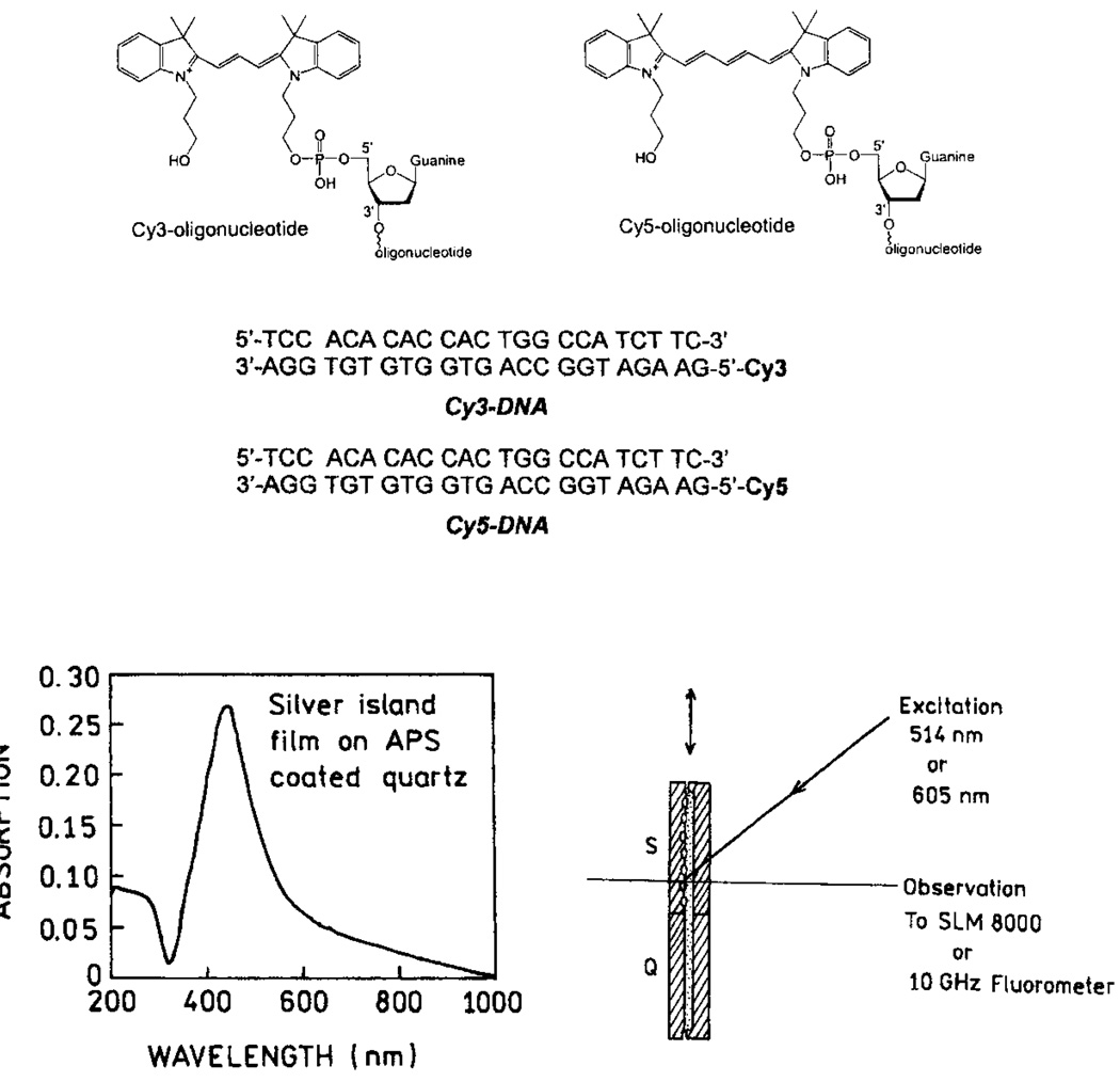
DNA structures and sample geometry. Structures and sequences of the labeled and unlabeled DNA oligomers (top). Absorption spectrum of silver islands on APS and experimental geometry (bottom).
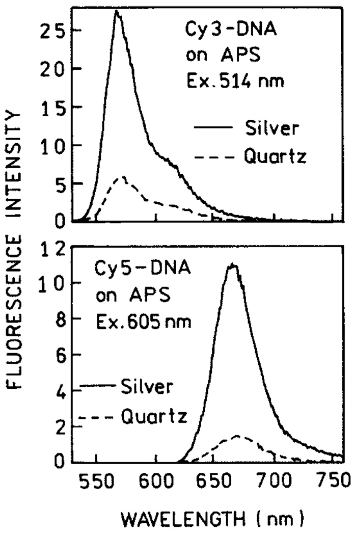
Emission spectra of Cy3-DNA (top) and Cy5-DNA (bottom) with and without silver island films.

Frequency-domain intensity decays of Cy3-DNA and Cy5-DNA with (●) and without (○) SIFs.
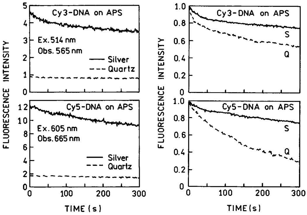
Photostability of Cy3-DNA and Cy5-DNA on APS-treated slides, with and without silver island films, for the same incident power (left), and the excitation intensity adjusted to yield the same emission intensities on quartz and silver (right). Figure 1–Figure 4 are adopted from [24].
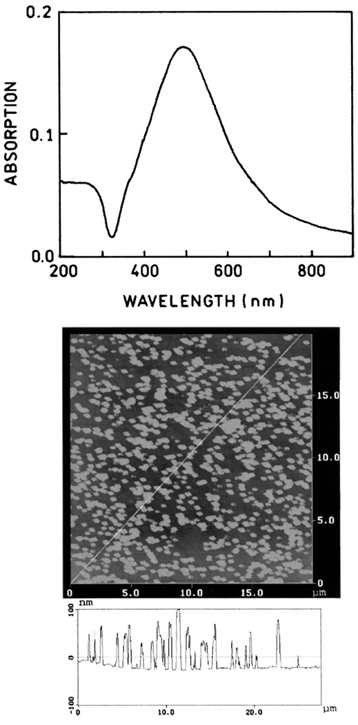
Absorption spectrum (top) and AFM image (bottom) of a representative SIFs.
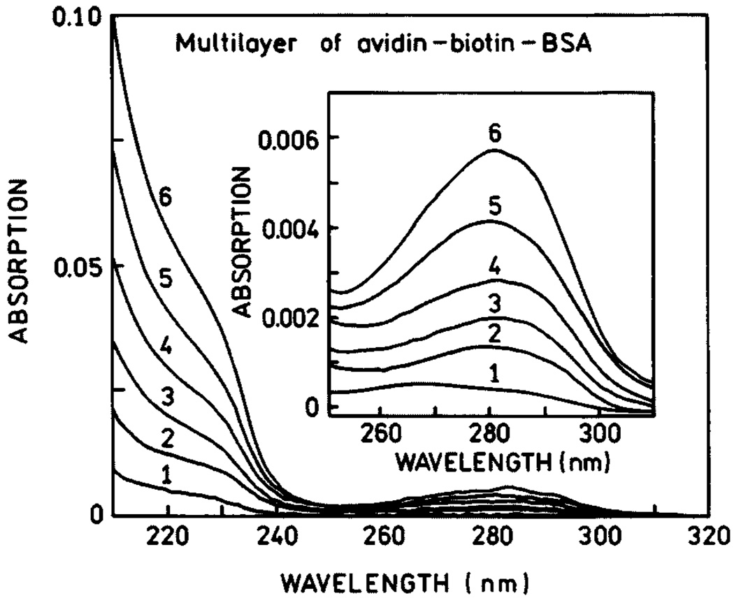
Absorption spectra of BSA-biotin-avidin layers.
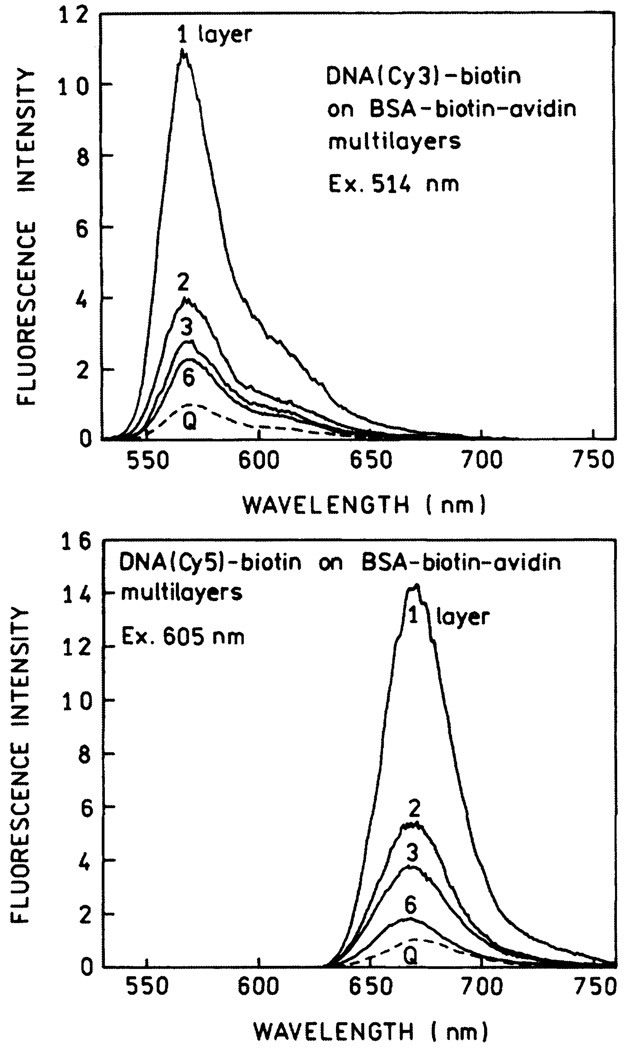
Emission spectra of DNA(Cy3)-biotin (top) and DNA(Cy5)-biotin (bottom) on BSA-biotin-avidin layers. Spectra are normalized to the spectrum on quartz (Q) with the same numbers of protein layers.
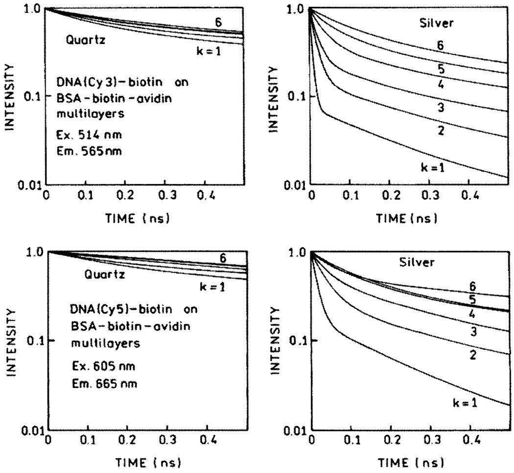
Time-domain representation of emission intensity decays of DNA(Cy3)-biotin (top) and DNA(Cy5)-biotin (bottom) on BSA-biotin-avidin multilayers deposited on quartz (left) and SIFs (right).
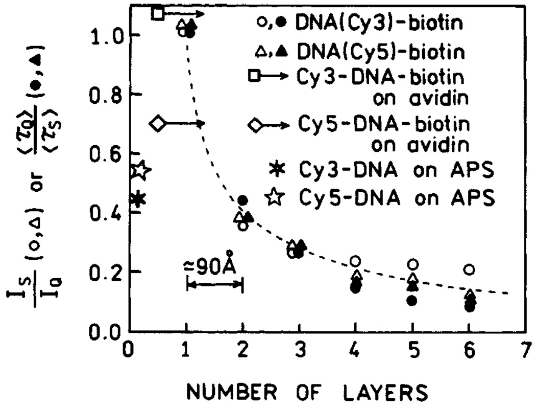
Fluorescence enhancements of Cy3- and Cy5-labeled oligomers for various distances from the silver surface. Also shown are the enhancements found on amine-coated slides which were treated with APS and slides with a single layer of avidin. Figure 5–Figure 9 are adopted from [31].
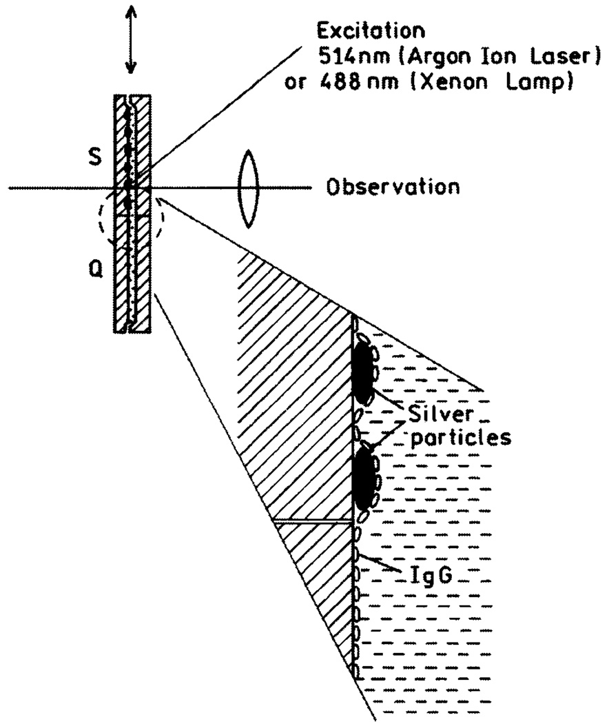
Experimental setup used in release of self-quenching study.
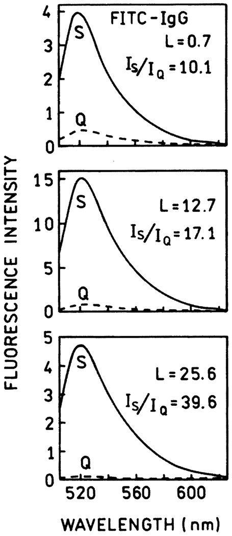
Emission spectra of Fl-IgG on quartz (---) and silver (—) for L = 0.7, 12.7 and 25.6. Excitation was 488 nm.
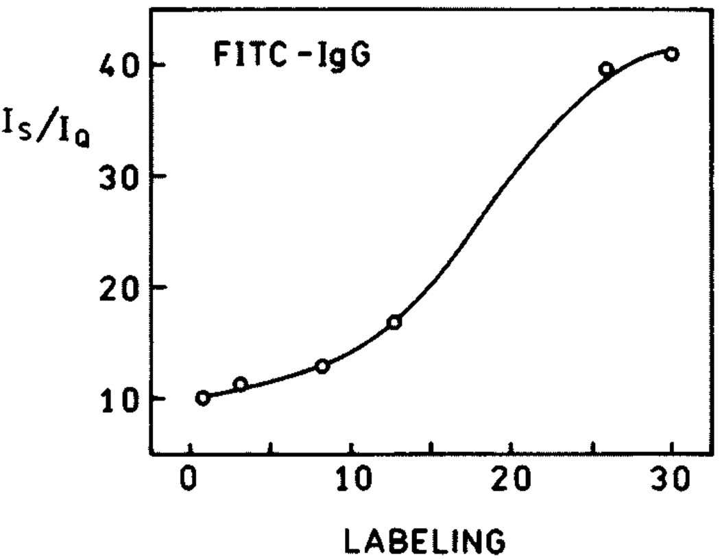
The dependence of brightness enhancement on labeling.
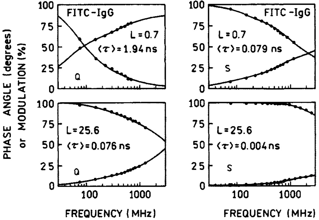
Frequency-domain intensity decays of Fl-IgG on quartz (left) and silver (right).
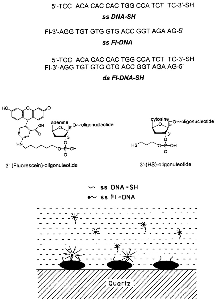
Structures of DNA oligomers. The lower panel shows schematic of the oligomers bound to silver particles.
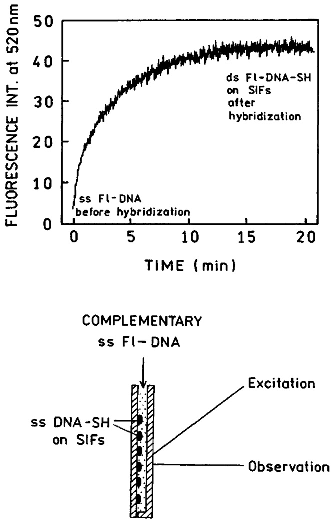
Time-dependent hybridization of ss Fl-DNA to ss DNA-SH. The lower panel shows the sample configuration.
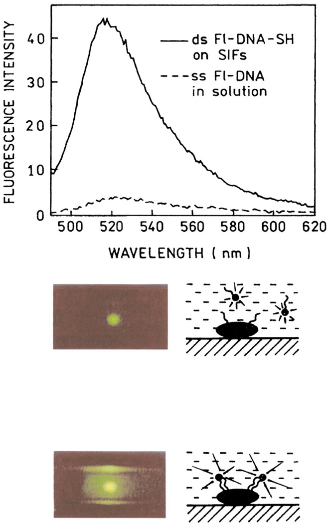
Emission spectra of ss Fl-DNA in solution (dashed line) and bound (solid line) to silver particles. Roughly the same number of molecules of ss Fl-DNA and ds Fl-DNA-SH was in the illuminated area. The lower panel shows photographs of ss Fl-DNA in solution (top) and ds Fl-DNA-SH on SIFs (bottom). Figure 14–Figure 16 are adopted from [58].
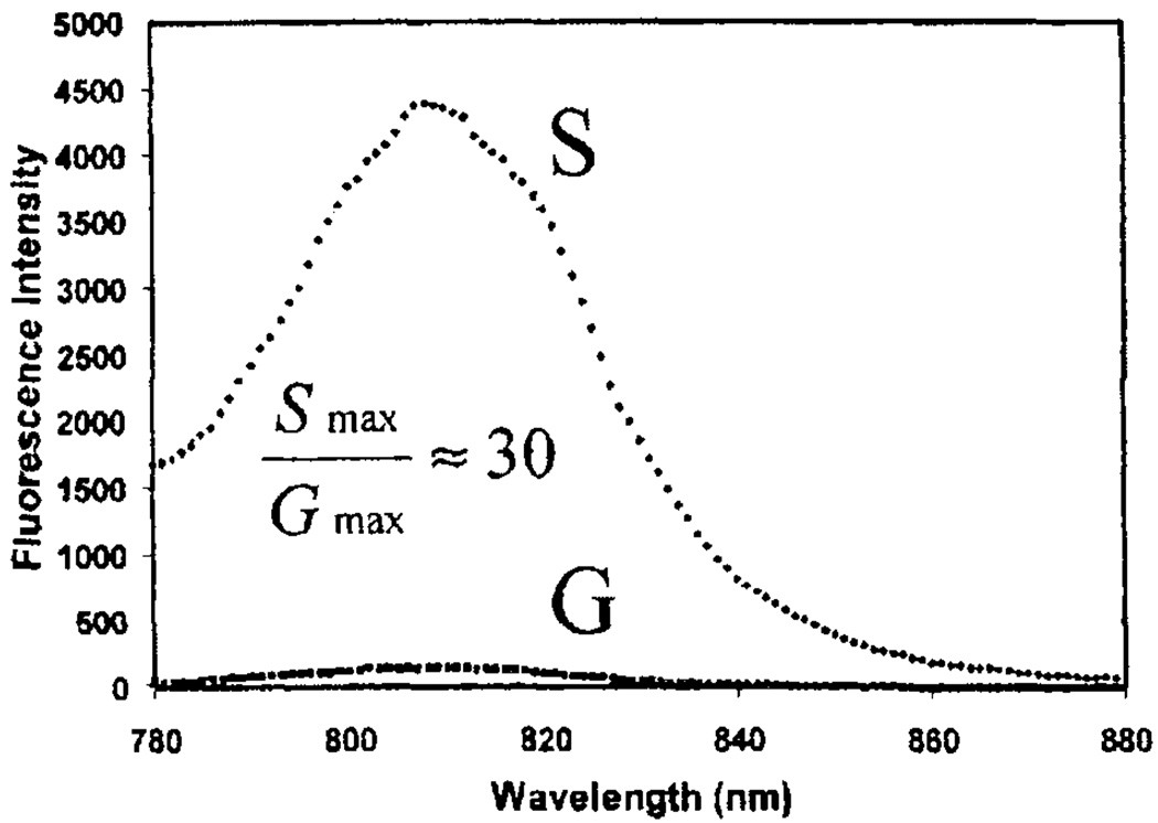
Fluorescence intensity of HSA-ICG coated glass, G, and silver colloids, S, Exc = 760 nm. Adopted from [66].
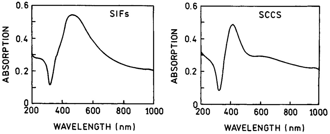
Absorption spctra of SIFs (left) and SCCS (right).
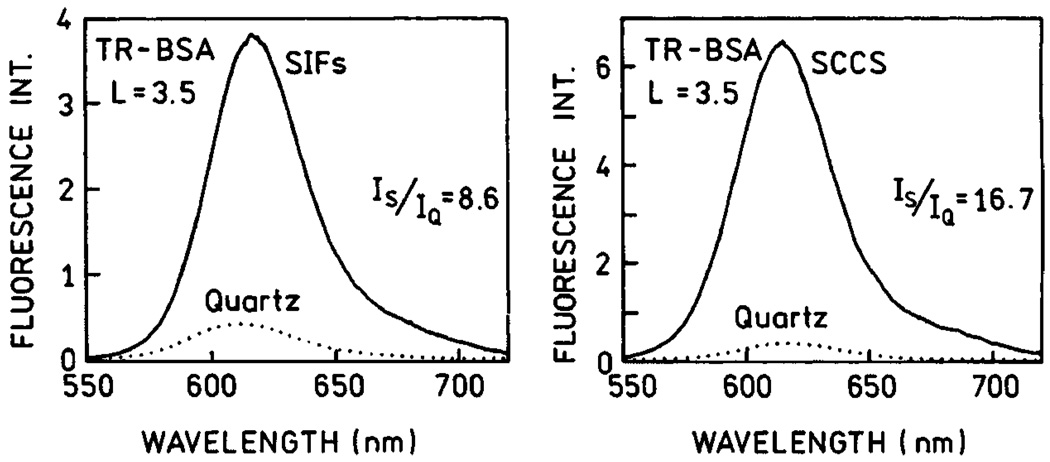
Emission spectra of Texas Red-labeled BSA (TR-BSA) measured on SIFs (left) and SCCS (right).
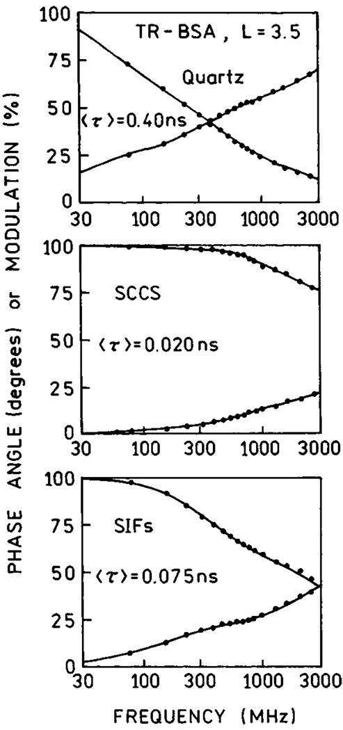
Frequency-domain lifetimes of TR-BSA on quartz (top), SCCS (middle) ans SIFs (bottom).
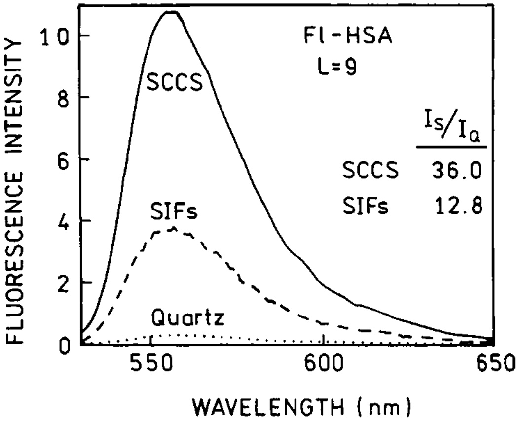
Emission spectra of highly labeled Fl-HSA measured on quartz (dotted line), SIFs (dashed line) and SCCS (solid line). Figure 18–Figure 21 are adopted from [67].
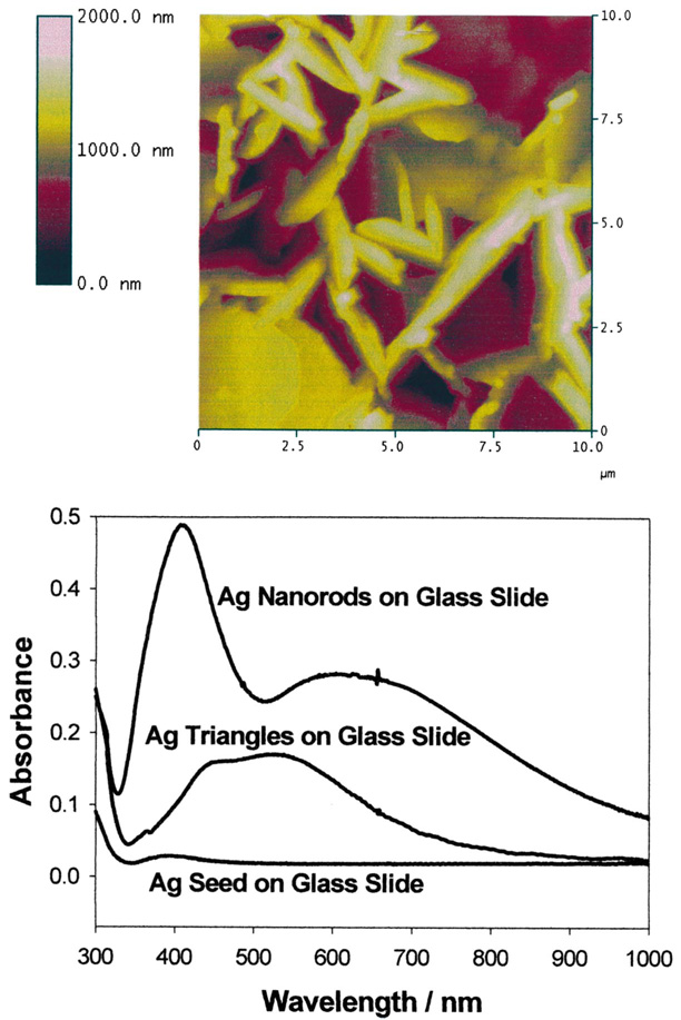
AFM image of silver nanorods deposited on glass substrate (top) and absorption spectra of silver nanorods, triangles and seeds deposited on glass substrate (bottom).
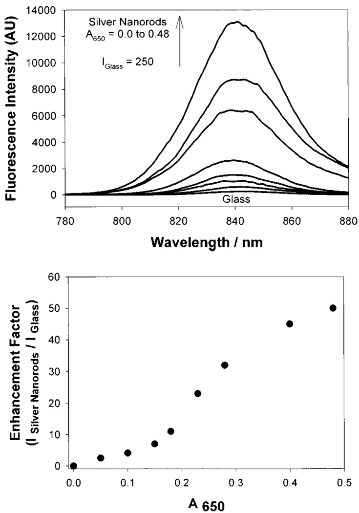
Fluorescence emission intensity of HSA-ICG on silver nanorods (top) and enhnacement factor vs the absorption of silver nanorods measured at 650 nm.
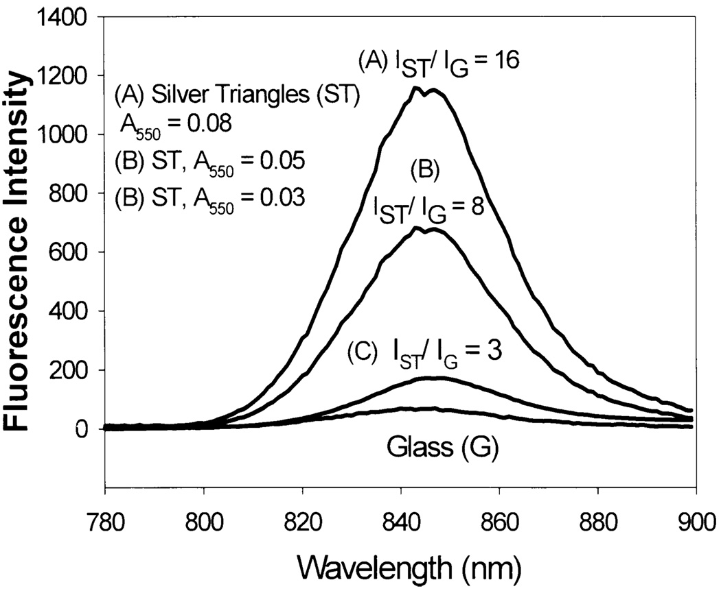
Fluorescence emission intensity of HSA-ICG on silver triangles.
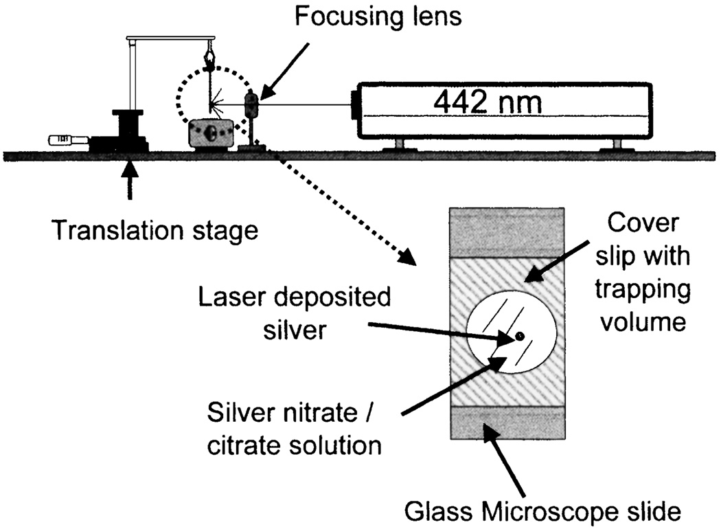
Experimental setup for laser deposition of silver on APS coated glass microscope slides. Adopted from [70].
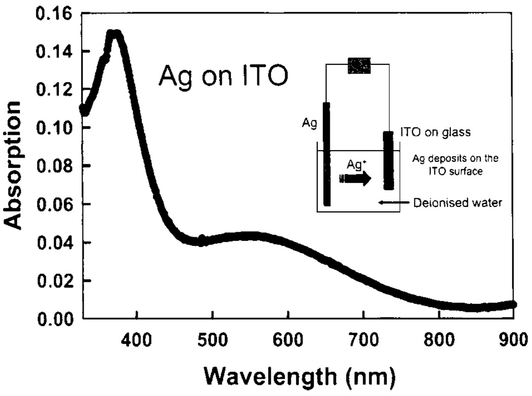
Absorption spectrum of silver-coated ITO produced via electrochemical deposition. ITO without silver was in the reference beam. The insert shows the scheme for electrochemical deposition of Ag on an ITO surface.
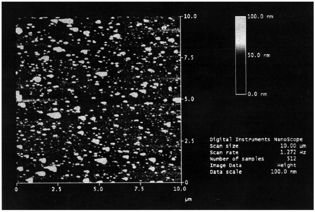
AFM image of the silver coated ITO surface produced by electrolysis. Figure 26–Figure 27 are adopted from [71].
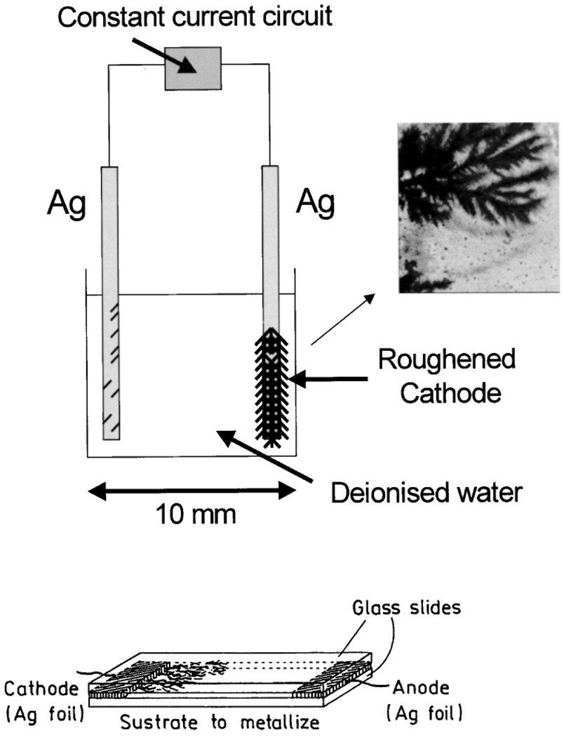
Constant current apparatus for electrode silver fractal growth (top). Silver growth on glass slide (bottom).
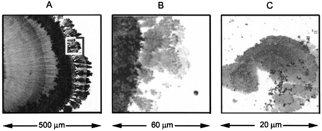
Silver nanostructures deposited on glass during electroplating (A). Panels B and C are consecutive magnification of the marked area on panel A. Bright field image.
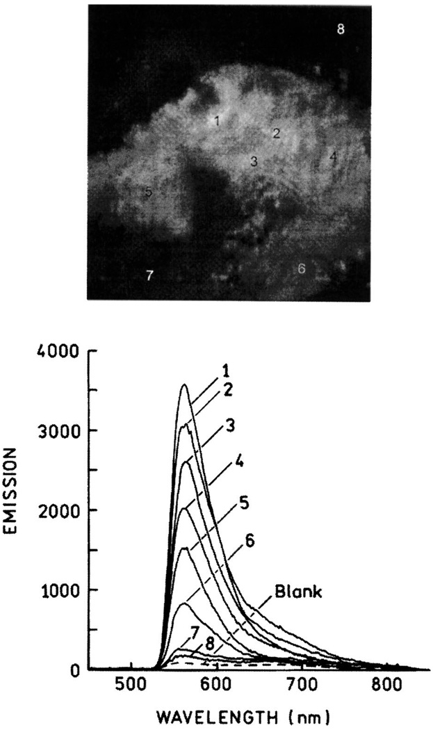
Fluorescence image of Fl-HSA deposited on the silver structure shown in Fig. 29C (top). Emission spectra of the numbered areas shown above (bottom). Figure 29–Figure 30 are adopted from [74].
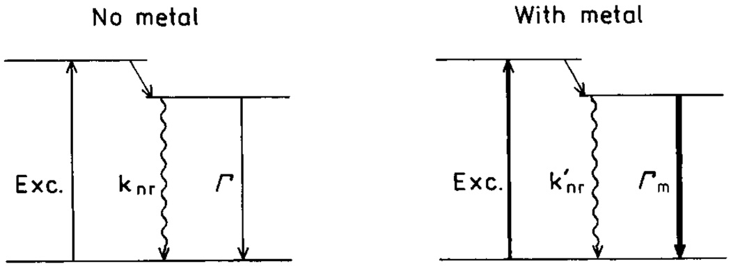
Jablonski diagram for molecules in absence (left) and presence (right) of metal particles.
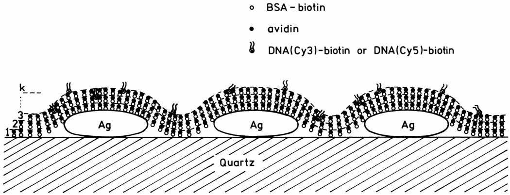
Schematic of BSA-avidin monolayers with labeled DNA.
References
-
- Drexhage KH. In: Progress in Optics. Wolfe E, editor. Amsterdam: North-Holland; 1974. pp. 161–323.
-
- Weitz DA, Garoff S, Hansen CD, Gramila TJ. Fluorescent lifetimes of molecules on silver-island films. Opt. Lett. 1982;7(2):89–91. - PubMed
-
- Aussenegg FR, Leitner A, Lippitsch ME, Reinish H, Reigler M. Novel aspects of fluorescence lifetime for molecules positioned close to metal surfaces. Surf. Sci. 1987;139:935–945.
-
- Leitner A, Lippitsch ME, Draxler S, Reigler M, Aussenegg FR. Fluorescence properties of dyes absorbed to silver islands, investigated by picosecond techniques. Appl. Phys. B. 1985;36:106–109.
-
- Gersten J, Nitzan A. Spectroscopic properties of molecules interacting with small dielectric particles. J. Chem. Phys. 1981;75(3):1139–1152.
Publication types
MeSH terms
Substances
Grants and funding
LinkOut - more resources
Full Text Sources
Other Literature Sources