. 2005 Jan;73(1):494-503.
doi: 10.1128/IAI.73.1.494-503.2005.

Differences in patterns of infection and inflammation for corticosteroid treatment and chemotherapy in experimental invasive pulmonary aspergillosis

Affiliations

Differences in patterns of infection and inflammation for corticosteroid treatment and chemotherapy in experimental invasive pulmonary aspergillosis

Viviane Balloy et al. Infect Immun. 2005 Jan.

Abstract

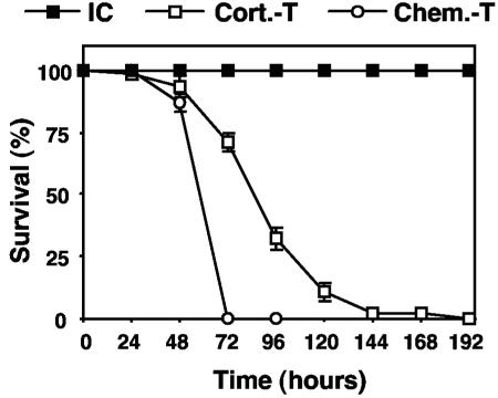
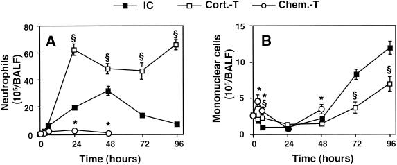
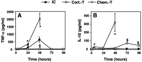
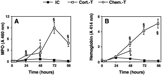
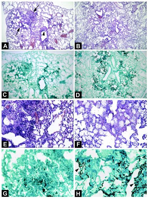
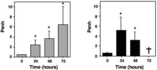
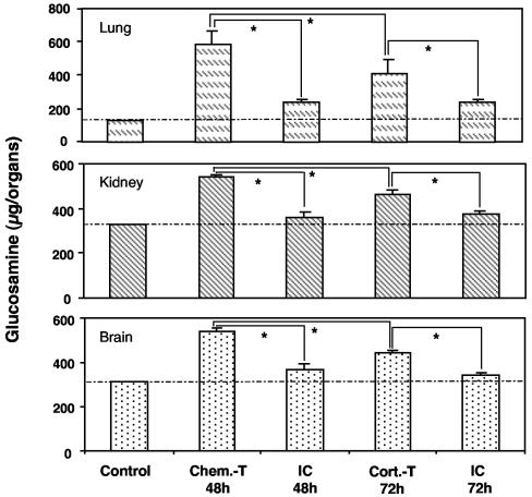
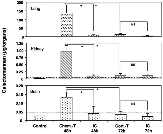
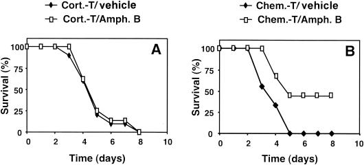
Aspergillus fumigatus causes invasive pulmonary aspergillosis (IPA). This disease is one of the most life-threatening opportunistic infections in immunocompromised patients. The type of immunosuppressive regimen under which IPA occurs has rarely been investigated. In this study, we evaluated various parameters of the innate immune response during the progression of murine IPA induced by the intratracheal administration of A. fumigatus conidia as a function of two immunosuppressive treatments: a corticosteroid and a chemotherapeutic agent. We compared host responses various times after infection in terms of survival, pulmonary production of pro- and anti-inflammatory cytokines, cellular trafficking in the airways, lung injury, respiratory distress, and fungal development. We found that IPA pathogenesis involved predominantly fungal development in mice treated by chemotherapy and an adverse host response in mice treated with a corticosteroid. These previously unrecognized differences should be taken into account in evaluations of the pathogenesis of IPA in animal models.


Figures

FIG. 1.
FIG. 1.
Survival of immunocompetent and immunosuppressed mice after intratracheal infection with conidia of A. fumigatus. Corticosteroid-treated (Cort.-T) mice were subjected to immunosuppression by administration of cortisone acetate (10 mg/mouse) 3 days before infection, on the day of infection, and on days 2 and 4 after infection. Chemotherapy-treated (Chem.-T) mice were subjected to immunosuppression by administration of vinblastine (5 mg/kg) 66 h before infection. Control immunocompetent (IC) mice were left untreated. Ten animals per group were infected with 107 conidia in four independent experiments. The values presented are the means and SEMs for 40 mice. The difference between the survival of corticosteroid-treated mice and the survival of chemotherapy-treated mice was statistically significant (P < 0.001), with median survival times of 4.13 ± 0.16 and 2.88 ± 0.05 days, respectively.
FIG. 2.
FIG. 2.
Leukocyte counts in BALF of immunocompetent and immunosuppressed mice infected with A. fumigatus. Mice were killed, and their lungs were washed out at 3, 6, 24, 48, 72, and 96 h after infection with 107 conidia. Neutrophils (A) and mononuclear cells (B) were counted as described in Materials and Methods. Abbreviations are as defined in the legend to Fig. 1. The values presented are the means and SEMs for at least five mice. Section signs and asterisks indicate statistically significant differences (P < 0.05) between corticosteroid- and chemotherapy-treated mice and immunocompetent mice, respectively.
FIG. 3.
FIG. 3.
TNF-α and IL-10 concentrations in BALF of immunocompetent and immunosuppressed mice infected with A. fumigatus. TNF-α (A) and IL-10 (B) concentrations in BALF were determined at 3, 6, 24, 48, 72, and 96 h after infection with 107 conidia as described in Materials and Methods. Abbreviations are as defined in the legend to Fig. 1. The values presented are the means and SEMs for at least five mice. Section signs and asterisks indicate statistically significant differences (P < 0.05) between corticosteroid- and chemotherapy-treated mice and immunocompetent mice, respectively.
FIG. 4.
FIG. 4.
MPO and hemoglobin levels in BALF of immunocompetent and immunosuppressed mice infected with A. fumigatus. MPO (A) and hemoglobin (B) levels in BALF were determined at 3, 6, 24, 48, 72, and 96 h after infection with 107 conidia as described in Materials and Methods. Abbreviations are as defined in the legend to Fig. 1. The values presented are the means and SEMs for at least five mice. Section signs and asterisks indicate statistically significant differences (P < 0.05) between corticosteroid- and chemotherapy-treated mice and immunocompetent mice, respectively.
FIG. 5.
FIG. 5.
Lung histological features of corticosteroid- and chemotherapy-treated mice infected with A. fumigatus. (A, C, E, and G) Lung sections from a corticosteroid-treated mouse 72 h after intratracheal inoculation with 107 conidia. (A) Hematoxylin-eosin stain; magnification, ×60. (E) Hematoxylin-eosin stain; magnification, ×250. A large focus of pneumonia (A, arrows) and exudative bronchiolitis (A, arrowhead) with the destruction of bronchi and alveolae can be seen. Hemorrhagic necrosis and numerous inflammatory cells, especially PMN, also can be seen (E). (C) Methenamine silver stain; magnification, ×60. (G) Methenamine silver stain; magnification, ×400. A few conidia and very few hyphae within the lesions of necrosis can be seen. (B, D, F, and H) Lung sections from a chemotherapy-treated mouse 48 h after intratracheal inoculation with 107 conidia. (B) Hematoxylin-eosin stain; magnification, ×60. (F) Hematoxylin-eosin stain; magnification, ×250. Lesions of bronchiolitis and diffuse pneumonia with edema and congestion within alveolae (B) but without an inflammatory exudate of any cell type, including PMN, can be seen. (D) Methenamine silver stain; magnification, ×60. (H) Methenamine silver stain; magnification, ×400. Several foci of bronchocentric necrosis containing numerous hyphae (H, arrowheads) can be seen. Invasion of alveolae by numerous hyphae of A. fumigatus also can be seen. Note the absence of an inflammatory exudate.
FIG. 6.
FIG. 6.
Measurement of respiratory distress in corticosteroid-treated (left) or chemotherapy-treated (right) mice infected with A. fumigatus. Penh was determined in conscious mice just before infection (time zero) and every day after infection with 107 conidia until the death of the animals. The values presented are the means and SEMs for at least five mice. A dagger indicates that no chemotherapy-treated mice survived to day 3. Asterisks indicate statistically significant differences (P < 0.05) between various time points and time zero.
FIG. 7.
FIG. 7.
Chitin determination in lungs, kidneys, and brains of immunocompetent and immunosuppressed mice infected with A. fumigatus. Organs of corticosteroid- and chemotherapy-treated mice were collected 72 and 48 h, respectively, after infection with 107 conidia. Chitin content is expressed in glucosamine equivalents. Abbreviations are as defined in the legend to Fig. 1. The values presented are the means and SEMs for at least five mice. Asterisks indicate statistically significant differences (P < 0.05) between the various values. The dotted line indicates the mean of the background values obtained with uninfected control mice.
FIG. 8.
FIG. 8.
Galactomannan determination in the lungs, kidneys, and brains of immunocompetent and immunosuppressed mice infected with A. fumigatus. Organs of corticosteroid- and chemotherapy-treated mice were collected 72 and 48 h, respectively, after infection with 107 conidia. Abbreviations are as defined in the legend to Fig. 1. The values presented are the means and SEMs for at least five mice. Asterisks indicate statistically significant differences (P < 0.05) between the various values; NS, not significant. The dotted line indicates the background value (see Materials and Methods) below which the various means were considered negative.
FIG. 9.
FIG. 9.
Effect of antifungal treatment on the survival of immunosuppressed mice infected with A. fumigatus. Corticosteroid-treated (A) and chemotherapy-treated (B) mice were treated 24 h after infection and every 24 h for 5 days with intravenous amphotericin B (1 mg/kg) (Amph. B). The survival of 10 animals per group was monitored after infection with 3 × 10⁶ conidia. Abbreviations are as defined in the legend to Fig. 1. One experiment representative of two is shown. The effect of the antifungal agent was statistically significant (P < 0.02) only for the chemotherapy-treated group.

Similar articles

Cited by

References

    1. Balloy, V., J. M. Sallenave, B. Crestani, M. Dehoux, and M. Chignard. 2003. Neutrophil DNA contributes to the antielastase barrier during acute lung inflammation. Am. J. Respir. Cell Mol. Biol. 28:746-753. - PubMed
    1. Becker, M. J., S. de Marie, M. H. Fens, H. A. Verbrugh, and I. A. Bakker-Woudenberg. 2003. Effect of amphotericin B treatment on kinetics of cytokines and parameters of fungal load in neutropenic rats with invasive pulmonary aspergillosis. J. Antimicrob. Chemother. 52:428-434. - PubMed
    1. Berenguer, J., M. C. Allende, J. W. Lee, K. Garrett, C. Lyman, N. M. Ali, J. Bacher, P. A. Pizzo, and T. J. Walsh. 1995. Pathogenesis of pulmonary aspergillosis. Granulocytopenia versus cyclosporin and methylprednisolone-induced immunosuppression. Am. J. Respir. Crit. Care Med. 152:1079-1086. - PubMed
    1. Brieland, J. K., C. Jackson, F. Menzel, D. Loebenberg, A. Cacciapuoti, J. Halpern, S. Hurst, T. Muchamuel, R. Debets, R. Kastelein, T. Churakova, J. Abram, R. Hare, and A. O'Garra. 2001. Cytokine networking in lungs of immunocompetent mice in response to inhaled Aspergillus fumigatus. Infect. Immun. 69:1554-1560. - PMC - PubMed
    1. Cenci, E., A. Mencacci, C. F. d'Ostiani, G. Del Sero, P. Mosci, C. Montagnoli, A. Bacci, and L. Romani. 1998. Cytokine- and T helper-dependent lung mucosal immunity in mice with invasive pulmonary aspergillosis. J. Infect. Dis. 178:1750-1760. - PubMed

MeSH terms