. 2005 Jan;73(1):523-31.
doi: 10.1128/IAI.73.1.523-531.2005.

Mucosal FOXP3-expressing CD4+ CD25high regulatory T cells in Helicobacter pylori-infected patients

Affiliations

Mucosal FOXP3-expressing CD4+ CD25high regulatory T cells in Helicobacter pylori-infected patients

Anna Lundgren et al. Infect Immun. 2005 Jan.

Abstract

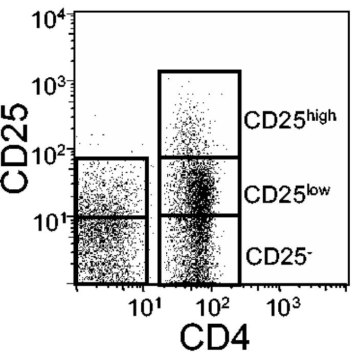
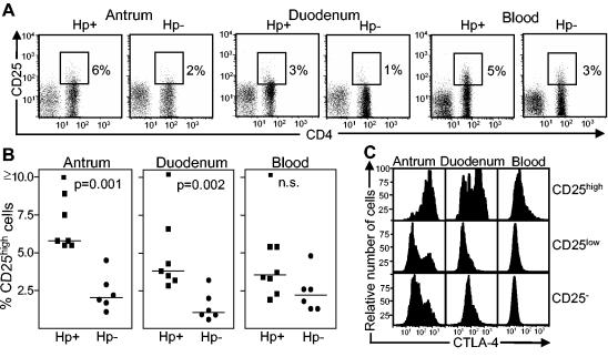
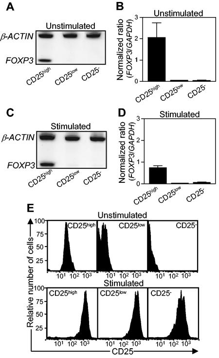
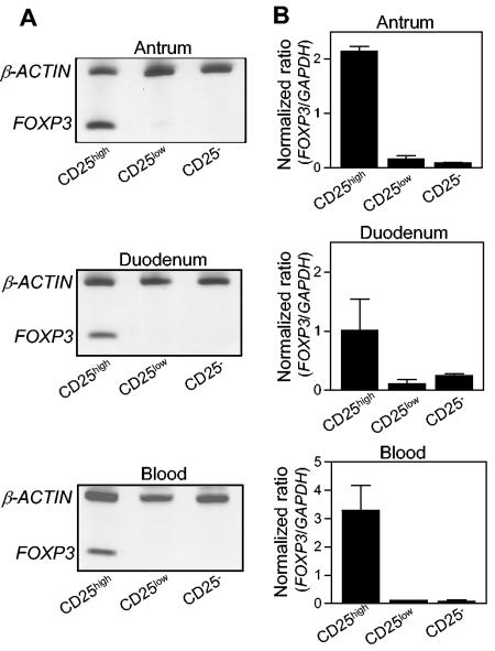
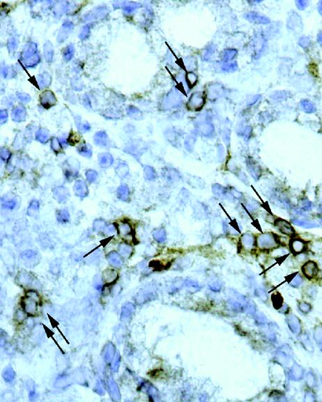
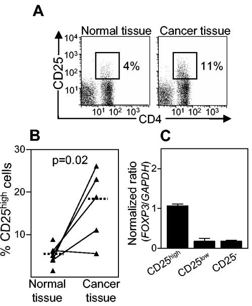
Helicobacter pylori chronically colonizes the stomach and duodenum and causes peptic ulcers or gastric adenocarcinoma in 10 to 20% of infected individuals. We hypothesize that the inability of patients to clear H. pylori infections is a consequence of active suppression of the immune response. Here we show that H. pylori-infected individuals have increased frequencies of CD4(+) CD25(high) T cells in both the stomach and duodenal mucosa compared to uninfected controls. These cells have the phenotype of regulatory T cells, as they express FOXP3, a key gene for the development and function of regulatory T cells, as well as high levels of the cytotoxic T lymphocyte-associated antigen 4 (CTLA-4) protein. In contrast, mucosal CD4(+) CD25(low) and CD4(+) CD25(-) cells express little FOXP3 mRNA and low levels of the CTLA-4 protein. Mucosal CD4(+) CD25(high) T cells are present in individuals with asymptomatic H. pylori infections as well as in duodenal ulcer patients. The frequencies of CD4(+) CD25(high) cells are also increased in the stomachs of H. pylori-infected patients with gastric adenocarcinoma, particularly in cancer-affected tissues. These findings suggest that regulatory T cells may suppress mucosal immune responses and thereby contribute to the persistence of H. pylori infections.


Figures

FIG. 1.
FIG. 1.
Gating approach for discrimination of CD25high, CD25low, and CD25 cells. Cells were labeled with antibodies against CD4, CD8, and CD25 and then analyzed by FCM. For this example, the CD25 expression on CD4+ and CD8+ T cells from the antral mucosa of an H. pylori-positive individual was analyzed in a single plot. The gates for the CD25high and CD25low populations were set by comparing the CD25 expression levels of CD4+ and CD8+ cells, as described in Materials and Methods.
FIG. 2.
FIG. 2.
Expression of CD25 and CTLA-4 on T cells isolated from the antral and duodenal lamina propriae and peripheral blood of H. pylori-positive asymptomatic individuals and H. pylori-negative controls. (A) CD25 expression detected by FCM on CD4+ and CD8+ T cells. CD25high cells were gated as indicated in the figure, and the frequencies of CD4+ cells expressing CD25high are shown by the numbers outside the gates. The results shown are for one representative experiment of seven experiments with H. pylori-positive individuals and six experiments with H. pylori-negative individuals. (B) Presence of CD4+ CD25high cells in the antral and duodenal lamina propriae and peripheral blood of H. pylori-positive and -negative individuals. Each symbol represents the percentage of CD25high cells among the CD4+ lymphocytes for one individual, and median values are indicated by horizontal bars. (C) Intracellular expression of CTLA-4 on CD4+ CD25high, CD25low, and CD25 cells from an H. pylori-positive individual. The results shown are from one representative experiment of three total experiments. Five thousand or more CD4+ cells and ≥250 CD4+ CD25high cells were analyzed for each sample.
FIG. 3.
FIG. 3.
Expression of FOXP3 and CD25 by CD4+ T cells from peripheral blood. Peripheral blood T cells were sorted into CD4+ CD25high, CD4+ CD25low, and CD4+ CD25 cell fractions, and the expression of FOXP3 and CD25 before and after stimulation with anti-CD3 and anti-CD28 antibodies was determined. (A and C) FOXP3 mRNA as well as mRNA for the housekeeping gene β-actin was detected by RT-PCR, and the PCR products were visualized in silver-stained polyacrylamide gels. Results for one representative experiment of three total experiments are shown. (B and D) Expression levels of FOXP3 were determined by real-time PCR and were related to the level of the housekeeping gene GAPDH. The values shown are arithmetic means plus standard errors of the means (SEM) (n = 3). (E) CD25 expression on sorted cells, as determined by FCM. The results shown are from one representative experiment of three total experiments.
FIG. 4.
FIG. 4.
FOXP3 expression by CD4+ T cells isolated from antral and duodenal lamina propriae and peripheral blood from asymptomatic H. pylori-positive individuals. T cells were sorted into CD4+ CD25high, CD4+ CD25low, and CD4+ CD25 cell fractions, and the expression of FOXP3 mRNA was determined. (A) FOXP3 mRNA as well as mRNA for the housekeeping gene β-actin was detected by RT-PCR, and the PCR products were visualized in silver-stained polyacrylamide gels. Results for one experiment of two total experiments are shown. (B) Expression levels of FOXP3 were determined by real-time PCR and were related to the level of the housekeeping gene GAPDH. The values shown are arithmetic means plus SEM (n = 2).
FIG. 5.
FIG. 5.
CD25 expression in the antrum of an H. pylori-positive asymptomatic individual, as detected by IHC. Cryosections were stained for CD25 and analyzed under a microscope. Arrows indicate CD25+ MNCs. Original magnification, ×400.
FIG. 6.
FIG. 6.
Expression of CD25 and FOXP3 by lamina propria T cells isolated from histologically normal and cancer-affected stomach mucosae of H. pylori-positive gastric adenocarcinoma patients. (A) CD25 expression on gated CD4+ and CD8+ T cells. The results shown are from one representative experiment each of seven experiments with healthy tissue and five experiments with cancer tissue. (B) Presence of CD4+ CD25high cells in healthy and cancer-affected stomach mucosae. Each symbol represents the percentage of CD25high cells among the CD4+ lymphocytes from one individual, and median values are indicated by horizontal bars. For FCM, ≥3,500 CD4+ cells and ≥150 CD4+ CD25high cells were analyzed for each sample. (C) Expression levels of FOXP3 in sorted CD4+ CD25high, CD4+ CD25low, and CD4+ CD25 cells isolated from histologically normal stomach mucosa were determined by real-time PCR and then related to the level of the housekeeping gene GAPDH. The values shown are arithmetic means plus SEM (n = 2).

References

    1. Aandahl, E. M., J. Michaelsson, W. J. Moretto, F. M. Hecht, and D. F. Nixon. 2004. Human CD4+ CD25+ regulatory T cells control T-cell responses to human immunodeficiency virus and cytomegalovirus antigens. J. Virol. 78:2454-2459. - PMC - PubMed
    1. Baecher-Allan, C., J. A. Brown, G. J. Freeman, and D. A. Hafler. 2001. CD4+CD25high regulatory cells in human peripheral blood. J. Immunol. 167:1245-1253. - PubMed
    1. Belkaid, Y., C. A. Piccirillo, S. Mendez, E. M. Shevach, and D. L. Sacks. 2002. CD4+CD25+ regulatory T cells control Leishmania major persistence and immunity. Nature 420:502-507. - PubMed
    1. Cao, D., V. Malmstrom, C. Baecher-Allan, D. Hafler, L. Klareskog, and C. Trollmo. 2003. Isolation and functional characterization of regulatory CD25bright CD4+ T cells from the target organ of patients with rheumatoid arthritis. Eur. J. Immunol. 33:215-223. - PubMed
    1. Chen, W., W. Jin, N. Hardegen, K. J. Lei, L. Li, N. Marinos, G. McGrady, and S. M. Wahl. 2003. Conversion of peripheral CD4+CD25− naive T cells to CD4+CD25+ regulatory T cells by TGF-beta induction of transcription factor Foxp3. J. Exp. Med. 198:1875-1886. - PMC - PubMed

Publication types

MeSH terms