. 2005 Jan;73(1):552-62.
doi: 10.1128/IAI.73.1.552-562.2005.

Cytolethal distending toxin from Shiga toxin-producing Escherichia coli O157 causes irreversible G2/M arrest, inhibition of proliferation, and death of human endothelial cells

Affiliations

Cytolethal distending toxin from Shiga toxin-producing Escherichia coli O157 causes irreversible G2/M arrest, inhibition of proliferation, and death of human endothelial cells

Martina Bielaszewska et al. Infect Immun. 2005 Jan.

Abstract

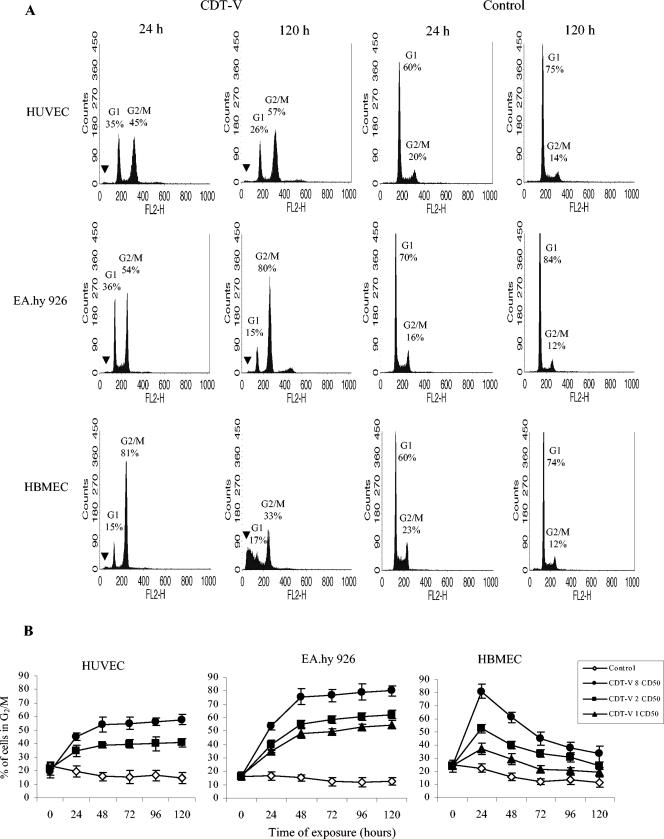
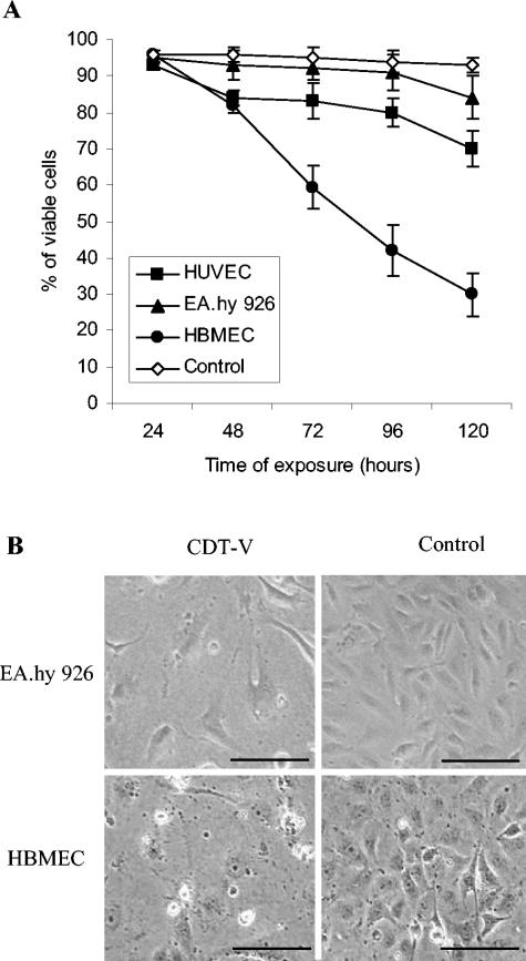
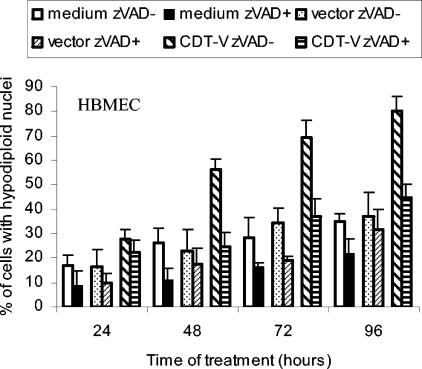
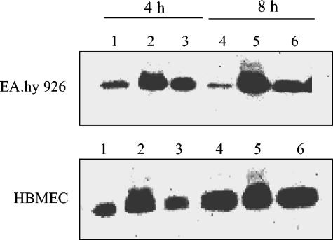
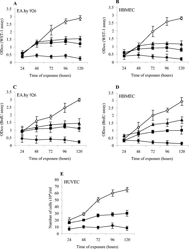
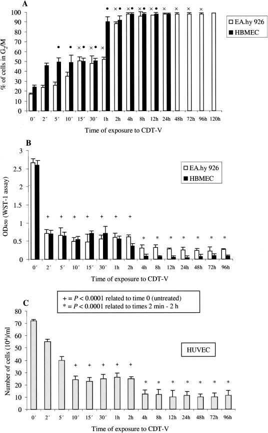
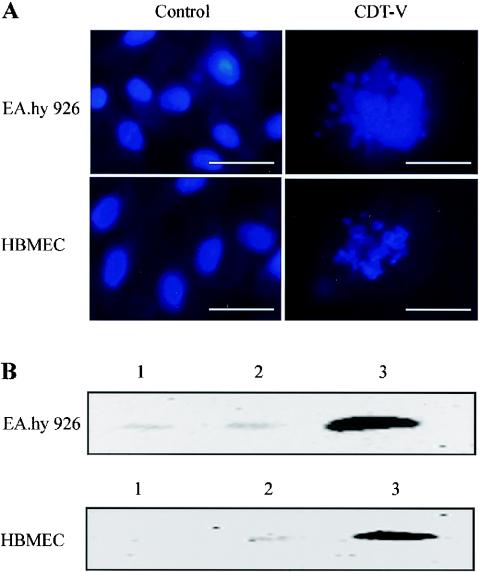
Recently, cytolethal distending toxin V (CDT-V), a new member of the CDT family, was identified in Shiga toxin-producing Escherichia coli (STEC) O157 and particular non-O157 serotypes. Here we investigated the biological effects of CDT-V from STEC O157:H(-) (strain 493/89) on human endothelial cells, which are believed to be major pathogenetic targets in severe STEC-mediated diseases. CDT-V caused dose-dependent G(2)/M cell cycle arrest leading to distension, inhibition of proliferation, and death in primary human umbilical vein endothelial cells (HUVEC) and two endothelial cell lines, EA.hy 926 cells (HUVEC derived) and human brain microvascular endothelial cells (HBMEC). The cell cycle effects of CDT-V were cell type specific. In HUVEC and EA.hy 926 cells, CDT-V caused a slowly developing but persistent G(2)/M block which resulted in delayed nonapoptotic cell death. In contrast, in HBMEC, CDT-V induced a rapidly evolving but transient G(2)/M block which was followed by progressive, mostly apoptotic cell death. In both HBMEC and EA.hy 926 cells, G(2)/M arrest was preceded by the early accumulation of a phosphorylated inactive form of cdc2 kinase. Significant G(2)/M arrest and inhibition of proliferation in both HUVEC and each of the endothelial cell lines were induced by 2 to 15 min of exposure to CDT-V, indicating that the effects of the toxin are irreversible. CDT-V-treated HBMEC and EA.hy 926 cells displayed fragmented nuclei and expressed phosphorylated histone protein H2AX, indicative of DNA damage followed by a DNA repair response. Our data demonstrate that CDT-V causes irreversible damage to human endothelial cells and thus may contribute to the pathogenesis of STEC-mediated diseases.


Figures

FIG. 1.
FIG. 1.
CDT-V causes G2/M arrest in human endothelial cells. (A) Flow cytometric analysis of HUVEC, EA.hy 926 cells, and HBMEC after 1 and 5 days of treatment with 8 CD50 of CDT-V/ml or with the control preparation. The proportions of cells in G1 (2n DNA) and G2/M (4n DNA) are indicated. Black arrowheads indicate the sub-G1 population. Data represent one of three independent experiments. (B) HUVEC, EA.hy 926 cells, and HBMEC were treated with 1 (triangles), 2 (squares), or 8 (circles) CD50 of CDT-V/ml or with the control preparation (diamonds) for the indicated times. The proportions of cells in G2/M were determined by flow cytometry. Time zero indicates basal numbers of cells in G2/M, which were obtained by exposing the cells to CDT-V or the control preparation for 5 min, followed by immediate flow cytometric analysis. Data are means and standard deviations from three independent experiments.
FIG. 2.
FIG. 2.
G2/M arrest of endothelial cells results in cell distension and ultimately cell death. (A) Viability of cells arrested in G2/M. Cells analyzed for G2/M arrest were stained with trypan blue. Proportions of viable cells were determined at each time point microscopically. Numbers of viable control cells did not differ significantly in the three cell cultures and are shown combined as “Control.” Data are means and standard deviations from three independent experiments. (B) Photomicrographs of EA.hy 926 cell and HBMEC cultures exposed for 5 and 4 days, respectively, to 8 CD50 of CDT-V/ml or to the control preparation. CDT-V caused the formation of giant cells that were four- to eightfold larger than control cells and had nuclei that were two- to threefold larger. Magnification, ×100; bars, 200 μm.
FIG. 3.
FIG. 3.
CDT-V induces apoptotic cell death in HBMEC. Cells were incubated with 8 CD50 of CDT-V/ml, the vector preparation, or cell culture medium in the absence or the presence of zVAD-fmk (50 μM). Apoptosis was determined by flow cytometry as a proportion of hypodiploid nuclei. Data are means and ranges from two experiments.
FIG. 4.
FIG. 4.
CDT-V induces the accumulation of phosphorylated, inactive cdc2 kinase. EA.hy 926 cells and HBMEC were treated with 8 CD50 of CDT-V/ml (lanes 2 and 5), the vector preparation (lanes 1 and 4), or nocodazole (lanes 3 and 6) for 4 h (lanes 1 to 3) or 8 h (lanes 4 to 6). Isolated total cellular proteins were analyzed by immunoblotting with anti-cdc2 antibody. The strong signals displayed by cells treated with CDT-V (lanes 2 and 5) were double bands which were not completely separated on the gels.
FIG. 5.
FIG. 5.
CDT-V inhibits the proliferation of human endothelial cells. EA.hy 926 cells (A and C), HBMEC (B and D), and HUVEC (E) were treated with 1 (triangles), 2 (squares), or 8 (circles) CD50 of CDT-V/ml or with the vector preparation (diamonds) for the indicated times. Cells then were processed in the WST-1 (A and B), BrdU (C and D), and trypan blue (E) assays. Data are means and standard deviations from three independent experiments.
FIG. 6.
FIG. 6.
Minimum length of exposure to CDT-V required for G2/M arrest and inhibition of proliferation in endothelial cells. Cells were exposed to CDT-V (8 CD50/ml) for the indicated times, the toxin then was removed, and cells were washed and incubated in medium without CDT-V. G2/M arrest (A) was analyzed by flow cytometry 24 h (HBMEC) or 120 h (EA.hy 926 cells) after the addition of CDT-V. The percentages of the total numbers of cells arrested in G2/M after 24 h (HBMEC) or 120 h (EA.hy 926 cells) are shown, and the exposure times which resulted in G2/M arrest in significant proportions (≥50%) of EA.hy 926 cells (×) and HBMEC (•) also are shown. Cell proliferation was measured by the WST-1 assay (EA.hy 926 cells and HBMEC) (B) or trypan blue exclusion (HUVEC) (C) 96 h after the addition of the toxin. Cells at 0 min were not exposed to CDT-V and were cultured in medium only. Differences in mean optical densities at 450 nm (OD450) (EA.hy 926 cells and HBMEC) and cell numbers (HUVEC) were compared with the paired t test, and the P values are shown. All data are means and standard deviations from three independent experiments.
FIG. 7.
FIG. 7.
CDT-V induces nuclear fragmentation and the formation of γ-H2AX in endothelial cells. (A) DAPI staining of EA.hy 926 cells and HBMEC treated for 5 days with the vector preparation or with 8 CD50 of CDT-V/ml. A Zeiss fluorescence microscope with a ×40 objective lens was used; magnification, ×400; bars, 50 μm. (B) Immunoblot analysis with a γ-H2AX-specific monoclonal antibody of EA.hy 926 cells and HBMEC treated for 21 h with 8 CD50 of CDT-V/ml (lanes 3) or the vector preparation (lanes 2) or left untreated (lanes 1).

References

    1. Aragon, V., K. Chao, and L. A. Dreyfus. 1997. Effect of cytolethal distending toxin on F-actin assembly and cell division in Chinese hamster ovary cells. Infect. Immun. 65:3774-3780. - PMC - PubMed
    1. Bantel, H., B. Sinha, W. Domschke, G. Peters, K. Schulze-Osthoff, and R. U. J.änicke. 2001. α-Toxin is a mediator of Staphylococcus aureus-induced cell death and activates caspases via the intrinsic death pathway independently of death receptor signaling. J. Cell Biol. 155:637-648. - PMC - PubMed
    1. Bielaszewska, M., and H. Karch. 2000. Non-O157:H7 Shiga toxin (verocytotoxin)-producing Escherichia coli strains: epidemiological significance and microbiological diagnosis. World J. Microbiol. Biotechnol. 16:711-718.
    1. Bielaszewska, M., M. Fell, L. Greune, R. Prager, A. Fruth, H. Tschäpe, M. A. Schmidt, and H. Karch. 2004. Characterization of cytolethal distending toxin genes and expression in Shiga toxin-producing Escherichia coli strains of non-O157 serogroups. Infect. Immun. 72:1812-1816. - PMC - PubMed
    1. Bitzan, M., and D. M. W. M. Te Loo. 2003. Interaction of Shiga toxin with endothelial cells. Methods Mol. Med. 73:243-262. - PubMed

Publication types

LinkOut - more resources