. 2005 Mar 4;280(9):7729-38.
doi: 10.1074/jbc.M409056200. Epub 2004 Dec 23.

Phorbol 12-myristate 13-acetate induces epidermal growth factor receptor transactivation via protein kinase Cdelta/c-Src pathways in glioblastoma cells

Affiliations

Phorbol 12-myristate 13-acetate induces epidermal growth factor receptor transactivation via protein kinase Cdelta/c-Src pathways in glioblastoma cells

Samson Amos et al. J Biol Chem. .

Abstract

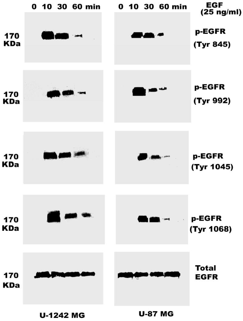
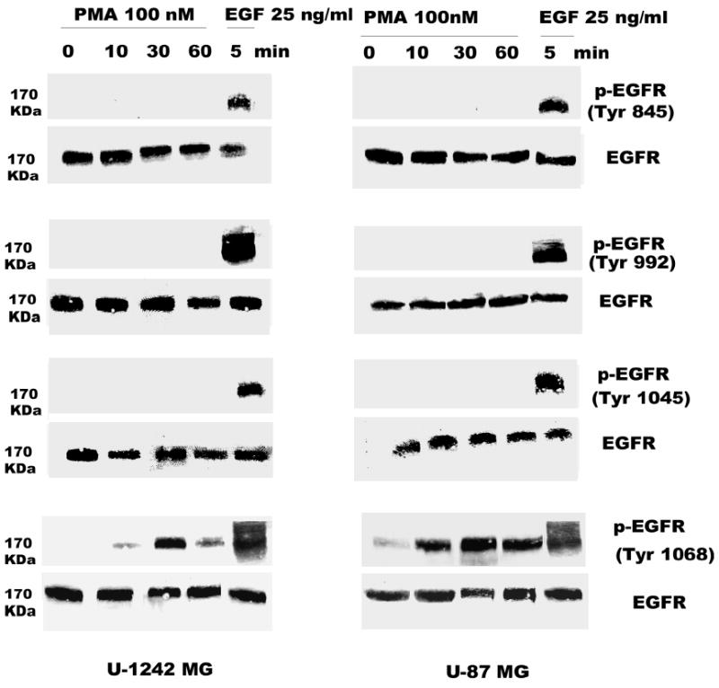
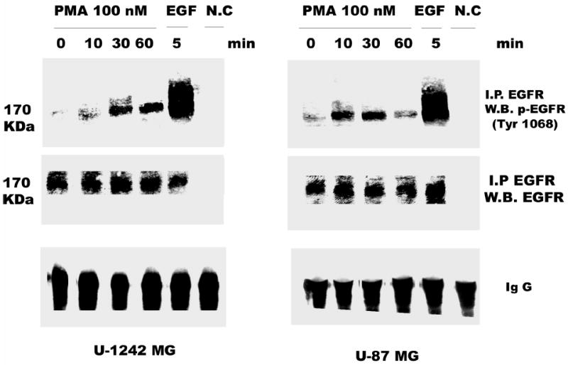
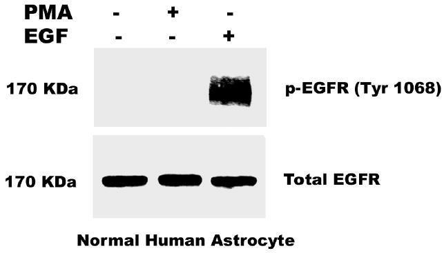
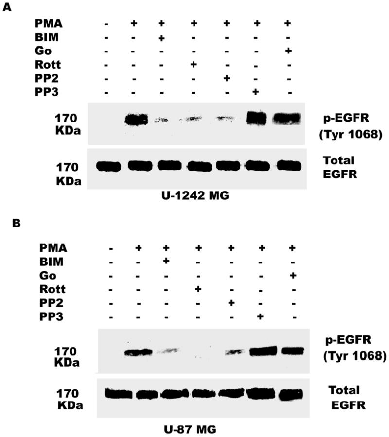
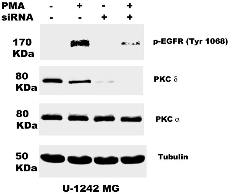
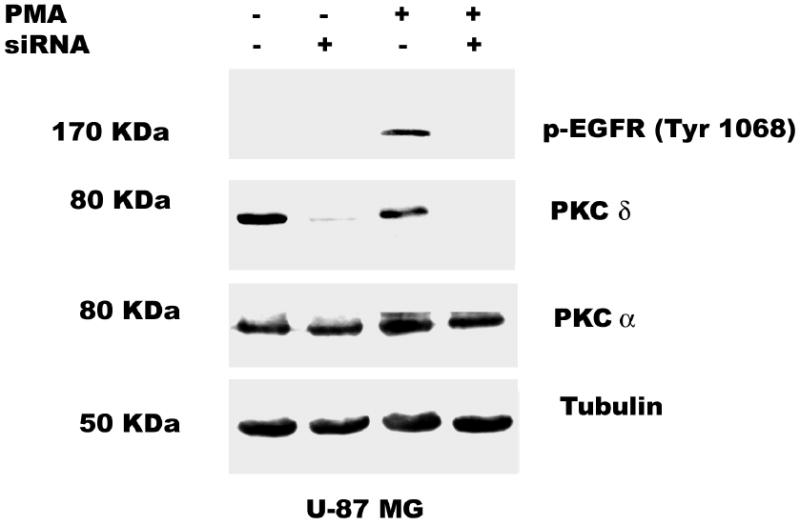
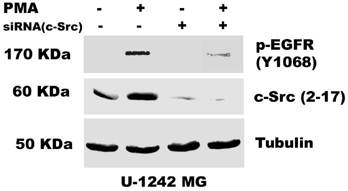
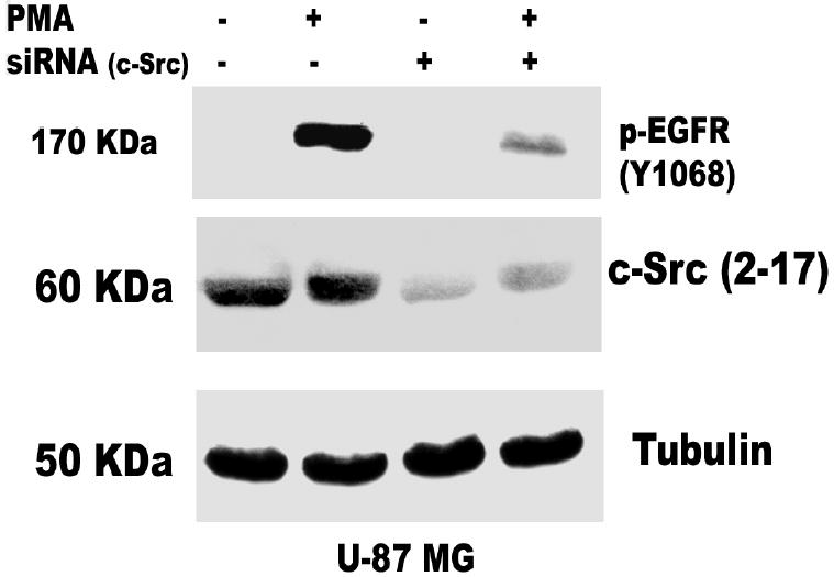
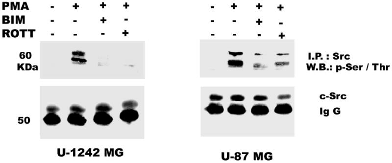
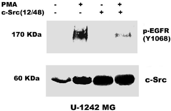
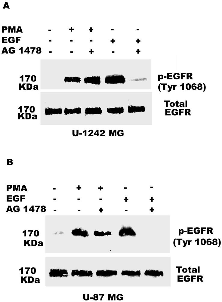
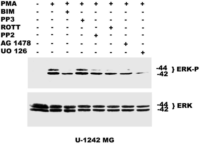
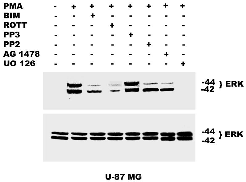
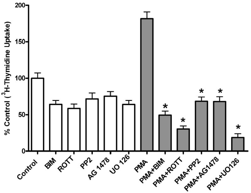
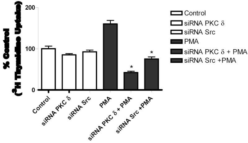
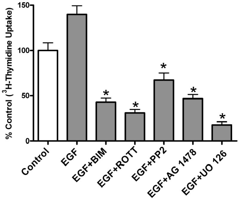
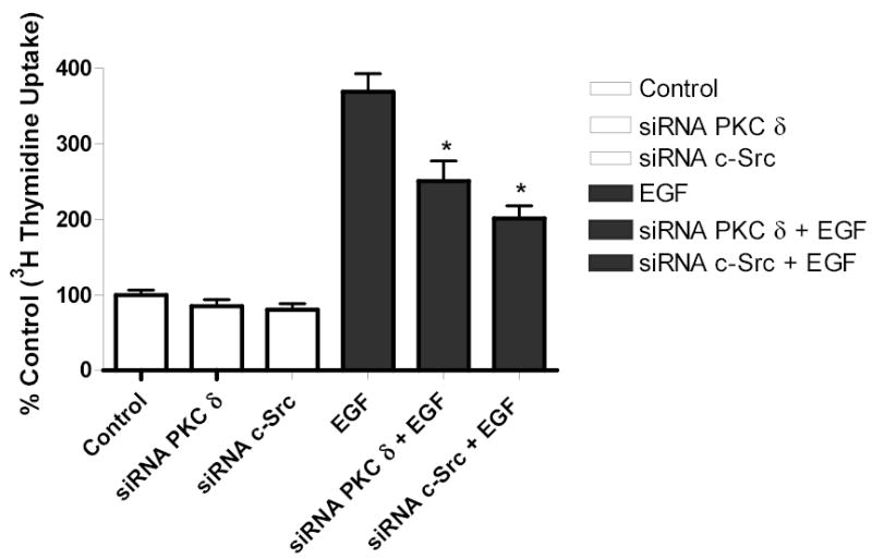
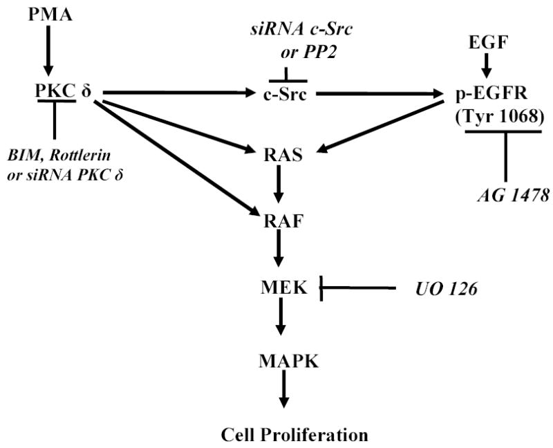
Both the epidermal growth factor receptor (EGFR) and protein kinase C (PKC) play important roles in glioblastoma invasive growth; however, the interaction between the EGFR and PKC is not well characterized in glioblastomas. Treatment with EGF stimulated global phosphorylation of the EGFR at Tyr(845), Tyr(992), Tyr(1068), and Tyr(1045) in glioblastoma cell lines (U-1242 MG and U-87 MG). Interestingly, phorbol 12-myristate 13-acetate (PMA) stimulated phosphorylation of the EGFR only at Tyr(1068) in the two glioblastoma cell lines. Phosphorylation of the EGFR at Tyr(1068) was not detected in normal human astrocytes treated with the phorbol ester. PMA-induced phosphorylation of the EGFR at Tyr(1068) was blocked by bisindolylmaleimide (BIM), a PKC inhibitor, and rottlerin, a PKCdelta-specific inhibitor. In contrast, Go 6976, an inhibitor of classical PKC isozymes, had no effect on PMA-induced EGFR phosphorylation. Furthermore, gene silencing with PKCdelta small interfering RNA (siRNA), siRNA against c-Src, and mutant c-Src(S12C/S48A) and treatment with a c-Src inhibitor (4-amino-5-(4-chlorophenyl)-7-(t-butyl) pyrazolo[3,4-d]pyrimidine) abrogated PMA-induced EGFR phosphorylation at Tyr(1068). PMA induced serine/threonine phosphorylation of Src, which was blocked by both BIM and rottlerin. Inhibition of the EGFR with AG 1478 did not significantly alter PMA-induced EGFR Tyr(1068) phosphorylation, but completely blocked EGF-induced phosphorylation of the EGFR. The effects of PMA on MAPK phosphorylation and glioblastoma cell proliferation were reduced by BIM, rottlerin, the MEK inhibitor U0126, and PKCdelta and c-Src siRNAs. Taken together, our data demonstrate that PMA transactivates the EGFR and increases cell proliferation by activating the PKCdelta/c-Src pathway in glioblastomas.


Figures

Fig. 1
Fig. 1
Time-dependent phosphorylation of EGFR in glioblastoma cells. Cells were serum-starved for 24 h after reaching 80–100% confluence by replacing the media with serum free α-MEM media. The cells were treated with EGF (25 ng/ml) at different time points (0, 10, 30, 60 min). Protein (200 μg/ lane) was fractionated by 8% SDS-PAGE and then transferred on to nitrocellulose. The nitrocellulose was reacted with primary antibody to EGFR (Tyr-845, Tyr-992, Tyr-1045, Tyr- 1068). Final detection was carried out using the enhanced chemuluminescence reagent (ECL). Blots were stripped and reprobed for EGFR total. The lower panel shows the total EGFR
Fig. 2
Fig. 2
PMA induces phosphorylation of EGFR (Tyr-1068). Serum-starved U-1242 MG and U-87 MG cells were treated with PMA (100 nM) at different time points (0, 10, 30, and 60 min). Triton solubilized astrocytic tumor cell lysates (200 μg/lane) were separated using 8% polyacrylamide gels and electroblotted on to nitrocellulose. The blots were probed with antibodies to phospho-EGFR (Tyr 1068) and total EGFR.
Fig. 3
Fig. 3
PMA stimulates tyrosine phosphorylation of EGFR (Tyr 1068). Serum- starved U-1242 MG and U-87 MG were stimulated with PMA for the indicated time periods. These cells were lysed using 1% Trition lysis buffer after washing with ice-cold PBS. Cell lysates were immunoprecipitated with anti EGFR antibody and then immunoblotted (IB) with antiphospho EGFR (Tyr 1068) or antibody to total EGFR.
Fig. 4
Fig. 4
Effect of PMA and EGF on Normal Human Astrocytes (NHA). NHA cells grown to 80–100% confluence were serum-starved overnight. The cells were then treated with PMA (100nM) for 30 min and EGF (25 ng/ml) for 10 min. Protein lysates (200 μg/lane) were fractionated using 8% SDS-PAGE gels and was electroblotted onto nitrocellulose. The blots were probed with antibody to EGFR (Tyr 1068). The membrane was stripped and reprobed for total EGFR.
Fig. 5
Fig. 5
Effect of BIM (PKC inhibitor), rottlerin (PKC δ inhibitor), PP2 (Src inhibitor) and Gö 6976 on EGFR (Tyr 1068) activation by PMA. Serum-starved U-1242 MG and U-87 MG cells were incubated with inhibitors for 60 min and then treated with PMA (100 nM) for 30 min. After washing twice with ice-cold PBS, cells were solubized in 1% Triton lysis buffer, analyzed and immunoblotted for EGFR (Tyr-1068). Blots were stripped and reprobed for total EGFR.
Fig. 6
Fig. 6
PKC δ mediates PMA-induced EGFR (Tyr 1068) phosphorylation. RNA interference with siRNA transfection was performed as described under “Experimental Procedure”. 24 h after transfection, cells were quiesced for 24 h. A, U-1242 MG cells and B, U-87 MG were transiently transfected with siRNA PKC δ and treated with or without PMA. Cells were then lysed and cell lysates of equal concentration of proteins (200 μg/lane) were separated on 8% SDS-PAGE gels and immunoblotted for phospho EGFR (Tyr 1068), PKC δ (total), anti PKCα and tubulin.
Fig. 6
Fig. 6
PKC δ mediates PMA-induced EGFR (Tyr 1068) phosphorylation. RNA interference with siRNA transfection was performed as described under “Experimental Procedure”. 24 h after transfection, cells were quiesced for 24 h. A, U-1242 MG cells and B, U-87 MG were transiently transfected with siRNA PKC δ and treated with or without PMA. Cells were then lysed and cell lysates of equal concentration of proteins (200 μg/lane) were separated on 8% SDS-PAGE gels and immunoblotted for phospho EGFR (Tyr 1068), PKC δ (total), anti PKCα and tubulin.
Fig. 7
Fig. 7
Role of c-Src in PMA-induced EGFR transactivation. siRNA against c-Src was transfected into A, U-1242 MG cells and B, U-87 MG cells and treated with PMA for 30 min. Cell were lysed using 1% Triton lysis buffer after washing with ice-cold PBS. Cell lysates were immunoblotted against EGFR (Y1068) and tubulin as loading control. C, Serum starved U-1242 MG and U-87 MG were pretreated with BIM (1 μM) and rottlerin (5 μM) for 60 min, after which PMA (100 nM) was added for 30 min. The cell lysates were immunoprecipitated with anti- c-Src (–17) and the precipitates were immunoblotted with antibodies against phospho-serine/ threonine and total c- Src protein. D, Mutant c-Src (ser12cys/ser48ala) transfected into U-1242 MG cells were treated with PMA (100 nM) for 30 min. Cells were lysed using 1% Triton lysis buffer after washing with ice-cold PBS. Cell lysates were immunoblotted against EGFR (Y1068).
Fig. 7
Fig. 7
Role of c-Src in PMA-induced EGFR transactivation. siRNA against c-Src was transfected into A, U-1242 MG cells and B, U-87 MG cells and treated with PMA for 30 min. Cell were lysed using 1% Triton lysis buffer after washing with ice-cold PBS. Cell lysates were immunoblotted against EGFR (Y1068) and tubulin as loading control. C, Serum starved U-1242 MG and U-87 MG were pretreated with BIM (1 μM) and rottlerin (5 μM) for 60 min, after which PMA (100 nM) was added for 30 min. The cell lysates were immunoprecipitated with anti- c-Src (–17) and the precipitates were immunoblotted with antibodies against phospho-serine/ threonine and total c- Src protein. D, Mutant c-Src (ser12cys/ser48ala) transfected into U-1242 MG cells were treated with PMA (100 nM) for 30 min. Cells were lysed using 1% Triton lysis buffer after washing with ice-cold PBS. Cell lysates were immunoblotted against EGFR (Y1068).
Fig. 8
Fig. 8
Inhibition of the epidermal growth factor receptor (EGFR) kinase activity did not affect PMA-induced phosphorylation of EGFR (Tyr 1068). Panel A, U-1242 MG cells were pretreated with EGFR kinase inhibitor AG 1478 (100 nM) for 60 min prior to treatment with media alone or EGF (25 ng/ml) or PMA (100 nM) for 10 min and 30 min, respectively. Cellular proteins were extracted and subjected to Western blotting using phospho-specific antibody against Tyr 1068. Panel B, Similarly, U-87 MG cells were pretreated with EGFR kinase inhibitor AG 1478 (100 nM) for 60 min prior to treatment with media alone, EGF (25 ng/ml) or PMA (100 nM) for 10 min and 30 min, respectively. Cellular proteins were extracted and subjected to Western blotting using phospho-specific antibody against Tyr 1068. Blots were stripped and reprobed for total EGFR.
Fig. 9
Fig. 9
Effect of pharmacological inhibitors on ERK activation by PMA. Panel A, Serum starved U-1242 MG were pretreated with BIM (1 μM) and rottlerin (5 μM), PP2 (5 μM), AG 1478 (100 nM) or UO 126 (5 μM) for 60 min, before the addition of PMA (100 nM) for 30 min. ERK activation by PMA was attenuated in presence of different inhibitors. Panel B, Serum starved U-87 MG were pretreated with BIM (1 μM) and rottlerin (5 μM), PP2 (5 μM), AG 1478 (100 nM) and UO 126 (10 μM) for 60 min, before PMA (100 nM) addition. Proteins were subjected to Western blotting on a 10% SDS-PAGE gel and probed with phosphospecific antibody to dually phosphorylated extracellular signal regulated kinase –1 and –2 (ERK 1 / ERK 2) and subsequently reprobed for ERK 1 /ERK2 total.
Fig. 9
Fig. 9
Effect of pharmacological inhibitors on ERK activation by PMA. Panel A, Serum starved U-1242 MG were pretreated with BIM (1 μM) and rottlerin (5 μM), PP2 (5 μM), AG 1478 (100 nM) or UO 126 (5 μM) for 60 min, before the addition of PMA (100 nM) for 30 min. ERK activation by PMA was attenuated in presence of different inhibitors. Panel B, Serum starved U-87 MG were pretreated with BIM (1 μM) and rottlerin (5 μM), PP2 (5 μM), AG 1478 (100 nM) and UO 126 (10 μM) for 60 min, before PMA (100 nM) addition. Proteins were subjected to Western blotting on a 10% SDS-PAGE gel and probed with phosphospecific antibody to dually phosphorylated extracellular signal regulated kinase –1 and –2 (ERK 1 / ERK 2) and subsequently reprobed for ERK 1 /ERK2 total.
Fig. 10
Fig. 10
[3H] Thymidine incorporation in astroctytic tumor cells. A, Effect of PMA on cell proliferation and B, PKC δ and c-Src inhibition abrogates PMA-induced cell proliferation. Cells were transfected with PKC δ siRNA or c-Src siRNA, serum straved overnight and the incubated in the presence of PMA. C, Effect of EGF and D, PKC δ and c-Src inhibition abrogates EGF-induced [3H] thymidine incorporation in U-87 MG cells. Quiescent U-87 MG cells were preincubated with BIM (1 μM ), Rottlerin (5 μM), UO 126 (10 μM) for 60 min. Cells were incubated in the presence and absence of PMA or EGF for 18 h. Cells were then pulse-labeled with [3H] thymidine for 6 h, and thymidine incorporation was measured by Beckman scintillation counter. Quantitative analysis from three independent experiments (means ± SEM). Cell proliferation was calculated as % of control. The data in the graph are the mean ± SEM of at least 2 independent experiments with each experiment performed in quadruplicate.
Fig. 10
Fig. 10
[3H] Thymidine incorporation in astroctytic tumor cells. A, Effect of PMA on cell proliferation and B, PKC δ and c-Src inhibition abrogates PMA-induced cell proliferation. Cells were transfected with PKC δ siRNA or c-Src siRNA, serum straved overnight and the incubated in the presence of PMA. C, Effect of EGF and D, PKC δ and c-Src inhibition abrogates EGF-induced [3H] thymidine incorporation in U-87 MG cells. Quiescent U-87 MG cells were preincubated with BIM (1 μM ), Rottlerin (5 μM), UO 126 (10 μM) for 60 min. Cells were incubated in the presence and absence of PMA or EGF for 18 h. Cells were then pulse-labeled with [3H] thymidine for 6 h, and thymidine incorporation was measured by Beckman scintillation counter. Quantitative analysis from three independent experiments (means ± SEM). Cell proliferation was calculated as % of control. The data in the graph are the mean ± SEM of at least 2 independent experiments with each experiment performed in quadruplicate.
Fig. 11
Fig. 11
Schematic representation of the signaling pathways involved in PMA transactivation of EGFR (Tyr 1068) in glioblastoma cell lines.

References

    1. Collins VP. Cancer Surv. 1998;32:37–51. - PubMed
    1. Rasheed BK, Wiltshire RN, Bigner SH, Bigner DD. Curr Opin Oncol. 1999;11:162–167. - PubMed
    1. Kleihues, P., Burger, P.C., Collins V.P., Newcomb E.W., Ohgaki, H., and Cavene, W.K., (2000). Glioblastoma. Tumors of the Nervous System. Kleihues, P., and Cavene, W.K. (eds) IARC Press: Lyon, pp 29–39.
    1. Libermann TA, Nusbaum HR, Razon N, Kris R, Lax I, Soreq H, Whittle N, Waterfield MD, Ullrich A, Schlessinger J. Nature. 1985;313:144–147. - PubMed
    1. Ekstrand AJ, Sugawa N, James CD, Collins VP. Proc Natl Acad Sci USA. 1992;89:1169–1174. - PMC - PubMed

Publication types

MeSH terms

LinkOut - more resources