. 2005 Mar 1;563(Pt 2):597-610.
doi: 10.1113/jphysiol.2004.076034. Epub 2004 Dec 23.

Candidate interneurones mediating group I disynaptic EPSPs in extensor motoneurones during fictive locomotion in the cat

Affiliations

Candidate interneurones mediating group I disynaptic EPSPs in extensor motoneurones during fictive locomotion in the cat

M J Angel et al. J Physiol. .

Abstract

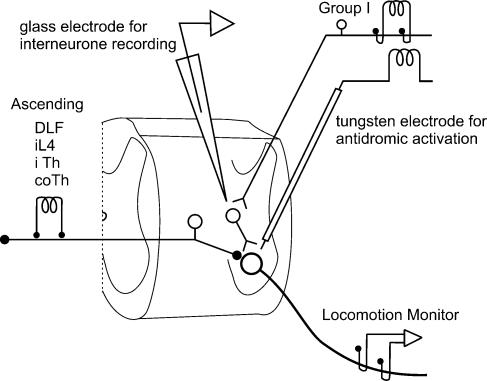
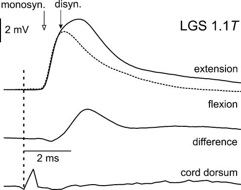
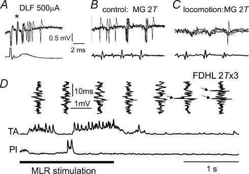
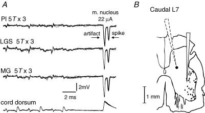
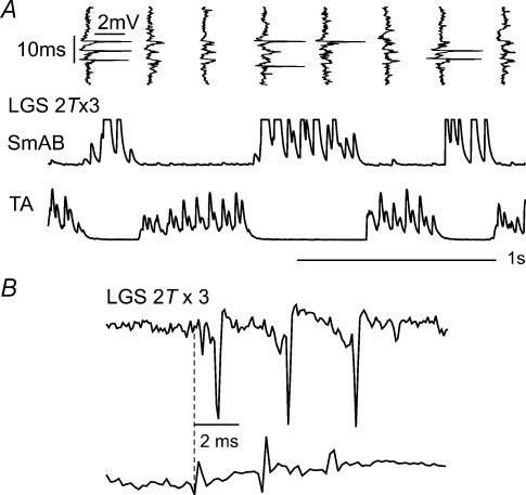
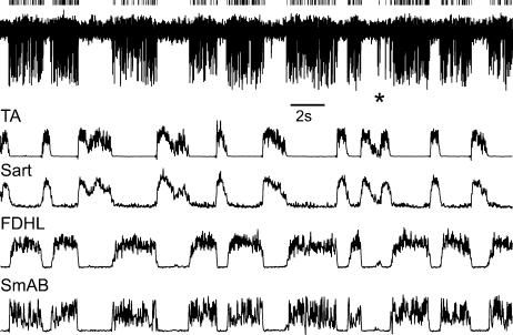
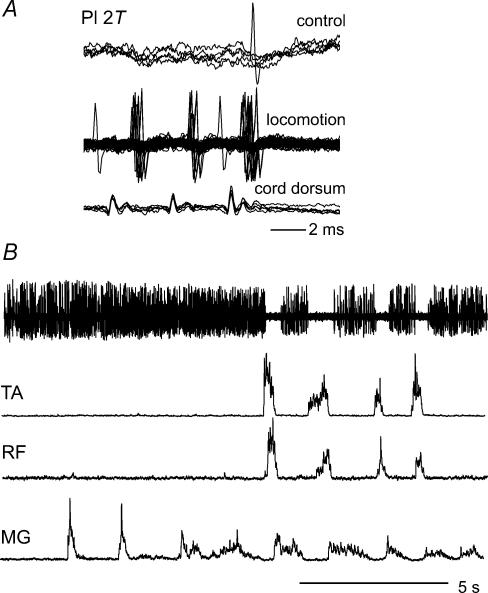
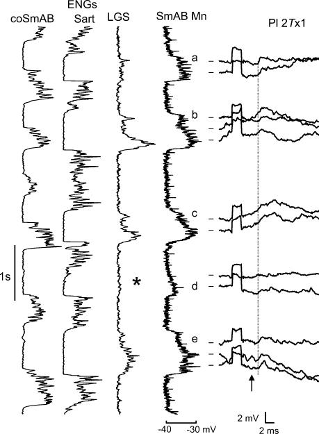
In the present study we sought to find interneurones responsible for the group I-evoked disynaptic excitation of hindlimb extensor motoneurones that occurs during fictive locomotion. Locomotion was produced by stimulation of the mesencephalic locomotor region (MLR) in decerebrate paralysed cats in which activation of ankle extensor group I afferents evoked a disynaptic excitation of motoneurones during the extension phase of fictive locomotion. Extracellular recordings were used to locate interneurones fulfilling all, or five of the six following criteria: (i) weak or no response to stimulation of extensor group I afferents in the absence of locomotion; (ii) strong group I activation during locomotion; (iii) group I activation at monosynaptic latencies; (iv) strong group I activation during only the extensor phase of locomotion; and (v) antidromic activation from the extensor motor nuclei; but (vi) no antidromic activation from rostral spinal segments. Candidate excitatory interneurones were located in mid to caudal parts of the L7 segments in areas where monosynaptic field potentials were evoked by group I afferents, within 2 mm of the stimulation site in the ventral horn from which they were antidromically activated. All were activated during extension by stimulation of group I afferents in extensor nerves. In the absence of peripheral nerve stimulation, six of the seven candidate excitatory interneurones were rhythmically active with maximal activation during the extension phase of fictive locomotion. Rhythmic activity during extension was also seen in five additional interneurones located near candidate interneurones but not activated by group I strength stimulation of the tested nerves. We suggest that the lumbosacral interneurones located in the intermediate laminae that can be activated by extensor group I afferents during the extension phase are a previously unknown population of interneurones, and may mediate group I-evoked disynaptic excitation of extensor motoneurones. Their rhythmic activity suggests that they also provide central excitatory drive to extensor motoneurones during locomotion.


Figures

Figure 1
Figure 1. Experimental arrangement used to locate premotor interneurones in pathways from group I afferents
The glass recording microelectrode and tungsten stimulating electrodes were inserted through the dorsal columns and the lateral funiculus, respectively. Within the grey mater the stimulating and recording microelectrode tips were often separated by < 1 mm in the rostro-caudal plane. Pairs of stimulating electrodes were placed on the ipsilateral and contralateral DLF at L4 and on both ipsilateral and contralateral thoracic dorsal columns for antidromic activation of laminae V–VI inhibitory cells and ascending tract cells.
Figure 2
Figure 2. Example of disynaptic EPSPs in extensor motoneurones indicates the preparation is suitable for locating relevant interneurones during extension
Top traces are superimposed averaged records of EPSPs evoked in a LGS motoneurone by near-threshold (1.1 T) stimulation of group I afferents in the LGS nerve during extension (continuous line) and flexion (dashed line) phases of the locomotor cycle. The difference between the two records is shown underneath. Open arrow indicates the onset of monosynaptic EPSPs. The filled arrow indicates the onset of disynaptic EPSPs which were evoked only during the extension phase. Bottom trace shows the afferent volley.
Figure 3
Figure 3. Inhibition during locomotion of responses of two interneurones which probably mediate group I non-reciprocal inhibition of motoneurones
AC, records from an interneurone antidromically activated by stimulation of the L4 DLF (A, *), activated by MG group I afferents at monosynaptic latencies in the absence of locomotion (B) and only weakly activated by the same stimuli during MLR-evoked locomotion (C). D, another interneurone monosynaptically activated by group I afferents at rest, which failed to respond to FDHL stimulation during locomotion, but became responsive to the same stimuli (arrows) within 1 s after terminating MLR stimulation (horizontal bar).
Figure 4
Figure 4. Examples of records from a candidate excitatory interneurone which is unresponsive to group I stimulation in the absence of locomotion
A, overlayed extracellular recordings (n = 5) from an interneurone that was antidromically activated by stimuli applied in the ankle extensor motor nuclei (latency, 0.6 ms) but failed to respond to stimulation supramaximal for group I afferents (5T × 3, 300 Hz). Bottom trace is the cord dorsum recording of the afferent volleys. B, camera lucida reconstruction of the recording microelectrode track (dotted outline) indicating the estimated location (filled circle) of the interneurone illustrated in Figs 4A, 5, 6 and 8A. The solid outline to the right is the track of the tungsten stimulating electrode. The scale bar takes into account a 10% shrinkage due to mounting. This interneurone was located in the intermediate nucleus.
Figure 5
Figure 5. Activation of a candidate excitatory interneurone by group I afferents during fictive locomotion
A, top traces are vertically orientated extracellular records from the interneurone illustrated in Figs 4 and 6, showing its responses to stimulation of the LGS nerve during locomotion. The records were obtained at the same time as the horizontally displayed discharges in the extensor (SmAB) and flexor (TA) nerves during locomotion. B, records obtained during fictive locomotion at an expanded time base, showing the latency of activation of the interneurone (0.9 ms) with respect to the incoming volleys.
Figure 6
Figure 6. Spontaneous activity of a candidate excitatory interneurone during extension
Top trace, activity of the interneurone illustrated in Figs 4 and 5 (largest spikes; represented by small vertical ticks) during MLR-evoked fictive locomotion in the absence of peripheral nerve stimulation. The responses are superimposed on responses of at least two other interneurones. The remaining four traces show discharges in two flexor (TA, Sart) and two extensor (FDL, SmAB) muscle nerves evoked simultaneously with responses of the interneurones.
Figure 7
Figure 7. Tonic activity of a candidate interneurone in the absence of flexor nerve activity
A, a comparison of responses of an interneurone to group I afferents before and during fictive locomotion (at a minimal latency of 0.8 ms from the third incoming volley). B, activity of the same interneurone (largest spikes) during MLR-evoked fictive locomotion in the absence of peripheral nerve stimulation superimposed on spike potentials of at least one other interneurone. Note that tonic activation of the interneurones occurred when MLR stimulation produced rhythmic activation of only extensors (MG). The interneurones discharge rhythmically only when activation of flexors started to alternate with activation of extensors.
Figure 8
Figure 8. Relationship between disynaptic EPSPs and activity of the extensor rhythm generator
Vertically orientated records from an ipsilateral flexor (Sart) and extensor (LGS) nerves are plotted alongside a low gain intracellular record from a SmAB motoneurone during MLR-evoked fictive locomotion. Horizontally plotted high gain intracellular records from this SmAB motoneurone show disynaptic EPSPs evoked by stimulation of Pl nerve during five extension phases (a–e). The arrow indicates arrival of group I afferent volleys. The dashed vertical line indicates onset of disynaptic EPSPs following these volleys (latency, 1.4 ms). Calibration pulse, 2 mV, 2 ms. Notice that the disynaptic EPSPs are largest when Pl stimulation occurs during large locomotor drive potentials (a, b, e). In contrast, when there is no activity in the extensor nerve (*) and the locomotor drive potential is small, the Pl-evoked disynaptic EPSPs are absent (d).
Figure 9
Figure 9. Evidence for spatial facilitation of synaptic actions of neurones activated by the MLR stimulation and of group I afferents onto interneurones mediating locomotion-related disynaptic EPSPs in motoneurones
A (top traces), superimposed records (n = 51) of spikes of two or three interneurones locked to MLR stimuli during fictive locomotion; the traces were collected during the extension phase and aligned to the MLR stimulation indicated by the dashed vertical line. A (bottom traces), discharges recorded in the LGS peripheral nerve. B, three averaged intracellular records of potentials evoked in a SmAB motoneurone: disynaptic EPSPs evoked by Pl group I afferents (n = 25, continuous line) preceded by MLR stimuli by 1–10 ms (see text); disynaptic EPSPs evoked by the same stimuli (n = 187, dashed line) which were not preceded by MLR stimulation; EPSPs evoked by MLR stimulation alone without peripheral nerve stimulation (n = 17, dotted line). C, as in B but for EPSPs evoked by selective activation of group Ia triceps surae and Pl afferents by muscle stretch. Calibration pulse, 2 mV, 2 ms.

References

    1. Angel MJ, Guertin P, Jiménez I, McCrea DA. Group I extensor afferents evoke disynaptic EPSPs in cat hindlimb extensor motorneurons during fictive locomotion. J Physiol. 1996;494:551–561. - PMC - PubMed
    1. Brink E, Harrison PJ, Jankowska E, McCrea DA, Skoog B. Post-synaptic potentials in a population of motoneurones following activity of single interneurones in the cat. J Physiol. 1983a;343:341–359. - PMC - PubMed
    1. Brink E, Jankowska E, McCrea DA, Skoog B. Inhibitory interactions between interneurones in reflex pathways from group Ia and group Ib afferents in the cat. J Physiol. 1983b;343:361–373. - PMC - PubMed
    1. Cavallari P, Edgley SA, Jankowska E. Postsynaptic actions of midlumbar interneurones on motoneurones of hindlimb muscles in the cat. J Physiol. 1987;389:675–690. - PMC - PubMed
    1. Conway BA, Hultborn H, Kiehn O. Proprioceptive input resets central locomotor rhythm in the spinal cord. Exp Brain Res. 1987;68:643–656. - PubMed

Publication types

LinkOut - more resources