. 2005 Jan 4;102(1):45-50.
doi: 10.1073/pnas.0408579102. Epub 2004 Dec 23.

The cAMP binding domain: an ancient signaling module

Affiliations

The cAMP binding domain: an ancient signaling module

Helen M Berman et al. Proc Natl Acad Sci U S A. .

Abstract

cAMP-binding domains from several different proteins were analyzed to determine the properties and interactions of this recognition motif. Systematic computational analyses, including structure-based sequence comparison, surface matching, affinity grid analysis, and analyses of the ligand protein interactions were carried out. These analyses show distinctive roles of the sugar phosphate and the adenine in the cAMP-binding module. We propose that the cAMP-binding regulatory proteins function by providing an allosteric system in which the presence or absence of cAMP produces a substantial structural change through the loss of hydrophobic interactions with the adenine ring and consequent repositioning of the C helix. The modified positioning of the helix in turn is recognized by a protein-binding event, completing the allostery.


Figures

Fig. 1.
Fig. 1.
Cyclic nucleotide-binding (CNB) domain. (a) The chemical structure of cAMP. (b) Tertiary structure of RIα(A) showing the secondary structural units. The image was created by using pymol (Delano Scientific) (32). (c) Sequence of RIα(A) showing the secondary structure color-coded as in a. The PBC is red.
Fig. 2.
Fig. 2.
Four representative CNB domain structures. Helices are colored cyan; sheets are red. (a) RIIβ (PDB ID 1CX4). There are two domains: A and B. The A domain is at the top, and the B domain is at the bottom. Each domain has one cAMP. (b)RIα (PDB ID 1NE6). There are two domains: A and B. The A domain is at the top, and the B domain is at the bottom. Each domain has one cAMP. (c) CAP (PDB ID 2CGP). There are two subunits related by 2-fold symmetry. For CAP in general each subunit can contain either one or two cAMP molecules. In this example there are two. The one in the N domain is the CNB domain. The other is in the domain that binds to DNA (shown in gray). (d) Cyclic nucleotide-gated channel (HCN) (PDB ID 1Q43). There are four subunits related by 4-fold symmetry. The C-terminal domain contains the cAMP. (e) A schematic of the four representative structures. Blue highlights the domains analyzed here. This image was created by using chimera (16).
Fig. 3.
Fig. 3.
Structure and sequence alignments of CNB domains. (a) Comparison of the A and B domains of RIIβ.(Upper) Structural superposition of the two domains. The A domain is yellow; the B domain is red. (Lower) The sequence alignment based on the results of the SAT comparisons. The residues that are the same are shown in red. The line above the sequences indicates those that align well structurally. The PBC is underlined in red. The αB and αC helices are indicated. (b) Comparison of the A domains in RIα (blue) and RIIβ (yellow). (Upper) Structural superposition. (Lower) The sequence comparison. The color codes for the sequence alignment are as in a.(c) Comparison of the B domains in RIα (magenta) and RIIβ (red). (Upper) Structural superposition. (Lower) The sequence comparison. The color codes for the sequence alignment are as in a. (d) Comparison of CAP (black), HCN (gray), and RIIβ(B) (red). (Upper) The structural superposition. (Lower) The sequence alignment. The gray line indicates the sequences in HCN that align well with RIIβ(B). The black line indicates the sequences in CAP that align well RIIβ(B). The color codes for the sequence alignment are as in a. This image was created by using chimera (16).
Fig. 4.
Fig. 4.
Examples of surface match graphs RIIβ(B) and CAP. The PBC is shown in red.
Fig. 5.
Fig. 5.
Molecular graphics showing the cAMP interactions. Conserved residues from surface matching are shown by using chemical colors. The domain in which the cAMP resides is cyan. The other domain is white. The PBC and the residue forming the hydrophobic interaction is in red. Conserved specific hydrogen bonds are in yellow. (a) RIIβ(A). The residue numbering for the conserved residues is shown. The B domain is white. The A domain is cyan. (b) RIIβ(B). (c)RIα(A). The B domain is white. (d)RIα(B). (e) CAP. (f) HCN. This image was created by using chimera (16).
Fig. 6.
Fig. 6.
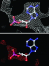
The affinity grids for cAMP in RIIβ. Contours are drawn at a level of -0.4 kcal/mol for carbon atoms (Upper in white) and oxygen/nitrogen atoms (Lower in red). The hydrogen affinity is not included, because it showed no features at this contour level. Fig. 8 shows more examples.
Fig. 7.
Fig. 7.
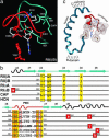
Conserved features of the cAMP binding domain. (a) Conserved features of the cAMP-binding site. The PBC is shown in red. The second conserved site is shown in cyan. The conserved hydrophobic residue is shown as a red ball-and-stick model. (b) Key residues in the four representative structures. The sequences are aligned to show the conserved residues from surface matching (gold). Residues involved in hydrogen bonding have a red border. Residues in hydrophobic contact with the adenine are enclosed in a red box. (c) The prototypic allosteric binding module. The example shown in a is for RIIβ(B) and in c is RIα(A).

References

    1. Canaves, J. M. & Taylor, S. S. (2002) J. Mol. Evol. 54, 17-29. - PubMed
    1. Diller, T. C., Madhusudan, Xuong, N. H. & Taylor, S. S. (2001) Structure (London) 9, 73-82. - PubMed
    1. McKay, D. B. & Steitz, T. A. (1981) Nature 290, 744-749. - PubMed
    1. Weber, I. T., Steitz, T. A., Bubis, J. & Taylor, S. S. (1987) Biochemistry 26, 343-351. - PubMed
    1. Weber, I. T., Shabb, J. B. & Corbin, J. D. (1989) Biochemistry 28, 6122-6127. - PubMed

Publication types

LinkOut - more resources