

Coexistence of lactating adenoma and invasive ductal adenocarcinoma of the breast in a pregnant woman
- PMID: 15623491
- PMCID: PMC1770558
- DOI: 10.1136/jcp.2004.018275
Coexistence of lactating adenoma and invasive ductal adenocarcinoma of the breast in a pregnant woman
Abstract
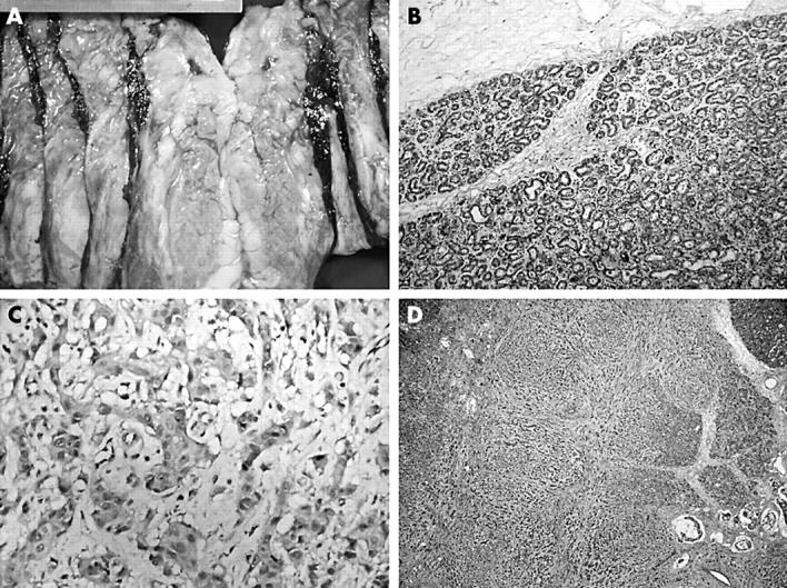
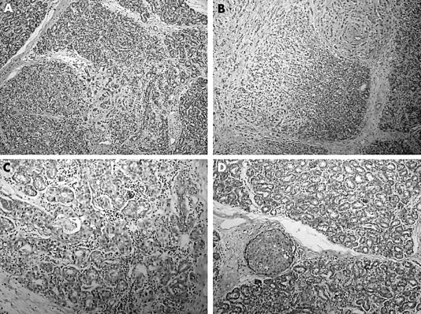
A 36 year old pregnant woman was admitted to hospital complaining of an enlarging mass in her left breast. Histopathological examination of the mastectomy specimen revealed a high grade infiltrating ductal adenocarcinoma intermixed with a lactating adenoma. Lactating adenomas are rare entities but are the most common masses that occur during pregnancy. Although they are not thought to carry an increased risk of cancer there are two other case reports in the literature of a lactating adenoma associated with an infiltrating carcinoma. In this case, areas where the lactating adenoma and the infiltrating carcinoma were intermixed could be identified. This case could simply be a collision tumour, although the possibility of an invasive carcinoma arising within a lactating adenoma cannot be ruled out. Because of the relative lack of experience with lactating adenomas, the question of an increased association with carcinoma development remains unclear.
Figures
References
-
- Reed W , Hannisdal E, Skovlund E, et al. Pregnancy and breast cancer: a population-based study. Virchows Arch 2003;443:44–50. - PubMed
-
- Baker TP, Lenert JT, Parker J, et al. Lactating adenoma: a diagnosis of exclusion. Breast J 2001;7:354–7. - PubMed
-
- Hertel BF, Zaloudek C, Kempson RL. Breast adenomas. Cancer 1976;37:2891–905. - PubMed
-
- Lishner M . Cancer in pregnancy. Ann Oncol 2003;14 (suppl 3) :iii31–6. - PubMed
Publication types
MeSH terms
LinkOut - more resources
Full Text Sources
Medical