

Primary pleural epithelioid haemangioendothelioma with metastases to the skin. A case report and literature review
- PMID: 15623498
- PMCID: PMC1770548
- DOI: 10.1136/jcp.2004.018937
Primary pleural epithelioid haemangioendothelioma with metastases to the skin. A case report and literature review
Abstract
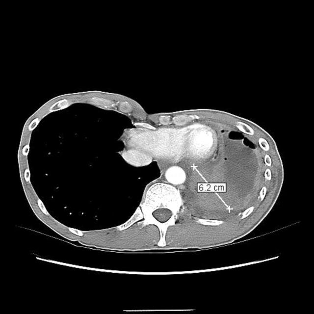
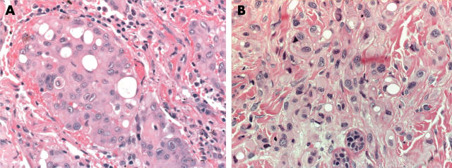
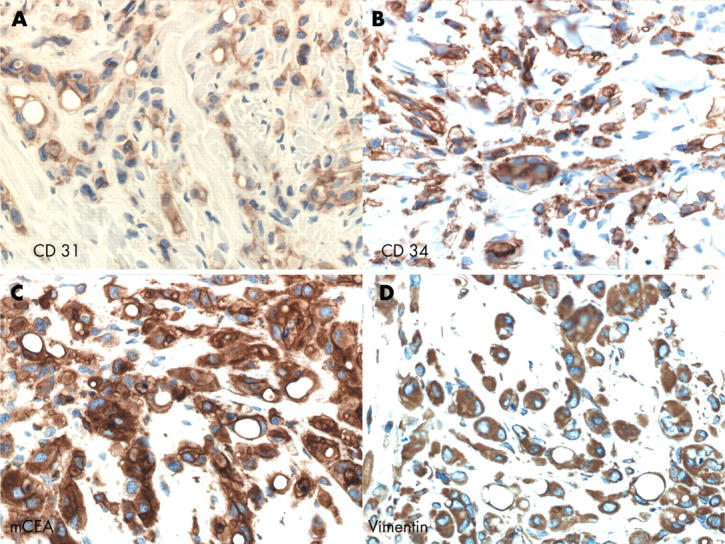
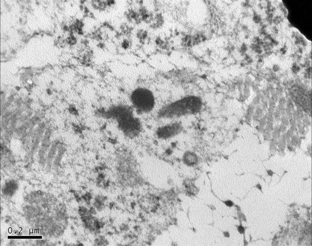
Epithelioid haemangioendothelioma (EHE) is a rare vascular tumour of intermediate behaviour. It can arise from various sites including the liver, spleen, pleura, or lung. Cutaneous EHE can be primary or secondary. This report describes the case of a 51 year old man who presented with a history of dry cough, shortness of breath, and pleural effusion, and who developed two cutaneous nodules in the anterior abdominal wall a few weeks later. He had a previous history of asbestos exposure. Computed tomography scan showed a left sided pleural effusion and nodular pleural mass. Histology of both the pleural and cutaneous lesions was compatible with EHE. Electron microscopic examination demonstrated the presence of Weibel-Palade bodies. The patient underwent elliptical excision of the metastatic cutaneous nodules after decortication of the primary pleural tumour and adjuvant treatment. A few reports have described metastasis of intrathoracic EHE to the skin. Despite treatment with interferon, the patient developed more cutaneous lesions two years after the initial diagnosis. Even though the tumour has the classic light histological and ultrastructural features of EHE, it behaved in an aggressive manner.
Figures
References
-
- Weiss SF, Enzinger FM. Epithelioid hemangioendothelioma. A vascular tumor often mistaken for a carcinoma. Cancer 1982;50:970–81. - PubMed
-
- Verbeken E , Beyls J, Moerman P, et al. Lung metastasis of malignant epithelioid hemangioendothelioma mimicking a primary intravascular bronchioalveolar tumor. A histologic, ultrastructural, and immunohistochemical study. Cancer 1985;55:1741–6. - PubMed
-
- Yousem SA, Hochholzer L. Unusual thoracic manifestations of epithelioid hemangioendothelioma. Arch Pathol Lab Med 1987;111:459–63. - PubMed
-
- Quante M , Patel NK, Hill S, et al. Epithelioid hemangioendothelioma presenting in the skin: a clinicopathologic study of eight cases. Am J Dermatopathol 1998;20:541–6. - PubMed
Publication types
MeSH terms
Substances
LinkOut - more resources
Full Text Sources
Medical