. 2005 Jan 11;102(2):473-8.
doi: 10.1073/pnas.0406458102. Epub 2004 Dec 27.

Acquired deficit of forebrain glucocorticoid receptor produces depression-like changes in adrenal axis regulation and behavior

Affiliations

Acquired deficit of forebrain glucocorticoid receptor produces depression-like changes in adrenal axis regulation and behavior

Maureen P Boyle et al. Proc Natl Acad Sci U S A. .

Abstract

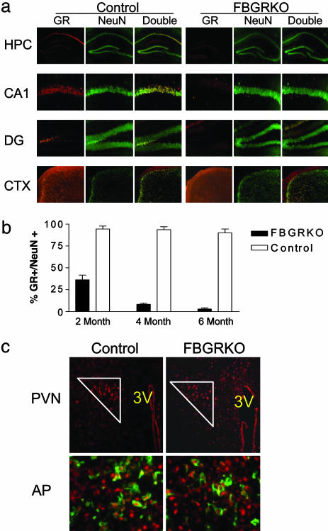
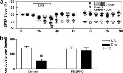
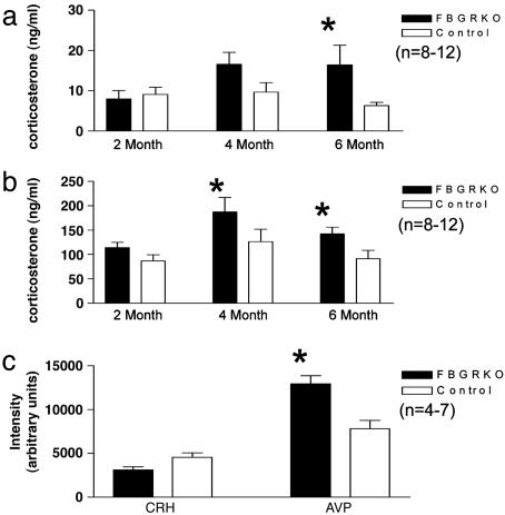
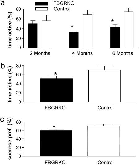
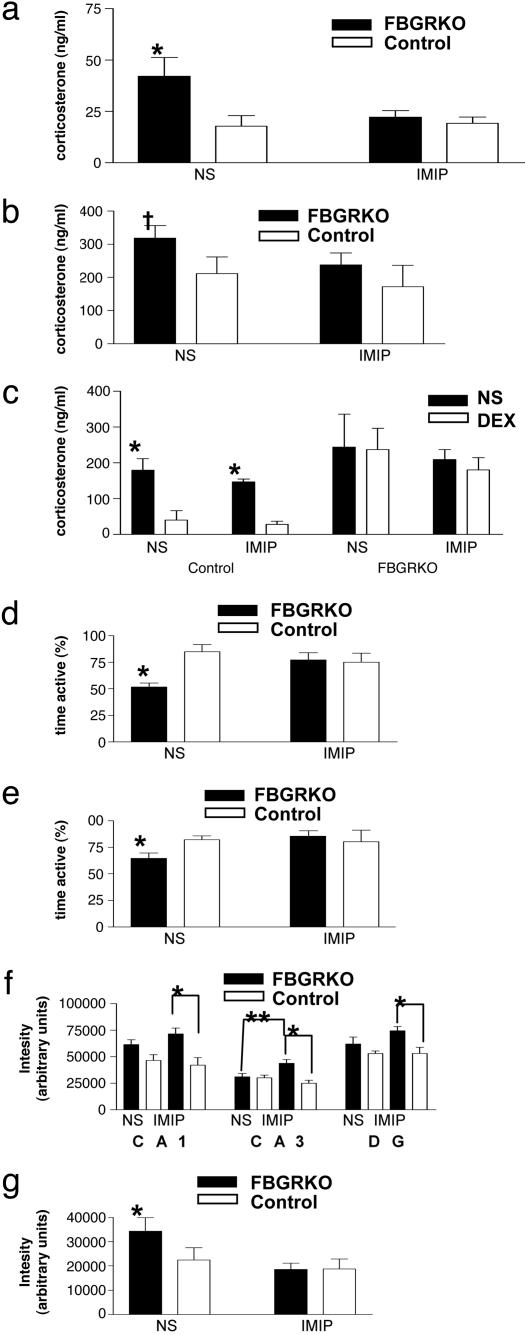
Dysregulation of the hypothalamic-pituitary-adrenal (HPA) axis is a hallmark of major depressive disorder. A number of studies have shown that this dysregulation is correlated with impaired forebrain glucocorticoid receptor (GR) function. To determine whether a primary, acquired deficit in forebrain GR signaling is an etiologic factor in the pathogenesis of depression, we generated a line of mice with time-dependent, forebrain-specific disruption of GR (FBGRKO). These mice develop a number of both physiological and behavioral abnormalities that mimic major depressive disorder in humans, including hyperactivity of the HPA axis, impaired negative feedback regulation of the HPA axis and, increased depression-like behavior. Importantly, a number of these abnormalities are normalized by chronic treatment with the tricyclic antidepressant, imipramine. Our findings suggest that imipramine's proposed activities on forebrain GR function are not essential for its antidepressant effects, and that alteration in GR expression may play a causative role in disease onset of major depressive disorder.


Figures

Fig. 1.
Fig. 1.
GR immunoreactivity is lost throughout the forebrain of the FBGRKO mice in a time-dependent manner. (a) Immunohistochemical analysis of GR expression in forebrain neurons of 6-month-old FBGRKO and control mice. Each panel shows immunoreactivity for GR (red), NeuN (green), and a merged image in which double labeled cells appear yellow. Top row shows the hippocampus (HPC) at ×100 magnification. Middle rows show area CA1 and the dentate gyrus (DG) at ×400 magnification. Bottom row shows a ×100 magnification image of cortex (CTX). (b) Quantification of the number of CA1 NeuN-positive GR-expressing neurons in FBGRKO and control mice at 2, 4, and 6 months of age (n = 4–6). (c) GR expression remains intact in the PVN and anterior pituitary. (Upper) Representative sections of the PVN from FRGRKO and control mice stained for GR (red). GR-positive PVN neurons are enclosed within the white triangle. (Lower) Representative sections of anterior pituitary (AP) stained for GR (red, nuclear) and proopiomelanocortin (green, cytoplasmic).
Fig. 2.
Fig. 2.
Functional glucocorticoid resistance in FBGRKO mice. (a) Low-frequency stimulation (LFS) of the commissural pathway in the presence of corticosterone (cort) elicits significant long-term depression of excitatory postsynaptic field potentials in area CA1 of control (P = 0.0076) but not FBGRKO mice. (b) Injection of DEX produces a significant suppression of corticosterone release in control mice (*, P < 0.0001) but produces no suppression in FBGRKO mice in the DST.
Fig. 3.
Fig. 3.
HPA axis regulation is altered in the FBGRKO mice. (a and b) Basal (a) and peak circadian (b) corticosterone release in FBGRKO and control mice at 2, 4, and 6 months of age. FBGRKO mice show a significant increase in basal corticosterone at 6 months of age, and in peak corticosterone at 4 and 6 months of age relative to control mice of the same age (*, P < 0.05). (c) Quantification of CRH and AVP mRNA expression by in situ hybridization. Densitometric analysis revealed a significant increase in AVP (*, P = 0.0035) but not CRH mRNA at baseline in the PVN of FBGRKO mice compared to controls.
Fig. 4.
Fig. 4.
FBGRKO mice show a time-dependent increase in depression-like behaviors. (a) FBGRKO mice show significantly less activity in the forced swim test (n = 6–9) at 4 (*, P = 0.01) and 6 (**, P = 0.01) months, but not at 2 months of age. (b) In the tail suspension test (n = 6–8), 6-month-old FBGRKO mice again showed significantly less activity (*, P = 0.04). (c) In the two-bottle sucrose preference test (n = 5), FBGRKO mice show significantly decreased sucrose preference (pref.) (*, P < 0.04).
Fig. 5.
Fig. 5.
Chronic treatment with imipramine reverses HPA axis hyperactivity and the behavioral despair phenotype of FBGRKO mice. (a and b) FBGRKO mice treated with vehicle show an increase in basal circadian corticosterone release (P < 0.05, n = 4–6) (a) and a trend toward increased peak corticosterone release (†, P = 0.08, n = 4–6) (b). After chronic treatment with imipramine, FBGRKO mice show no significant differences in basal or peak corticosterone release from control mice treated with either imipramine or normal saline (NS). (c) In the DST, imipramine does not reverse the impairment in negative feedback suppression of the HPA axis in the FBGRKO mice. (d and e) FBGRKO mice treated with NS showed decreased activity in both the tail suspension (P < 0.0001, n = 4–6) (d) and forced swim tests (P < 0.001, n = 4–6) (e) compared with controls. However, FBGRKO mice treated with imipramine showed no significant difference compared with controls (n = 3–6). (f) NS administration tended to increase MR mRNA expression in areas CA1 and DG of the FBGRKO, and imipramine treatment further augmented the differences between genotypes (*,P < 0.004, n = 4–6). In region CA3, increased MR mRNA was specific for imipramine treatment in FBGRKO mice (**, P = 0.02). (g) FBGRKO mice treated with NS express significantly more CRH mRNA than vehicle-treated controls (P = 0.015, n = 4–6); imipramine treatment decreased PVN CRH mRNA to control levels.

References

    1. Herman, J. P., Figueiredo, H., Mueller, N. K., Ulrich-Lai, Y., Ostrander, M. M., Choi, D. C. & Cullinan, W. E. (2003) Front. Neuroendocrinol. 24, 151-180. - PubMed
    1. Barden, N. (2004) J. Psychiatry Neurosci. 29, 185-193. - PMC - PubMed
    1. O'Toole, S. M., Sekula, L. K. & Rubin, R. T. (1997) Biol. Psychiatry 42, 85-89. - PubMed
    1. Calfa, G., Kademian, S., Ceschin, D., Vega, G., Rabinovich, G. A. & Volosin, M. (2003) Psychoneuroendocrinology 28, 687-701. - PubMed
    1. Gass, P., Reichardt, H. M., Strekalova, T., Henn, F. & Tronche, F. (2001) Physiol. Behav. 73, 811-825. - PubMed

Publication types