. 2005 Jan 3;168(1):41-54.
doi: 10.1083/jcb.200407182. Epub 2004 Dec 28.

Condensed mitotic chromatin is accessible to transcription factors and chromatin structural proteins

Affiliations

Condensed mitotic chromatin is accessible to transcription factors and chromatin structural proteins

Danyang Chen et al. J Cell Biol. .

Abstract

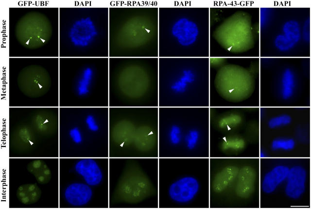
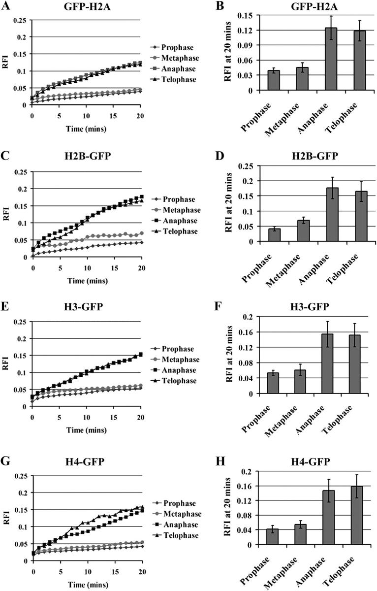
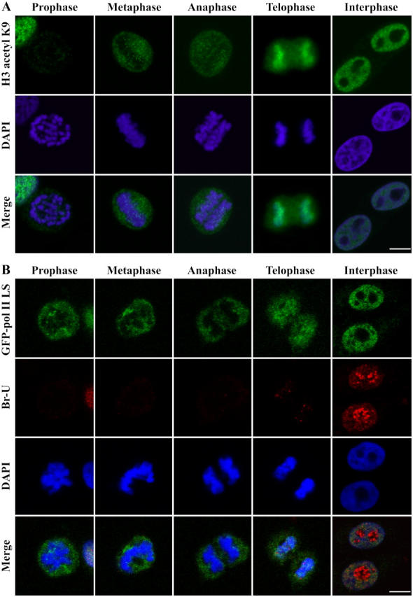
During mitosis, chromosomes are highly condensed and transcription is silenced globally. One explanation for transcriptional repression is the reduced accessibility of transcription factors. To directly test this hypothesis and to investigate the dynamics of mitotic chromatin, we evaluate the exchange kinetics of several RNA polymerase I transcription factors and nucleosome components on mitotic chromatin in living cells. We demonstrate that these factors rapidly exchange on and off ribosomal DNA clusters and that the kinetics of exchange varies at different phases of mitosis. In addition, the nucleosome component H1c-GFP also shows phase-specific exchange rates with mitotic chromatin. Furthermore, core histone components exchange at detectable levels that are elevated during anaphase and telophase, temporally correlating with H3-K9 acetylation and recruitment of RNA polymerase II before the onset of bulk RNA synthesis at mitotic exit. Our findings indicate that mitotic chromosomes in general and ribosomal genes in particular, although highly condensed, are accessible to transcription factors and chromatin proteins. The phase-specific exchanges of nucleosome components during late mitotic phases are consistent with an emerging model of replication independent core histone replacement.


Figures

Figure 1.
Figure 1.
Subcellular localization of GFP-fusion proteins of RNA pol I transcription factors in cells at various phases of cell cycle as labeled. Bar, 10 μM. Arrowheads indicate NOR-associated corresponding GFP-fusion proteins.
Figure 2.
Figure 2.
FRAP and FLIP of GFP-UBF1 during mitosis in HeLa cells. BL represents the first image acquired after photobleaching. The number at the bottom of each panel indicates the time after photobleaching. (A) Fluorescent images of the FRAP (top) and FLIP (bottom) processes. Box indicates the area of bleaching and fluorescence recovery. Bar, 10 μm. (B) Quantification of the fluorescence recoveries through changes in RFI over time for GFP-UBF1 in interphase and all phases of mitotic cells. (C) Graphic presentation of the time of 50% fluorescence recovery (t50) of GFP-UBF in cells at various mitotic phases and interphase. (D) FLIP of GFP-UBF1 at interphase and metaphase cells. Error bars represent averages from 12 to 16 cells ± SD.
Figure 3.
Figure 3.
FRAP of GFP-fusion proteins of RNA pol I subunits, RPA39/40 and RPA43 during anaphase–telophase transition. Bar, 10 μm. (A) Fluorescent images of the FRAP processes. Boxes indicate the areas of bleaching and fluorescence recovery. (B) Quantitation of the fluorescence recoveries through changes in RFI for GFP-RPA39/40 and (C) graphic presentation of t50 of GFP-RPA39/40 in interphase and mitotic cells. (D) FRAP of RPA43-GFP and (E) graphic presentation of t50 of RPA43-GFP. Error bars represent averages from 10 to 14 cells ± SD.
Figure 4.
Figure 4.
Deletion mutagenesis analyses demonstrate that DNA-binding capacity is required for the association of GFP-UBF1 with the mitotic NORs. (A) Schematic diagram of the mutants generated. (B) The localization of the UBF mutant GFP-fusion proteins on mitotic NORs (arrowheads). Bar, 10 μm.
Figure 5.
Figure 5.
H1c-GFP rapidly exchanges with mitotic chromosome and the dynamics of the process varies throughout different mitotic phases. FRAP analyses of GFP-tagged histone H1c at various phases of mitosis (A). Fluorescent images of FRAP of H1c-GFP. (B and C) Quantification of the fluorescence recoveries through changes in RFI over time. (D) Fluorescent images of the FLIP of H1c-GFP. (E) Quantitation of the FLIP reflected by FRI over time. Bars, 10 μm. Error bars represent averages from 10 to 12 cells ± SD.
Figure 6.
Figure 6.
FRAP analyses of H2B-GFP at all phases of mitosis. Cells were imaged before and during recovery after bleaching of chromosome area. For each M phase cell, top panel represents original images, bottom panel represents pseudocolored images. Bar, 10 μM.
Figure 7.
Figure 7.
FRAP analyses of core histones at all phases of mitosis. Quantification of the fluorescence recoveries through changes in RFI over time for GFP-H2A, H2B-GFP, H3-GFP, and H4-GFP, correspondingly. Error bars represent averages from 10 cells ± SD.
Figure 8.
Figure 8.
Western blot analyses of the presence of histones in mitotic cytoplasm. (A) Endogenous histones were detected in chromosome (Ch) and cytoplasmic fractions (Cy) prepared from both stably transfected and non transfected cells. (B) The GFP-fusion histones were detected in both the chromosome (Ch) fractions and the cytoplasmic fractions (Cy) by an anti-GFP antibody.
Figure 9.
Figure 9.
H3 acetylation and polymerase II association to chromosome take place at anaphase–telophase transition prior to the onset of bulk RNA synthesis. (A) Detection of histone H3 lysine 9 acetylation using a specific antibody at various mitotic phases and in interphase cells. (B) Simultaneous detection of GFP-pol II LS and Br-U incorporation through various phases mitosis. Bar, 10 μm.

References

    1. Ahmad, K., and S. Henikoff. 2002. a. Epigenetic consequences of nucleosome dynamics. Cell. 111:281–284. - PubMed
    1. Ahmad, K., and S. Henikoff. 2002. b. The histone variant H3.3 marks active chromatin by replication-independent nucleosome assembly. Mol. Cell. 9:1191–1200. - PubMed
    1. Barra, J.L., L. Rhounim, J.L. Rossignol, and G. Faugeron. 2000. Histone H1 is dispensable for methylation-associated gene silencing in Ascobolus immersus and essential for long life span. Mol. Cell. Biol. 20:61–69. - PMC - PubMed
    1. Bell, S.P., R.M. Learned, H.M. Jantzen, and R. Tjian. 1988. Functional cooperativity between transcription factors UBF1 and SL1 mediates human ribosomal RNA synthesis. Science. 241:1192–1197. - PubMed
    1. Belotserkovskaya, R., and D. Reinberg. 2004. Facts about FACT and transcript elongation through chromatin. Curr. Opin. Genet. Dev. 14:139–146. - PubMed

Publication types

MeSH terms