. 2005 Jan 11;102(2):343-8.
doi: 10.1073/pnas.0408438102. Epub 2004 Dec 29.

Cell cycle-dependent translocation of PRC1 on the spindle by Kif4 is essential for midzone formation and cytokinesis

Affiliations

Cell cycle-dependent translocation of PRC1 on the spindle by Kif4 is essential for midzone formation and cytokinesis

Changjun Zhu et al. Proc Natl Acad Sci U S A. .

Abstract

The spindle midzone, a conspicuous network of antiparallel interdigitating nonkinetochore microtubules between separating chromosomes, plays a crucial role in regulating the initiation and completion of cytokinesis. In this study, we report the use of time-lapse microscopy and a human kinesin endoribonucleases RNase III-prepared short interfering RNA (esiRNA) library to identify Kif4 as a motor protein that translocates PRC1, a spindle midzone-associated cyclin-dependent kinase substrate protein, to the plus ends of interdigitating spindle microtubules during the metaphase-to-anaphase transition. We show that Kif4 binds to PRC1 through its "stalk plus tail" domains and Kif4 and PRC1 colocalize on the spindle midzone/midbody during anaphase and cytokinesis. Suppression of Kif4 expression by Kif4 esiRNA results in the inhibition of PRC1 translocation, a block of the midzone formation, and a failure of cytokinesis. PRC1 translocation and midzone formation can be restored, and the cytokinetic defects can be rescued in Kif4 esiRNA-treated cells by coexpression of Kif4 but not its motor dead mutant Kif4md. Furthermore, we show that cyclin-dependent kinase phosphorylation of PRC1 controls the timing of PRC1 translocation by Kif4. These results, in light of the crucial role of PRC1 in midzone formation, indicate that cell cycle-dependent translocation of PRC1 by Kif4 is essential for midzone formation and cytokinesis.


Figures

Fig. 1.
Fig. 1.
Abnormal subcellular localization of PRC1 and failure of cytokinesis in HeLa cells treated with Kif4 esiRNA. HeLa cells grown on glass-bottom microwell dishes were transfected with pEYFP-PRC1 and pECFP-H2B together with 100 nM control (luciferase) (A) or Kif4 (B) esiRNA. Thirty-six hours posttransfection, cells were placed on a 37°C heated stage, and time-lapse images were collected every 2 min for 10 h with a Zeiss Axiovert 100M inverted fluorescence microscope and an automatic digital charge-coupled device camera. The movies were edited with slidebook 3.0 software. Representative images of the movies are shown (EYFP-PRC1, pseudocolored red and ECFP-H2B, pseudocolored green). Blue arrows indicate midzone/midbody localization of PRC1 in control esiRNA-treated cells, and black arrows indicate abnormal spindle localization of PRC1 in Kif4 esiRNA-treated cells. The numbers indicate time periods in minutes. For details, see Movies 1 and 2.
Fig. 2.
Fig. 2.
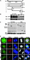
Kif4 and PRC1 interact with each other in vivo and colocalize at the mitotic spindle and spindle midzone/midbody during mitosis and cytokinesis. (A) A schematic illustration shows EGFP-tagged Kif4, its motor dead version, and a series of deletion mutant constructs used in coimmunoprecipitation assays (lanes a–g; for details see Materials and Methods). The asterisk indicates triple point mutations in the ATP-binding Walker A consensus site of the Kif4 motor domain (GQTGSGKTYSMG to GQTGSAAAYSMG). (B) Coimmunoprecipitation analysis of EGFP-tagged Kif4 or EGFP-tagged Kif4md (motor dead mutant) with FLAG-tagged PRC1. EGFP-tagged Kif4 (lanes a) or EGFP-tagged Kif4md (lanes b) were coexpressed with FLAG-tagged PRC1 in 293T cells. Cell lysates were made and immunoprecipitated with anti-FLAG antibody. Immunoprecipitates (IP) and 1/10 of whole-cell lysates (WCL) used in immunoprecipitations were subjected to SDS/PAGE, transferred to poly(vinylidene difluoride) membrane, and immunoblotted with anti-GFP antibody (Upper) or anti-FLAG antibody (Lower). (C) Coimmunoprecipitation analysis of EGFP-tagged Kif4 or its mutant proteins with FLAG-tagged PRC1. EGFP-tagged Kif4 or its mutant proteins (lanes a and c–g) were coexpressed with FLAG-tagged PRC1 in 293T cells. Cell lysates were made and immunoprecipitated with anti-FLAG antibody. Immunoprecipitates (IP) (Left) and 1/10 of whole-cell lysates (WCL) used in immunoprecipitations (Right) were subjected to SDS/PAGE, transferred to poly(vinylidene difluoride) membrane, and immunoblotted with anti-GFP antibody (Upper) or anti-FLAG antibody (Lower). (D) Colocalization of Kif4 and PRC1 on the mitotic spindle and spindle midzone/midbody during mitosis and cytokinesis is shown. Asynchronous HeLa cells grown on coverslips were fixed with 3% formaldehyde and then stained with rabbit anti-Kif4 antibodies (green), Alexa Fluor 594-conjugated anti-PRC1 antibodies (red), mouse anti-α-tubulin antibody (blue), and DAPI (DNA, white). Kif4 and PRC1 colocalized on mitotic spindle in early mitosis and spindle midzone and midbody in late mitosis. (Scale bar: 5 μm.)
Fig. 4.
Fig. 4.
Kif4 specifically binds to unphosphorylated PRC1. (A) HeLa cells were synchronized at the G2/M boundary by a thymidine/nocodazole treatment. Mitotic cells were collected by shake-off, released into fresh medium, and harvested at 10, 30, 50, 70, or 90 min after release. Percentages of cells at different stages of mitosis were determined by counting under a microscope. Whole-cell lysates (WCL) were made, subjected to SDS/PAGE, and immunoblotted with corresponding antibodies as indicated. (B) Lysates as in A were immunoprecipitated by anti-PRC1 or anti-Kif4 antibodies. The immunoprecipitates (IP) were subjected to SDS/PAGE and immunoblotted with anti-Kif4, anti-phospho-PRC1, or anti-PRC1 antibodies. (C) One microgram of bacterially expressed glutathione agarose-bound GST or GST-PRC1 was incubated with or without 0.5 μg of purified baculovirus-expressed Cdc2/cyclin B1 (K1/B1) in the presence of 100 μM ATP for 2 h at 30°C. (Middle) After washing, 1/10th of beads were subjected to SDS/PAGE and immunoblotted with anti-phospho-PRC1 antibodies. The rest of the beads were then incubated with 200 μg of early mitotic HeLa cell lysates (10 min release from a thymidine/nocodazole block) for 2 h at 4°C. (Top) After washing, bead-bound proteins were subjected to SDS/PAGE and immunoblotted with anti-Kif4 antibodies. (Bottom) One microgram of GST, GST-PRC1, and Cdk-phosphorylated GST-PRC1 was used in the pull-down assay on SDS/PAGE by Coommasie blue staining. (D) The proposed model for cell cycle-dependent translocation of PRC1 to the plus ends of interdigitating MTs by Kif4 during the metaphase-to-anaphase transition (for details, see text). APC, anaphase promoting complex.
Fig. 3.
Fig. 3.
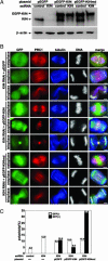
Restoration of the localization of PRC1 and rescue of cytokinetic defects in Kif4 esiRNA-treated cells by ectopic expression of GFP-Kif4. (A) HeLa cells grown in a 6-well plate were transfected with 400 ng of pEGFP, pEGFP-Kif4, or pEGFP-Kif4md. Twenty-four hours later, cells were transfected with 100 nM of control (luciferase) esiRNA or Kif4A esiRNA. Two days after esiRNA transfection, cells were lysed and lysates were subjected to SDS/PAGE and immunoblotted with anti-Kif4 antibody or anti-β-actin antibody. (B) HeLa cells grown on coverslips were transfected with plasmids and esiRNAs as in A. After transfection, cells were fixed and stained with anti-PRC1 antibodies (red), anti-α-tubulin antibody (blue), and DAPI (DNA, white). The expression of GFP proteins is shown in green. (Scale bar: 5 μm.) (C) HeLa cells grown on coverslips were transfected with plasmids and/or esiRNAs, fixed, and stained with antibodies and DAPI as in B. The percentages of polyploidy in GFP-negative or GFP-positive cells transfected with the indicated esiRNAs and/or plasmids were scored under a fluorescence microscope. More than 300 cells were counted in each experiment.

Similar articles

Cited by

References

    1. Glotzer, M. (2001) Annu. Rev. Cell Dev. Biol. 17, 351-386. - PubMed
    1. Wheatley, S. P., Hinchcliffe, E. H., Glotzer, M., Hyman, A. A., Sluder, G. & Wang, Y. (1997) J. Cell Biol. 138, 385-393. - PMC - PubMed
    1. Adams, R. R., Carmena, M. & Earnshaw, W. C. (2001) Trends Cell Biol. 11, 49-54. - PubMed
    1. Mollinari, C., Kleman, J. P., Jiang, W., Schoehn, G., Hunter, T. & Margolis, R. L. (2002) J. Cell Biol. 157, 1175-1186. - PMC - PubMed
    1. Matuliene, J. & Kuriyama, R. (2004) Mol. Biol. Cell 15, 3083-3094. - PMC - PubMed

Publication types

LinkOut - more resources