. 2005 Jan 11;102(2):425-30.
doi: 10.1073/pnas.0408357102. Epub 2004 Dec 29.

Induction, differentiation, and remodeling of blood vessels after transplantation of Bcl-2-transduced endothelial cells

Affiliations

Induction, differentiation, and remodeling of blood vessels after transplantation of Bcl-2-transduced endothelial cells

David R Enis et al. Proc Natl Acad Sci U S A. .

Abstract

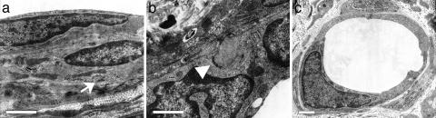
Implants of collagen-fibronectin gels containing Bcl-2-transduced human umbilical vein endothelial cells (Bcl-2-HUVECs) induce the formation of human endothelial cell (EC)/murine vascular smooth muscle cell (VSMC) chimeric vessels in immunodeficient mice. Microfil casting of the vasculature 60 d after implantation reveals highly branched microvascular networks within the implants that connect with and induce remodeling of conduit vessels arising from the abdominal wall circulation. Approximately 85% of vessels within the implants are lined by Bcl-2-positive human ECs expressing VEGFR1, VEGFR2, and Tie-2, but not integrin alpha(v)beta(3). The human ECs are seated on a well formed human laminin/collagen IV-positive basement membrane, and are surrounded by mouse VSMCs expressing SM-alpha actin, SM myosin, SM22alpha, and calponin, all markers of contractile function. Transmission electron microscopy identified well formed EC-EC junctions, chimeric arterioles with concentric layers of contractile VSMC, chimeric capillaries surrounded by pericytes, and chimeric venules. Bcl-2-HUVEC-lined vessels retain 70-kDa FITC-dextran, but not 3-kDa dextran; local histamine rapidly induces leak of 70-kDa FITC-dextran or India ink. As in skin, TNF induces E-selectin and vascular cell adhesion molecule 1 only on venular ECs, whereas intercellular adhesion molecule-1 is up-regulated on all human ECs. Bcl-2-HUVEC implants are able to engraft within and increase perfusion of ischemic mouse gastrocnemius muscle after femoral artery ligation. These studies show that cultured Bcl-2-HUVECs can differentiate into arterial, venular, and capillary-like ECs when implanted in vivo, and induce arteriogenic remodeling of the local mouse vessels. Our results support the utility of differentiated EC transplantation to treat tissue ischemia.


Figures

Fig. 1.
Fig. 1.
Microfil casting. Glycerol-cleared casts of abdominal-wall vasculature of mice bearing either a collagen/fibronectin gel implant containing Bcl-2-HUVEC (a, c, and d) or a mock implant (b). (c and d) Higher magnifications of cellularized implants that reveal heterogeneously sized, branched vessels within the graft, and induced remodeling of conduit vessels connecting the implants to the local mouse circulation Results representative of casts from six animals. (Scale bar, 1 mm.)
Fig. 2.
Fig. 2.
Cellular phenotyping of investing VSMCs. Recruited mouse cells express SMA (a), calponin (b), SM22α (c), and SM myosin (d) molecules indicative of VSMC contractile function. All staining is red with blue hematoxylin counterstain. Similar results were obtained from four specimens. (Scale bar, 100 μm.)
Fig. 3.
Fig. 3.
Transmission electron microscopy. Engrafted vessels have recruited multiple layers of investing cells (a) that contain networks of microfilaments organized around dense bodies and subsarcolemmal dense plaques (arrow), which is consistent with formation of an SMC contractile apparatus. EC junctional complexes (b, arrowhead) and basement membrane are also visible. Numerous simple capillaries (c) are also present in the implants. Images were collected from an analysis of four specimens. (Scale bars, 1 μm.)
Fig. 4.
Fig. 4.
Analysis of vascular permeability and histamine response. (a) Bcl-2-HUVECs [stained with i.v.-injected, human EC-binding lectin rhodamine-UEA-1 (red)] retain 70-kDa FITC-dextran (a, green) that remains within vessel lumena 30 min after injection (n = 5). (b) In contrast, 3-kDa FITC-dextran (b, green) diffuses throughout the interstitum within 3 min of i.v. injection (n = 3). (c) Histamine injection induces leak of circulating 70-kDa FITC-dextran within gels containing Bcl-2-HUVECs (c, green) (n = 6). (d) Extravasation of circulating colloidal carbon (arrows) by a subset Bcl-2-HUVEC-lined vessels (identified by UEA-1 staining, red) after local injection of histamine, but not saline (Inset) (n = 3). (Scale bars, 100 μm.)
Fig. 5.
Fig. 5.
Analysis of TNF response in vitro and in vivo. In vitro, Bcl-2-HUVECs uniformly up-regulate E-selectin in response to TNF, as noted by flow cytometry of E-selectin expression on 2D-cultured ECs (a) or immunohistochemistry of Bcl-2-HUVECs suspended in TNF-treated gels (b Inset, with saline-treated control gel). Specimens in b were double-stained with anti-E-selectin (blue) and UEA-1 (red). (d, f, h, and j) In vivo, Bcl-2-HUVECs are compared with vessels in human skin grafts harvested from the same animal (shown in c, e, g, and i). (Insets) Identically stained, saline-treated controls are shown. Double staining with anti-E-selectin (c and d), anti-VCAM-1 (e and f), and anti-ICAM-1 (g and h, all in blue) with UEA-1 (red) demonstrates that all human ECs in both the skin and Bcl-2-HUVEC implants respond to TNF by up-regulating ICAM-1, in contrast to a restricted induction of E-selectin and VCAM-1. E-selectin expression is generally confined to vessels with venular, and not arteriolar, specialization of the vessel wall, as indicated by double-staining with anti-SMA (blue) and anti-E-selectin (red) in both human skin (i) and Bcl-2-HUVECs (j). Similar results obtained in gel implants from four experiments. (Scale bar, 100 μm.)
Fig. 6.
Fig. 6.
Bcl-2-HUVEC implants engraft in ischemic hindlimb. After bilateral ligation of the femoral artery (a and b, arrowheads), implants containing Bcl-2-HUVECs (a) or a mock implant of collagen/fibronectin alone (b) were placed into the gastrocnemius muscle (a and b, black arrows). Hematoxylin/eosin staining demonstrates engraftment and formation of complex microvessels within the graft containing Bcl-2-HUVECs (c) but not mock (d). Images are representative of specimens harvested from six animals over two experiments. (White scale bar, 1 mm; black scale bar, 100 μm.)

References

    1. Jain, R. K. (2003) Nat. Med. 9, 685-693. - PubMed
    1. Choi, J., Enis, D., Koh, K., Shiao, S. & Pober, J. S. (2004) Annu. Rev. Immunol. 22, 683-709. - PubMed
    1. Tsurumi, Y., Takeshita, S., Chen, D., Kearney, M., Rossow, S. T., Passeri, J., Horowitz, J. R., Symes, J. F. & Isner, J. M. (1996) Circulation 94, 3281-3290. - PubMed
    1. Dor, Y., Djonov, V. & Keshet, E. (2003) Ann. N.Y. Acad. Sci. 995, 208-216. - PubMed
    1. de Muinck, E. D. & Simons, M. (2004) J. Mol. Cell. Cardiol. 36, 25-32. - PubMed

Publication types