. 2005 Jan 15;19(2):214-23.
doi: 10.1101/gad.1258705. Epub 2004 Dec 29.

Sustained Hedgehog signaling is required for basal cell carcinoma proliferation and survival: conditional skin tumorigenesis recapitulates the hair growth cycle

Affiliations

Sustained Hedgehog signaling is required for basal cell carcinoma proliferation and survival: conditional skin tumorigenesis recapitulates the hair growth cycle

Mark E Hutchin et al. Genes Dev. .

Abstract

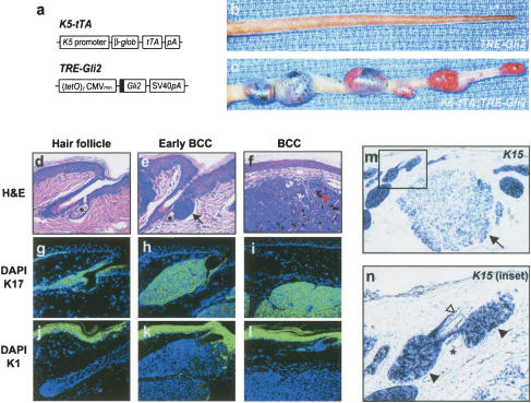
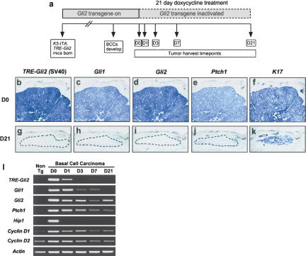
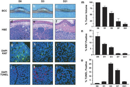
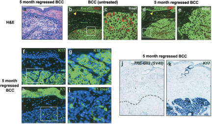
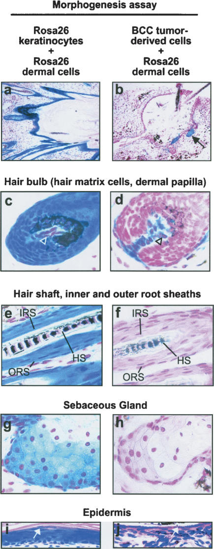
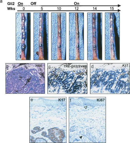
Temporally and spatially constrained Hedgehog (Hh) signaling regulates cyclic growth of hair follicle epithelium while constitutive Hh signaling drives the development of basal cell carcinomas (BCCs), the most common cancers in humans. Using mice engineered to conditionally express the Hh effector Gli2, we show that continued Hh signaling is required for growth of established BCCs. Transgene inactivation led to BCC regression accompanied by reduced tumor cell proliferation and increased apoptosis, leaving behind a small subset of nonproliferative cells that could form tumors upon transgene reactivation. Nearly all BCCs arose from hair follicles, which harbor cutaneous epithelial stem cells, and reconstitution of regressing tumor cells with an inductive mesenchyme led to multilineage differentiation and hair follicle formation. Our data reveal that continued Hh signaling is required for proliferation and survival of established BCCs, provide compelling support for the concept that these tumors represent an aberrant form of follicle organogenesis, and uncover potential limitations to treating BCCs using Hh pathway inhibitors.


Figures

Figure 1.
Figure 1.
Follicle-derived BCC development in K5–tTA;TRE–Gli2 bitransgenic mice. (a) Schematic showing transgenic constructs designed to yield bitransgenic mice harboring a skin-targeted tetracycline transactivator (K5–tTA) and the tetracycline response element–Gli2 transgene (TRE–Gli2). Sequence encoding Flag epitope tag is shown by black box. (b,c) Tails from single and bitransgenic mice, with BCCs in bitransgenic mouse only. (df) Photomicrographs (H&E staining) of normal hair follicle and early BCC arising from hair follicle (e, black arrow), with sebaceous glands marked by asterisk (d,e). (f) Established BCCs frequently contain focal BCC pigmentation (red arrowhead). (gl) Immunofluorescence for keratins K17 and K1, both visualized with FITC (green) with nuclei counterstained with DAPI (blue). Normal hair follicle as well as BCCs express K17, while abundant expression of K1 is detected in the epidermis. (m,n) In situ hybridization for the follicle marker K15 shows high expression in early tumors (n, inset, black arrowheads) and lower expression in larger BCCs (m, black arrow). A hair shaft (n, white arrowhead) is visible near the K15-positive early BCC, illustrating that early BCCs arise from hair follicles. Sebaceous gland marked by asterisk.
Figure 2.
Figure 2.
Trangene inactivation and hedge-hog target gene down-regulation following administration of doxycycline to BCC-bearing mice. (a) Schematic showing time course of transgene inactivation experiments. (bk) Transgene and hedgehog target gene expression in BCCs from untreated vs. doxycycline-treated (21 d) bitransgenic mice, assessed using digoxigenin-labeled antisense riboprobes. Efficient transgene inactivation was confirmed by undetectable levels of transgene-derived Gli2 mRNA (g), which was accompanied by strong down-regulation of Gli1, Gli2, and Ptch1 (hj). (k) The presence of K17-positive cells in treated tumors indicates the persistence of a subset of cells in the absence of transgene expression and verifies mRNA integrity. Dashed lines indicate residual tumor cells in Day 21 regressed samples. (d,i) Note that riboprobe for Gli2 recognizes both transgene-derived and endogenous Gli2. (l) Semiquantitative RT–PCR demonstrating inactivation kinetics of transgene-derived Gli2 (TRE–Gli2) and hedgehog target genes in tumor-bearing mice fed doxycycline. Note up-regulation of hedgehog target genes Gli1, Gli2, Ptch1, Hip1, and Cyclins D1 and D2 in Day 0 BCC compared to nontransgenic mouse skin (Non Tg), and down-regulation following transgene inactivation.
Figure 3.
Figure 3.
BCC regression following transgene inactivation is associated with reduced proliferation and increased apoptosis. (ac) Gross appearance of representative tail BCC demonstrating reduction in tumor size over the 21-d time course. (df) H&E-stained sections demonstrating histology of regressing BCCs. Note reduced cellularity of BCC at Day 21 (D21) relative to Day 0 (D0). (gi) Immunofluorescence for the proliferation marker Ki67 (green) and associated nuclear counterstain DAPI (blue) at various time points. Note the dramatic reduction in proliferative nuclei over the 21-day period of regression. (jl) TUNEL staining (red) as an indicator of apoptosis and nuclear counterstain DAPI (blue). Note the striking increase in apoptotic cells at Day 3 (D3) time point. (m) Quantitation of reduction in tumor volume over time. Each bar represents the average percentage of the initial (Day 0) tumor size for three BCCs from different mice. Standard errors indicated by bars. (n,o) Quantitation of the percentage of Ki67 or TUNEL-positive nuclei over time. Data are presented as average counts from three tumors per time point, with 10 high-powered fields counted per tumor for each time point. Standard errors indicated by bars.
Figure 4.
Figure 4.
Long-term persistence of nonproliferative cell population in regressed BCCs. (a) Photomicrograph illustrating typical morphology of persistent cell population in long-term (5-mo) regressed BCC (H&E stain). (be) Immunofluorescence for keratins (anti-pan-keratin; green) and Ki67 (red), demonstrating extensive proliferation in untreated BCCs and the absence of detectable proliferation in long-term treated tumors (5-mo regressed BCC). Note the presence of Ki67-positive nuclei in the basal layer of the epidermis in both conditions (yellow arrowheads). (fi) Immunofluorescent images demonstrating keratins K1 or K17 (green) and DAPI nuclear counterstain (blue) in long-term regressed BCCs. K17 expression is present in nearly all tumor cells, while K1 was present focally in a subset of tumors treated with doxycycline for 5 mo. (j,k) In situ hybridization illustrating the absence of Gli2 transgene expression [TRE–Gli2 (SV40)] and presence of K17 expression (blue) in residual tumor cells. Area of tumor in left panel delineated by black dashed line.
Figure 5.
Figure 5.
Regressing BCC tumor cells produce hair follicle and other epithelial lineages when combined with inductive dermal cells. Hair morphogenesis assays using inductive mesenchyme provided by newborn dermal cells from β-galactosidase-expressing Rosa26 mice. Epithelial cell component consisted either of newborn Rosa26 keratinocytes (left column) or BCC tumor-derived cells (right column). Mesenchymal-epithelial cell mixtures were injected subcutaneously into doxycycline-fed NOD-scid mice to inactivate Gli2 transgene expression, and grafts harvested 3 wk later. Tissue was whole-mount stained to detect β-galactosidase activity, and counterstained with either nuclear fast red (NFR, ah) or H&E (i,j). (a,b) Hair follicles project into cystic cavities in assays containing either control keratinocytes or BCC tumor-derived cells. Note that in control assay (left column) all epithelial cells stain deep blue, whereas in BCC-derived assay (right column) nearly all epithelial cells are β-galactosidase-negative, with the exception of focal staining (b, black arrow) consistent with the presence of a small number of contaminating Rosa26 keratinocytes in the dermal cell preparation. In BCC-derived assay, mature hair shafts are visible in the upper right corner of b.(c,d) Hair bulbs demonstrating deep-blue staining of follicular matrix cells and lighter staining of dermal papilla in control (c, arrowhead). In BCC-derived assays, the relationship of the β-galactosidase-negative epithelial cells to the β-galactosidase-positive dermal papilla cells (d, arrowhead) is preserved. (e,f) A mature-appearing hair shaft (HS) is present in both control and BCC-derived cell morphogenesis assays. In addition, inner and outer root sheath compartments (IRS and ORS) are present in controls (e) and BCC-derived follicles (f). (g,h) Sebaceous glands were evident in both control and tumor-derived cell assays. (i,j) Cyst lining showing keratohyaline granules characteristic of the epidermal granular cell layer (white arrows), in both control and BCC-derived assays.
Figure 6.
Figure 6.
Reactivation of transgene in regressed tumors results in resumption of tumor growth. (a) Gross appearance of tumor-bearing tail over a 15-wk observation period, beginning with 5 wk of doxycycline treatment (Gli2 Off, dashed line). Note reduction in tumor size following doxycycline treatment, with regrowth of several tumors (white arrow, panel on far right) following discontinuation of doxycycline (Gli2 On, solid line). (b) Histology of regressed-reactivated tumor, with pigment (black) surrounding tumor nodules. (c,d) In situ hybridization [TRE–Gli2 (SV40)] showing expression of transgene and K17 in regressed-reactivated tumor following transgene reactivation. (e,f) Immunohistochemistry for K17 and Ki67 in reactivated tumors. Tumors were positive for K17 expression. The basal layer of the epidermis was Ki67-positive (f, open arrowhead) as was the reactivated tumor epithelium (f, black arrowhead).

References

    1. Al Hajj M., Wicha, M.S., Benito-Hernandez, A., Morrison, S.J., and Clarke, M.F. 2003. Prospective identification of tumorigenic breast cancer cells. Proc. Natl. Acad. Sci. 100: 3983–3988. - PMC - PubMed
    1. Allen M., Grachtchouk, M., Sheng, H., Grachtchouk, V., Wang, A., Wei, L., Liu, J., Ramirez, A., Metzger, D., Chambon, P., et al. 2003. Hedgehog signaling regulates sebaceous gland development. Am. J. Pathol. 163: 2173–2178. - PMC - PubMed
    1. Aszterbaum M., Epstein, J., Oro, A., Douglas, V., LeBoit, P.E., Scott, M.P., and Epstein Jr., E.H. 1999. Ultraviolet and ionizing radiation enhance the growth of BCCs and trichoblastomas in patched heterozygous knockout mice. Nat. Med. 5: 1285–1291. - PubMed
    1. Bai C.B., Auerbach, W., Lee, J.S., Stephen, D., and Joyner, A.L. 2002. Gli2, but not Gli1, is required for initial Shh signaling and ectopic activation of the Shh pathway. Development 129: 4753–4761. - PubMed
    1. Bale A.E. and Yu, K.P. 2001. The hedgehog pathway and basal cell carcinomas. Hum. Mol. Genet. 10: 757–762. - PubMed

Publication types

MeSH terms