Comparative Study
. 2003 May;1(2):145-54.
doi: 10.1016/s1672-0229(03)01018-0.

The structure analysis and antigenicity study of the N protein of SARS-CoV

Affiliations
Comparative Study

The structure analysis and antigenicity study of the N protein of SARS-CoV

Jingqiang Wang et al. Genomics Proteomics Bioinformatics. 2003 May.

Abstract

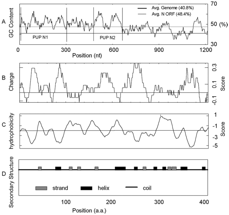
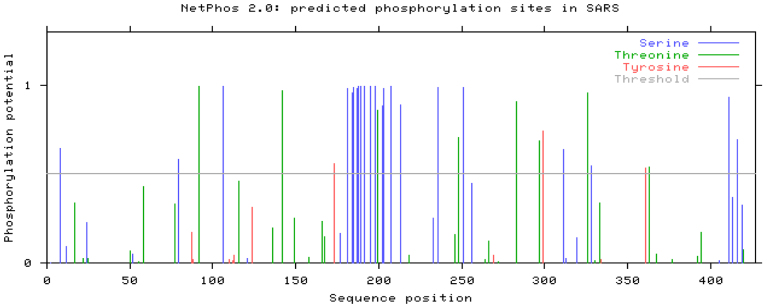
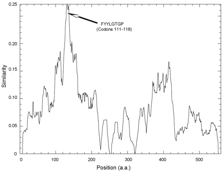
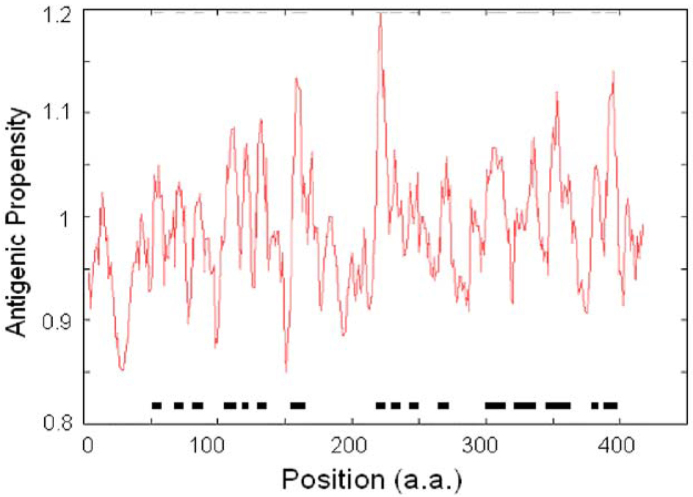
The Coronaviridae family is characterized by a nucleocapsid that is composed of the genome RNA molecule in combination with the nucleoprotein (N protein) within a virion. The most striking physiochemical feature of the N protein of SARS-CoV is that it is a typical basic protein with a high predicted pI and high hydrophilicity, which is consistent with its function of binding to the ribophosphate backbone of the RNA molecule. The predicted high extent of phosphorylation of the N protein on multiple candidate phosphorylation sites demonstrates that it would be related to important functions, such as RNA-binding and localization to the nucleolus of host cells. Subsequent study shows that there is an SR-rich region in the N protein and this region might be involved in the protein-protein interaction. The abundant antigenic sites predicted in the N protein, as well as experimental evidence with synthesized polypeptides, indicate that the N protein is one of the major antigens of the SARS-CoV. Compared with other viral structural proteins, the low variation rate of the N protein with regards to its size suggests its importance to the survival of the virus.


Figures

Fig. 1
Fig. 1
The predicted distributions of GC content (A), electric charge (B), hydrophobicity (C) and secondary structure (D) in the N protein of SARS-CoV.
Fig. 2
Fig. 2
The predicted phosphorylation sites on the N protein. We identified 33 potential phosphorylation sites in the N protein, including 22 serines, 8 threonines and 3 tyrosines. The average score of serines are significantly higher than that of the other two. The phosphorylation sites concentrate in the middle of the N protein.
Fig. 3
Fig. 3
The similarity chart of the N protein. Based on multi-alignment of totally nineteen coronavirus N proteins, two conserved regions were found around a.a. 81-140 and a.a. 270-320 (amino acid positions are all referred to the N protein of SARS-CoV, Isolate BJ01). The arrow indicates the most conserved domain, and its sequence and amino acid position are given. In contrast, the two termini of the N protein are more variable, particularly the C- terminal. The figure was generated by Plotcon in the EMBOSS package (http://www.hgmp.mrc.ac.uk/Software/EMBOSS/).
Fig. 4
Fig. 4
The possible antigenic sites of the N protein. We have predicted sixteen antigenic sites of the N protein, and found they are clustering in the middle and the C-terminal. There is a strong antigenic site (TALALLLLDR) located around Codons 218-227. In addition, another three strong antigenic sites are detected around Codons 156-166 (AATVLQLPQGT), Codons 347-363 (FKDNVILLNKHIDAYKT) and Codons 389-398 (KQPTVTLLPA).
Fig. 5
Fig. 5
The phylogenetic tree based on nineteen coronavirus N proteins. It reveals that SARS-CoV is closer to Group 2 than to Groups 1 and 3. Abbreviations: AIBV: avian infectious bronchitis virus; BCoV: bovine coronavirus; CCoV: canine coronavirus; CECoV: canine enteric coronavirus; ECoV: equine coronavirus; FCoV: feline coronavirus; FIPV: feline infectious peritonitis virus; HCoV-OC43: human coronavirus strain OC43; HCoV-229E: human coronavirus strain 229E; MHV: murine hepatitis virus; PEDV: porcine epidemic diarrhea virus; PHEV: porcine hemagglutmating encephalomyelitis virus; PRCoV: porcine respiratory coronavirus; PTGV: transmissible gastroenteritis virus; PV: puffinosis virus; RCoV: rat coronavirus; RSCoV: Rat sialodacryoadenitis coronavirus; SARS-CoV: human severe acute respiratory syndrome-associated coronavirus isolate BJ01; TCoV: turkey coronavirus.

References

    1. Ksiazek T.G. A Novel Coronavirus Associated with Severe Acute Respiratory Syndrome. N. Engl. J. Med. 2003;348:1953–1966. - PubMed
    1. Hiscox J.A. The coronavirus infectious bronchitis virus nucleoprotein localizes to the nucleolus. J. Virol. 2001;75:10506–10512. - PMC - PubMed
    1. Wootton S.K. Phosphorylation of the porcine reproductive and respiratory syndrome virus nucleocapsid protein. J. Virol. 2002;76:10569–10576. - PMC - PubMed
    1. Laity J.H. Zinc finger proteins: new insights into structural and functional diversity. Curr Opin Struct Biol. 2001;11:39–46. - PubMed
    1. Molenkamp R., Spaan W.J.M. Identification of a specific interaction between the coronavirus mouse hepatitis virus A59 nucleocapsid protein and packaging signal. Virology. 1997;239:78–86. - PMC - PubMed

Publication types