. 2004 Sep-Dec;25(5-6):243-51.
doi: 10.1159/000081387.

Surface expression of Hsp25 and Hsp72 differentially regulates tumor growth and metastasis

Affiliations

Surface expression of Hsp25 and Hsp72 differentially regulates tumor growth and metastasis

María A Bausero et al. Tumour Biol. 2004 Sep-Dec.

Abstract

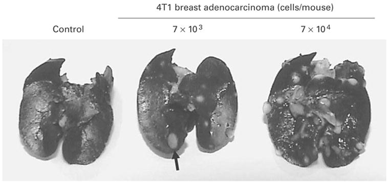
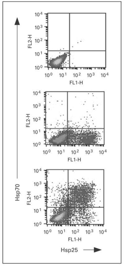
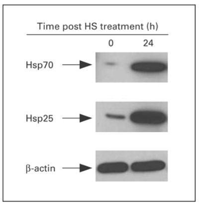
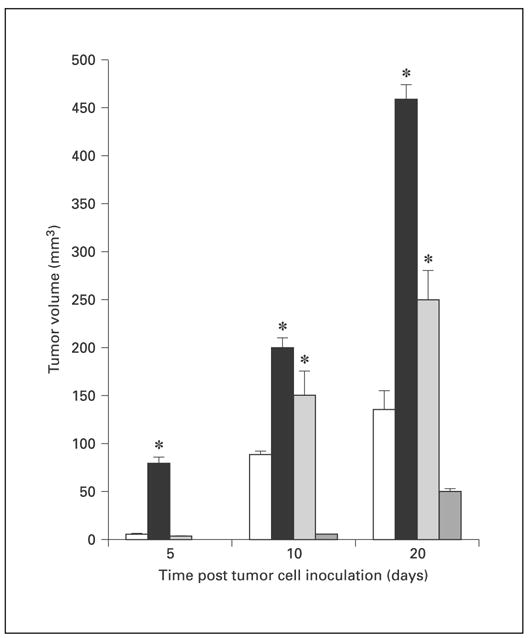
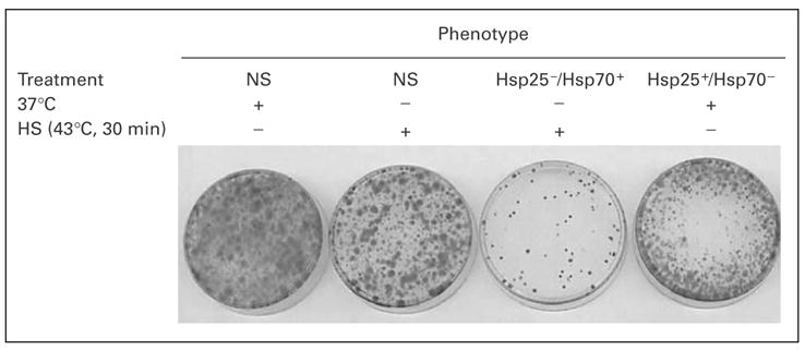
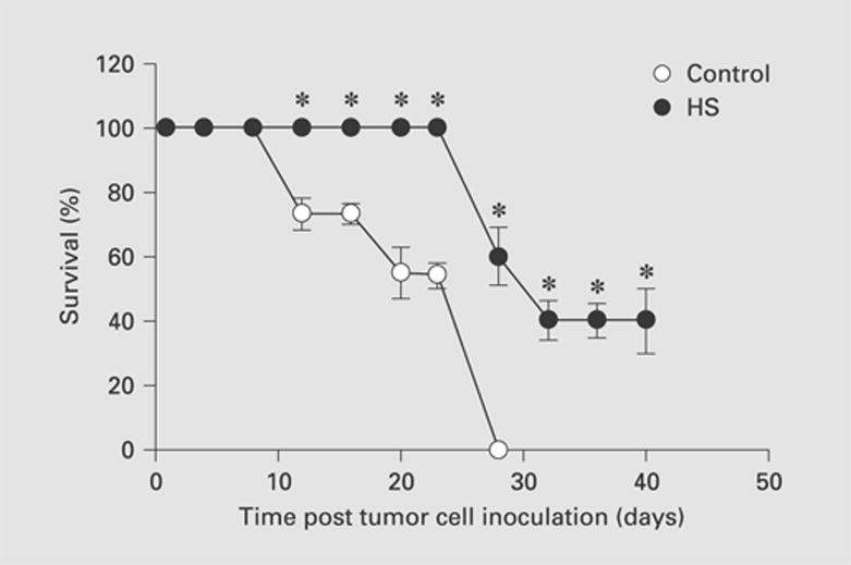
The expression of unique surface structures on tumors that allow for recognition and activation of host immunocompetent cells plays an important role in determining tumor growth and/or metastasis. Recent studies have identified an important role for heat shock proteins (Hsp) in antitumor surveillance; however, the exact role of Hsp expressed on the surface of tumors has not been fully addressed. In this study, we show that 4T1 mammary adenocarcinoma cells sorted for high Hsp25 surface expression (Hsp25(high)) grow significantly faster than cells sorted for intermediate Hsp25 surface expression (Hsp25(intermediate)) or wild-type 4T1 cells implanted into the abdominal breast gland of female BALB/c mice (p < 0.05). In addition, histological examination of lung tissues revealed that Hsp25(high) 4T1 cells metastasized to the lungs more aggressively than either Hsp25(intermediate) or wild-type 4T1 cells (p < 0.05). Exposure of 4T1 cells to nonlethal heat shock (43 degrees C, 30 min) induced the surface expression of Hsp72 and a concomitant reduction in Hsp25 surface expression. The growth and metastastic potential of Hsp72(+) 4T1 cells was significantly less than that of Hsp25(high), Hsp25(intermediate) or wild-type 4T1 cells (p < 0.05). Taken together, these studies identify an important role for expression of Hsp25 and Hsp72 during tumor growth and metastatic spread which might be helpful in the design of antimetastatic therapies.


Figures

Fig. 1
Fig. 1
Injection of 4T1 breast adenocarcinoma cells into the abdominal breast gland results in the development of lung metastasis. Female BALB/c mice (6–8 weeks old) were injected with 103 (middle panel) or 7 × 10⁴ (right panel) 4T1 cells, or with culture medium alone (left panel) subcutaneously into the abdominal breast gland. Twenty-one days after treatment, the animals were euthanized and the lungs were filled with a solution of India ink (1:6 in PBS) via the intratracheal route. The figures show lung metastasis (represented by a solid arrow) and are representative of three independently performed experiments with similar results.
Fig. 2
Fig. 2
Surface expression of Hsp is modulated by exposure of tumors to heat shock treatment. 4T1 breast adenocarcinoma cells (2 × 10⁶) were exposed to nonlethal heat shock treatment (43°C, 30 min; lower panel) or maintained at 37°C (middle panel) and allowed to recover at 37°C overnight. Cells were simultaneously stained with anti-Hsp72-PE, anti-Hsp25-Alexa Fluor or isotype control (top panel), and relative surface expression was analyzed by flow cytometry. Dot plots represent the relative percentage of cells expressing Hsp27 or Hsp72 from the gated population of cells and are representative of six independently performed experiments with similar results.
Fig. 3
Fig. 3
Kinetics of total expression of Hsp25 and Hsp72. Exponentially growing 4T1 cells were either exposed to nonlethal heat shock (HS) treatment (43°C, 30 min) and immediately lyzed (0 h) or allowed to recover for 24 h and lyzed (24 h). The extracted proteins were analyzed by 12% SDS-PAGE and immunoblot with antibodies specific for Hsp72 (upper panel) or Hsp25 (middle panel) and visualized with a coupled secondary antibody-chemiluminescence method. β-actin (bottom panel) was used as an internal control. Results are representative of two independently performed experiments with similar results.
Fig. 4
Fig. 4
Surface expression of Hsp25 and Hsp72 on 4T1 cells correlates with enhanced and suppressed tumor growth, respectively. Female BALB/c mice (6–8 weeks old) received an injection into the abdominal breast gland of 7 × 10⁴ 4T1 breast adenocarcinoma cells that were either unsorted (white bars) or sorted for Hsp25high/Hsp72-expressing (black bars), Hsp25intermediate/Hsp72-expressing (light-gray bars) or Hsp25/Hsp72+-expressing (dark-gray bars) cells, and tumor growth was measured using an electronic caliper. Data are mean tumor volume ± SE and represent the sum of two independently performed experiments with 5 mice per treatment group. tci = Tumor cell inoculation. * p < 0.05 versus unsorted cells.
Fig. 5
Fig. 5
Clonogenicity assay to measure the metastatic potential of 4T1 breast adenocarcinoma cells correlates with high Hsp25 surface expression. 4T1 breast adenocarcinoma cells (7 × 10⁴) that were either not sorted (NS) or sorted for Hsp25/Hsp72+ or Hsp25+/ Hsp72cells were injected into the abdominal breast gland of female BALB/c mice (6–8 weeks old). After 21 days, lungs were excised, digested and made into a single-cell suspension, then seeded in triplicate at 1,000 cells/60-mm3 petri dish and incubated at 37°C in a 5% CO2 air atmosphere. Ten to twelve days later, the plates were washed twice with PBS and colonies were stained with crystal violet. Results represent the cloning efficiencies following various treatment protocols and are representative of two independently performed experiments with similar results. HS = Heat shock.
Fig. 6
Fig. 6
Exposure of 4T1 breast adenocarcinoma cells to nonlethal heat shock treatment increased the survival of animals after tumor challenge. 4T1 breast adenocarcinoma cells (7 × 10⁴) that were either maintained at 37°C (open circles) or exposed to nonlethal heat shock treatment (HS; 43°C, 30 min) and allowed to recover for 24 h at 37°C (filled circles) were injected into the abdominal breast gland of female BALB/c mice (6–8 weeks old). Data are the mean percentage survival ± SEM and represent the sum of three independently performed experiments. * p < 0.05 versus control.

References

    1. Craig EA, Gross CA. Is hsp70 the cellular thermometer? Trends Biochem Sci. 1991;16:135–140. - PubMed
    1. Lindquist S, Craig EA. The heat-shock proteins. Annu Rev Genet. 1988;22:631–677. - PubMed
    1. Morimoto RI, Tissières A, Georgopoulos C. The Biology of Heat Shock Proteins and Molecular Chaperones. Cold Spring Harbor: Cold Spring Harbor Laboratory Press; 1994.
    1. Ciocca DR, Green S, Elledge RM, Clark GM, Pugh R, Ravdin P, Lew D, Martino S, Osborne CK. Heat shock proteins hsp27 and hsp70: Lack of correlation with response to tamoxifen and clinical course of disease in estrogen receptor-positive metastatic breast cancer (a Southwest Oncology Group Study) Clin Cancer Res. 1998;4:1263–1266. - PubMed
    1. Ciocca DR, Vargas-Roig LM. Hsp27 as a prognostic and predictive factor in cancer. Prog Mol Subcell Biol. 2002;28:205–218. - PubMed

Publication types

MeSH terms