Review
. 2005 Jan;132(1):1-26.
doi: 10.1016/j.jcpa.2004.09.005.

Attaching-effacing bacteria in animals

Affiliations
Review

Attaching-effacing bacteria in animals

A D Wales et al. J Comp Pathol. 2005 Jan.

Abstract

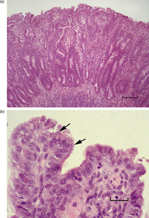
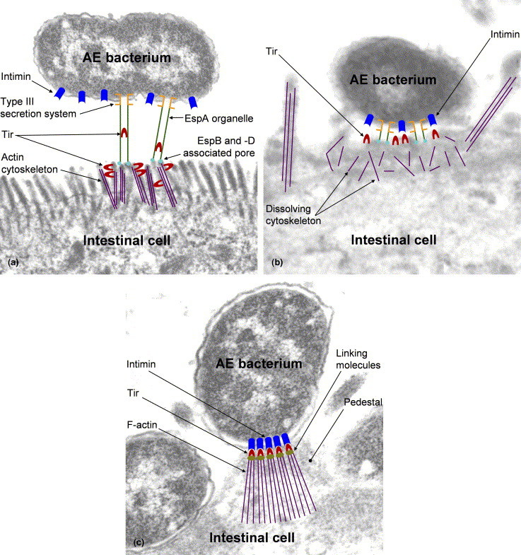
Enteric bacteria with a demonstrable or potential ability to form attaching-effacing lesions, so-called attaching-effacing (AE) bacteria, have been found in the intestinal tracts of a wide variety of warm-blooded animal species, including man. In some host species, for example cattle, pigs, rabbits and human beings, attaching-effacing Escherichia coli (AEEC) have an established role as enteropathogens. In other host species, AE bacteria are of less certain significance. With continuing advances in the detection and typing of AE strains, the importance of these bacteria for many hosts is likely to become clearer. The pathogenic effects of AE bacteria result from adhesion to the intestinal mucosa by a variety of mechanisms, culminating in the formation of the characteristic intimate adhesion of the AE lesion. The ability to induce AE lesions is mediated by the co-ordinated expression of some 40 bacterial genes organized within a so-called pathogenicity island, known as the "Locus for Enterocyte Effacement". It is also believed that the production of bacterial toxins, principally Vero toxins, is a significant virulence factor for some AEEC strains. Recent areas of research into AE bacteria include: the use of Citrobacter rodentium to model human AEEC disease; quorum-sensing mechanisms used by AEEC to modulate virulence gene expression; and the potential role of adhesion in the persistent colonization of the intestine by AE bacteria. This review of AE bacteria covers their molecular biology, their occurrence in various animal species, and the diagnosis, pathology and clinical aspects of animal diseases with which they are associated. Reference is made to human pathogens where appropriate. The focus is mainly on natural colonization and disease, but complementary experimental data are also included.


Figures

Fig. 1a–d
Fig. 1a–d
Ileum of calf infected with E. coli O26. (a) The mucosa has a flat appearance due to villous stunting and fusion. The surface epithelium has an irregular appearance. Haematoxylin and eosin (HE). Bar, 100 μm. (b) Adherent bacteria (arrows) are present on the surface of enterocytes. HE. Bar, 15 μm. (c) Adherent bacteria, labelled with an O26 antiserum, are present on the irregular epithelial surface. Immunoperoxidase. Bar, 20 μm. (d) Adherent bacteria are intimately attached to the surface of enterocytes and microvilli are effaced. Condensed actin filaments are seen at cell apex (arrow). TEM. Bar, 500 nm.
Fig. 1a–d
Fig. 1a–d
Ileum of calf infected with E. coli O26. (a) The mucosa has a flat appearance due to villous stunting and fusion. The surface epithelium has an irregular appearance. Haematoxylin and eosin (HE). Bar, 100 μm. (b) Adherent bacteria (arrows) are present on the surface of enterocytes. HE. Bar, 15 μm. (c) Adherent bacteria, labelled with an O26 antiserum, are present on the irregular epithelial surface. Immunoperoxidase. Bar, 20 μm. (d) Adherent bacteria are intimately attached to the surface of enterocytes and microvilli are effaced. Condensed actin filaments are seen at cell apex (arrow). TEM. Bar, 500 nm.
Fig. 2a–c
Fig. 2a–c
Composite, schematic diagram illustrating proposed mechanisms in the formation of the attaching-effacing lesion. (a) Non-intimate adhesion and protein translocation. EspA, -B and -D are exported via a type III secretion system and form a bi-functional organelle permitting adhesion of the AE bacterium to the host cell and translocation of Tir into the host cell. Esp, E. coli secreted protein;Tir, translocated intimin receptor. (b) Signal transduction. The host cell cytoskeleton is dissolved locally, leading to effacement of microvilli. Tir is inserted in the host cell membrane. Tir, translocated intimin receptor. (c) Intimate adhesion. Tir focuses filamentous (F) actin, forming a pedestal, and binds intimin in the bacterial outer membrane. Tir, translocated intimin receptor.

References

    1. Abe A., Heczko U., Hegele R.G., Finlay B.B. Two enteropathogenic Escherichia coli type III secreted proteins, EspA and EspB, are virulence factors. Journal of Experimental Medicine. 1998;188:1907–1916. - PMC - PubMed
    1. Acres S.D. Enterotoxigenic Escherichia coli infections in newborn calves: a review. Journal of Dairy Science. 1985;68:229–256. - PMC - PubMed
    1. Adams L.M., Simmons C.P., Rezmann L., Strugnell R.A., Robins-Browne R.M. Identification and characterization of a K88- and CS31A-like operon of a rabbit enteropathogenic Escherichia coli strain which encodes fimbriae involved in the colonization of rabbit intestine. Infection and Immunity. 1997;65:5222–5230. - PMC - PubMed
    1. Adu-Bobie J., Frankel G., Bain C., Goncalves A.G., Trabulsi L.R., Douce G., Knutton S., Dougan G. Detection of intimins alpha, beta, gamma, and delta, four intimin derivatives expressed by attaching and effacing microbial pathogens. Journal of Clinical Microbiology. 1998;36:662–668. - PMC - PubMed
    1. Aktan I., Sprigings K.A., La Ragione R.M., Faulkner L.M., Paiba G.A., Woodward M.J. Characterisation of attaching-effacing Escherichia coli isolated from animals at slaughter in England and Wales. Veterinary Microbiology. 2004;102:43–53. - PubMed

MeSH terms

LinkOut - more resources