. 2005 Jan;187(2):488-97.
doi: 10.1128/JB.187.2.488-497.2005.

Nature of the promoter activated by C.PvuII, an unusual regulatory protein conserved among restriction-modification systems

Affiliations

Nature of the promoter activated by C.PvuII, an unusual regulatory protein conserved among restriction-modification systems

Dieter Knowle et al. J Bacteriol. 2005 Jan.

Abstract

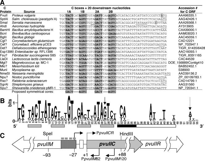
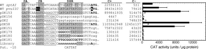
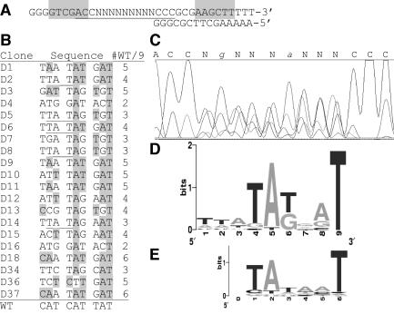
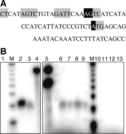
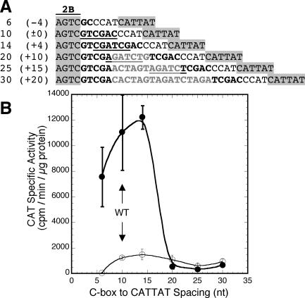
A widely distributed family of small regulators, called C proteins, controls a subset of restriction-modification systems. The C proteins studied to date activate transcription of their own genes and that of downstream endonuclease genes; this arrangement appears to delay endonuclease expression relative to that of the protective methyltransferase when the genes enter a new cell. C proteins bind to conserved sequences called C boxes. In the PvuII system, the C boxes have been reported to extend from -23 to +3 relative to the transcription start for the gene for the C protein, an unexpected starting position relative to a bound activator. This study suggests that transcript initiation within the C boxes represents initial, C-independent transcription of pvuIICR. The major C protein-dependent transcript appears to be a leaderless mRNA starting farther downstream, at the initiation codon for the pvuIIC gene. This conclusion is based on nuclease S1 transcript mapping and the effects of a series of nested deletions in the promoter region. Furthermore, replacing the region upstream of the pvuIIC initiation codon with a library of random oligonucleotides, followed by selection for C-dependent transcription, yielded clones having sequences that resemble -10 promoter hexamers. The -35 hexamer of this promoter would lie within the C boxes. However, the spacing between C boxes/-35 and the apparent -10 hexamer can be varied by +/-4 bp with little effect. This suggests that, like some other activator-dependent promoters, PpvuIICR may not require a -35 hexamer. Features of this transcription activation system suggest explanations for its broad host range.


Figures

FIG. 1.
FIG. 1.
Control region for the PvuII RM system. (A) Comparison of regions upstream of known and some hypothetical C protein-encoding genes. These genes were identified by using the Entrez BLink feature with both C.PvuII and C.SmaI as seeds and screening for proteins with predicted recognition helices similar to that of C.PvuII (HRTYI) (59). A question mark in the protein name indicates an open reading frame (ORF) that has not yet been formally named. Sequences upstream of the C open reading frames are shown; in all cases, the initiation codon is to the right (underlined where shown; others are farther downstream). The sequences are alphabetical by C open reading frame name, except for the top three: PvuII, the focus of this study; SptAI, which is very closely related to PvuII (43); and SmaI, for which the start of transcription has been determined (20). The shaded nucleotides indicate actual or predicted C boxes; the proposed symmetrical core (3, 60) is shown at the bottom. For C.PvuII (top line), the underlined nucleotides in box 2B indicate the previously identified transcript starts (60) while the shaded A of the C.PvuII initiation codon (far right) was identified in this study as a transcript start. (B) Sequence logo derived from the sequences in part A. The height of each stack reflects the extent of conservation at that position, while the height of symbols within the stack indicates the relative frequency of each nucleotide (56). The logo was generated by the server at http://weblogo.berkeley.edu. Gray bars under the numbers indicate the C boxes, the black bar delineates a region shown in this study to be dispensable for C-dependent transcription, while the white bar shows the regions found in this study to be required (at least in part) for C-dependent transcription. Vertical arrows indicate the transcript starts found previously (60) (solid) or in this study (dotted). (C) Genetic map of the PvuII RM system. Numbering is relative to the initiation codon of pvuIIC. The hatched area represents the previously identified promoter area for the pvuIICR genes, and the small gray rectangles above indicate the positions of the C boxes (the sequences shaded in panel A). The white rectangle between pvuIIM and pvuIIC corresponds to the white rectangle on the right of panel B; the two pvuIIC transcript starts (rightward bent arrows) indicate the one previously identified (solid, −27) and the one identified in this study (dotted, +1). Two promoters for the pvuIIM gene are also shown (leftward bent arrows) (60). Salm., Salmonella.
FIG. 2.
FIG. 2.
Effect of replacing DNA downstream of the C boxes on pvuIICR promoter activity. Promoter clones, all having the 5′ end at −93 relative to the pvuIIC initiation codon, were placed upstream of the promoterless cat gene in pKK232-8. Only the sequence 3′ of C box 2A is shown. To readily replace portions of the DNA, the nucleotides immediately following C box 2B were altered to generate restriction sites (underlined). Lowercase letters indicate vector DNA. These plasmids were used to transform E. coli already containing a compatible plasmid expressing either WT C.PvuII (black bars) or a defective variant (white bars), and CAT activity was determined in triplicate as described in Materials and Methods. Error bars indicate the standard deviation. For comparison, the sequence of the SptAI RM system is also shown (top line); the genes of this system are extremely closely related to those of PvuII, and their transcription is activated by C.PvuII (43), but even in this case conservation of the region between the C boxes and pvuIIC initiation codon is limited. Also for comparison, the bottom lines show the sequence context into which the randomized sequences were placed in other experiments (Library) and the location of the putative (Put.) −10 hexamer identified as a result.
FIG. 3.
FIG. 3.
Selection of functional sequences from a randomized library. (A) Two synthetic oligonucleotides were acquired. The 28-mer includes nine consecutive positions (N) that were synthesized in the presence of an equal mixture of all four phosphorothioate nucleotides. The second strand was synthesized by using the 12-mer shown and extending with Klenow polymerase. The shaded areas indicate restriction sites used for subsequent cloning. In the resulting plasmids, the pvuIICR promoter and C boxes are upstream of the sequence shown while downstream is a promoterless cat gene. (B) The plasmid library was used to transform an E. coli strain that already carried a compatible plasmid producing C.PvuII, and the transformants were plated onto agar containing CM to select for functional sequences. Twenty of these were sequenced as described in Materials and Methods; the 9-nt randomized region is shown with the native PvuII sequence at the bottom. Shading indicates a match to the native sequence, with number of matching positions indicated to the right; underlining reveals shifted occurrences of a TAT motif. In all 20 clones, the 9-nt region was preceded by ACC and followed by CCC. (C) Sequencing trace of the pooled randomized plasmid library before selection (portion underlined in panel A). The lower strand was sequenced, so the image has been reversed to match other portions of the figure and the complementary bases indicated above the trace. In the unselected randomized region, two positions showed enough bias for the sequencing software to call them as shown. (D) Logo analysis (56) of sequences shown in panel B. The logo was generated at weblogo.berkeley.edu and is aligned with the corresponding positions of panel C. (E) Logo analysis of 350 E. coli −10 promoter hexamers generated by the Computational Genomics Research Group, Department of Plant and Microbial Biology, University of California at Berkeley, and available at weblogo.berkeley.edu.
FIG. 4.
FIG. 4.
Determination of pvuIIC mRNA 5′ terminus via S1 nuclease digestion. (A) Sequence complementary to the 77-nt probe used in S1 mapping analyses. Gray boxes indicate the C box elements and the ATG initiation codon of pvuIIC. Transcript starts identified previously (60) and in this study are shown in black boxes. The distances from the labeled 5′ end of the probe (corresponding to the 3′ end of the sequence shown) are 30 nt to the ATG start and 56 or 57 nt to the AGTC start. In addition, a 51-nt probe was used, the 5′ end of which is complementary to the position 4 nt upstream of the ATG start codon (GTCT) and that extends 7 nt upstream of the entire sequence shown. (B) S1-digested products were resolved on acrylamide gels as described in Materials and Methods; autoradiograms are shown. The two marker lanes (M) show a 10-nt ladder, with the bands shown ranging from 20 to 100 nt. Unless noted otherwise, all reaction mixtures included the 77-nt probe. Lanes: 1, RNA from the strain used as background, E. coli TOP10; 2 and 3, 50 and 25 μg of RNA from TOP10 carrying pPvuRM3.4 (WT PvuII RM system); 4, same as lane 3 but with no nuclease S1 added; 5, same as lane 4 but with both 77- and 51-nt probes present; 6 and 7, 50 and 25 μg of RNA from TOP10 carrying pPvuRM3.4; 8 and 9, 50 and 25 μg of RNA from P. vulgaris, the native host for the PvuII RM system; 10 and 11, same as lanes 6 and 7 but with the 51-nt probe; 12 and 13, same as lanes 8 and 9 but with the 51-nt probe.
FIG. 5.
FIG. 5.
Effect of altered spacing between the C boxes and putative −10 hexamer on pvuIICR transcription. (A) Promoter variants tested. Each promoter began at −93 relative to the pvuIIC initiation codon; only sequences 3′ of C box 2B are shown. All include nt −23 to +6 of the native PvuII DNA and were inserted into pKK232-8 upstream of the promoterless cat gene. The nucleotides from −24 to −19 were replaced to generate a restriction site (underlined), and the spacing between the putative −10 hexamer for the ATG transcription start and the C boxes/promoter was varied by cleaving the restriction site and digesting the single-stranded tails, filling in the single-stranded regions, or inserting oligonucleotide duplexes of various lengths prior to ligation. The putative −10 hexamer, CATTAT, is −14 to −9 relative to the pvuIIC initiation codon. WT spacing of 10 nt between C box 2B and the putative −10 hexamer is indicated by ±0. (B) The sequences of all variants were confirmed, and they were used to transform E. coli already containing a compatible plasmid expressing either WT C.PvuII (closed circles) or a defective C variant (open circles); CAT activity was determined in triplicate as described in Materials and Methods. The bars indicate standard errors.
FIG. 6.
FIG. 6.
Model for interaction between C proteins and region 4 of RpoD. The lower half shows sequences of the predicted HTH motifs of selected C proteins. The sequence above C.PvuII is that of cI protein from bacteriophage lambda, which has been structurally characterized and has a known activation mechanism (24). The shading indicates the nearly complete conservation of the upstream α helix between λcI and C.PvuII (one Val → Leu substitution out of 11 consecutive amino acids), including the key activation residue (Glu shown in white on black). In the case of λcI, this Glu activates by contacting a conserved Arg in the HTH motif of region 4.2 at the carboxyl end of RpoD (σ70; upper half). This Arg, in turn, is coordinated with a conserved Glu (both shown in white on black), and the Glu contacts position −33 in the promoter −35 hexamer (see text for details). In the RpoD sequences, gray shading indicates amino acids that differ from those in the E. coli ortholog. The RpoD sequences shown were chosen to match the native hosts of the C protein sequences in the lower half. Sequences are from the GenBank database, except for RpoD of P. mirabilis, which was kindly made available from the partially complete genome sequence (http://www.sanger.ac.uk/Projects/P_mirabilis/).

Similar articles

Cited by

References

    1. Anton, B. P., D. F. Heiter, J. S. Benner, E. J. Hess, L. Greenough, L. S. Moran, B. E. Slatko, and J. E. Brooks. 1997. Cloning and characterization of the BglII restriction-modification system reveal a possible evolutionary footprint. Gene 187:19-27. - PubMed
    1. Balbas, P., X. Soberon, E. Merino, M. Zurita, H. Lomeli, F. Valle, N. Flores, and F. Bolivar. 1986. Plasmid vector pBR322 and its special-purpose derivatives—a review. Gene 50:3-40. - PubMed
    1. Bart, A., J. Dankert, and A. van der Ende. 1999. Operator sequences for the regulatory proteins of restriction modification systems. Mol. Microbiol. 31:1277-1278. - PubMed
    1. Beutel, B. A., and M. T. Record, Jr. 1990. E. coli promoter spacer regions contain nonrandom sequences which correlate to spacer length. Nucleic Acids Res. 18:3597-3603. - PMC - PubMed
    1. Blumenthal, R. M., S. A. Gregory, and J. S. Cooperider. 1985. Cloning of a restriction-modification system from Proteus vulgaris and its use in analyzing a methylase-sensitive phenotype in Escherichia coli. J. Bacteriol. 164:501-509. - PMC - PubMed

Publication types

MeSH terms

Substances

LinkOut - more resources