. 2005 Jan;187(2):522-33.
doi: 10.1128/JB.187.2.522-533.2005.

Intimin-mediated export of passenger proteins requires maintenance of a translocation-competent conformation

Affiliations

Intimin-mediated export of passenger proteins requires maintenance of a translocation-competent conformation

Thorsten M Adams et al. J Bacteriol. 2005 Jan.

Abstract

Intimins from pathogenic bacteria promote intimate bacterial adhesion to epithelial cells. Several structurally similar domains form on the bacterial cell surface an extended rigid rod that exposes the carboxy-terminal domain, which interacts with the translocated intimin receptor. We constructed a series of intimin-derived fusion proteins consisting of carboxy-terminally truncated intimin and the immunoglobulin light-chain variable domain REIv, ubiquitin, calmodulin, beta-lactamase inhibitor protein, or beta-lactamase. By systematically investigating the intimin-mediated cell surface exposure of these passenger domains in the presence or absence of compounds that interfere with outer membrane stability or passenger domain folding, we acquired experimental evidence that intimin-mediated protein export across the outer membrane requires, prior to export, the maintenance of a translocation-competent conformation that may be distinct from the final protein structure. We propose that, during export, competition exists between productive translocation and folding of the passenger domain in the periplasm into a stable conformation that is not compatible with translocation through the bacterial outer membrane. These results may expand understanding of the mechanism by which intimins are inserted into the outer membrane and expose extracellular domains on the cell surface.


Figures
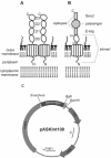
FIG. 1.
FIG. 1.
(A) Schematic representation of an EHEC intimin dimer, each monomer consisting of a transmembrane region and an extracellular rod formed by three immunoglobulin-like domains (D0 to D2) and one lectin-like domain (D3). (B) Schematic representation of a C-terminally truncated intimin (intimin′) monomer (ranging from amino acids 1 to 659) used in this study for the display of heterologous passenger domains. The passenger domain is flanked at its amino-terminal and carboxy-terminal ends by E epitope (GAPVPYPDPLEPR) and Send epitope (DGSLGDIEPYDSS) sequences, respectively, and fused to C-terminally truncated intimin. Residue 550 is predicted to be the last transmembrane residue (30). (C) Schematic representation of display vector pASKInt100 harboring structural gene eaeA′. f1, f1 replication origin; cat, chloramphenicol resistance marker; tetR, tetracycline repressor-encoding gene; colE1, ColE1 replication origin; eaeA′, truncated eaeA gene of EHEC O157:H7 (codons 1 to 659); E, E epitope sequence; S, 13-residue Send epitope sequence. Unique SmaI/AvaI and BglII restriction sites allowed the in-frame fusion of genes encoding various passenger domains.
FIG. 2.
FIG. 2.
Western blot analysis with anti-Send epitope antibody of a whole-membrane preparation of induced recombinant 71-18 cellsharboring pASKInt100-REI, pASKInt100-Ubi, pASKInt100-Bla,pASKInt100-BLIP, pASKInt100-Cal, or pASKInt100-ΔP and grown in the presence or absence of 20 mM 2-ME or 20 mM EGTA. M, marker proteins (pencil marked after Ponceau S staining); sizes (in thousands) are indicated. Arrowheads indicate full-length proteins.
FIG. 3.
FIG. 3.
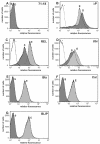
C-terminally truncated intimin-mediated cell surface display of passenger proteins. FACS histograms of E. coli 71-18 cells harboring no plasmid (A), pASKInt100-ΔP (B), pASKInt100-REI (C), pASKInt100-Ubi (D), pASKInt100-Bla (E), pASKInt100-Cal (F), or pASKInt100-BLIP (G) are shown. Induced cells were incubated with anti-Send epitope antibody (S) or anti-E epitope antibody (E), biotinylated anti-mouse antibody, and streptavidin-R-PE conjugate. Unlabeled 71-18 cells served as a control (−) in panels B to D.
FIG. 4.
FIG. 4.
(A) Model of fusion of C-terminally truncated intimin with a trapped passenger domain. Truncated intimin (intimin′; broken lines) is shown as a dimer, where the left extracellular domain is omitted for the sake of clarity. Domain D0 and the E epitope are located on the cell surface, whereas the passenger and the Send epitope are trapped within the periplasm. Residue 550 is the last amino acid of the transmembrane region, and amino acid 659 is the last amino acid of truncated intimin. Dimensions are not proportional. (B) Western blot analysis with anti-E epitope antibody of periplasmic protein preparations from induced recombinant 71-18 cells that harbored pASKInt100-REI, pASKInt100-Cal, or pASKInt100-Bla and that were treated with trypsin prior to osmotic shock. M, marker proteins (pencil marked after Ponceau S staining); sizes (in thousands) are indicated. (C) Linear representation of truncated intimin-Bla (top) and truncated intimin-Cal (bottom) fusion proteins. The putative trypsin cleavage site, as deduced from panel B, is indicated by scissors. tm, transmembrane region; E, E epitope; S, Send epitope.
FIG. 5.
FIG. 5.
C-terminally truncated intimin-based cell surface display of BLIP. A FACS histogram of recombinant 71-18 or 71-18dsbA cells harboring pASKInt100-BLIP is shown. Induced cells were harvested and successively incubated with anti-Send epitope antibody or anti-E epitope antibody, biotinylated anti-mouse antibody, and streptavidin R-PE conjugate. IS, 71-18(pASKInt100-BLIP) cells grown in dYT and labeled with anti-Send epitope antibody. IIS, 71-18(pASKInt100-BLIP) cells grown in dYT containing 20 mM 2-ME and labeled with anti-Send epitope antibody. IIIS, 71-18dsbA(pASKInt100-BLIP) cells grown in dYT and labeled with anti-Send epitope antibody. IVS, 71-18dsbA(pASKInt100-BLIP) cells grown in dYT containing 20 mM 2-ME and labeled with anti-Send epitope antibody; VE, 71-18(pASKInt100-BLIP) cells grown in dYT and labeled with anti-E epitope antibody. Unlabeled 71-18 cells served as a control (−).
FIG. 6.
FIG. 6.
C-terminally truncated intimin-mediated cell surface display of human calmodulin. (A and B) E. coli 71-18 cells harboring pASKInt100-Cal were grown in the absence (A) or presence (B) of 20 mM EDTA. Induced cells were harvested, successively incubated with anti-Send epitope antibody (S) or anti-E epitope antibody (E), biotinylated anti-mouse antibody, and streptavidin-R-PE conjugate, and analyzed by FACS. Unlabeled 71-18 cells served as a control (−). (C and D) Binding of an MykBlaSend fusion protein to surface-exposed calmodulin. Induced cells grown in the absence (C) or presence (D) of 20 mM EGTA were washed, resuspended in 50 mM CaCl2, and incubated with MykBlaSend, consisting of a calmodulin-binding segment of myosin kinase fused to β-lactamase and tagged with a Send epitope. Binding of the fusion protein to calmodulin was detected by fluorescence microscopy with anti-Send epitope antibody, biotinylated anti-mouse antibody, and streptavidin-R-PE conjugate for cell staining.
FIG. 7.
FIG. 7.
C-terminally truncated intimin-based cell surface display of β-lactamase variants (Bla). A FACS histogram of recombinant 71-18 cells harboring wild-type plasmid pASKInt100-Bla (I) or the C52R (II), C98R (III), or C98Y (IV) variant is shown. Induced cells were harvested and successively incubated with anti-Send epitope antibody, biotinylated anti-mouse antibody, and streptavidin-R-PE conjugate.

References

    1. Adu-Bobie, J., G. Frankel, C. Bain, A. G. Goncalves, L. R. Trabulsi, G. Douce, S. Knutton, and G. Dougan. 1998. Detection of intimins α, β, γ, and δ, four intimin derivatives expressed by attaching and effacing microbial pathogens. J. Clin. Microbiol. 36:662-668. - PMC - PubMed
    1. Akiyama, Y., S. Kamitani, N. Kusukawa, and K. Ito. 1992. In vitro catalysis of oxidative folding of disulfide-bonded proteins by the Escherichia coli dsbA (ppfA) gene product. J. Biol. Chem. 267:22440-22445. - PubMed
    1. Bardwell, J. C., K. McGovern, and J. Beckwith. 1991. Identification of a protein required for disulfide bond formation in vivo. Cell 67:581-589. - PubMed
    1. Batchelor, M., S. Prasannan, S. Daniell, S. Reece, I. Connerton, G. Bloomberg, G. Dougan, G. Frankel, and S. Matthews. 2000. Structural basis for recognition of the translocated intimin receptor (Tir) by intimin from enteropathogenic Escherichia coli. EMBO J. 19:2452-2464. - PMC - PubMed
    1. Benz, I., and M. A. Schmidt. 1989. Cloning and expression of an adhesin (AIDA-I) involved in diffuse adherence of enteropathogenic Escherichia coli. Infect. Immun. 57:1506-1511. - PMC - PubMed

Publication types

MeSH terms

LinkOut - more resources