. 2005 Jan 1;19(1):104-13.
doi: 10.1101/gad.1262905.

Mammalian poly(A)-binding protein is a eukaryotic translation initiation factor, which acts via multiple mechanisms

Affiliations

Mammalian poly(A)-binding protein is a eukaryotic translation initiation factor, which acts via multiple mechanisms

Avak Kahvejian et al. Genes Dev. .

Abstract

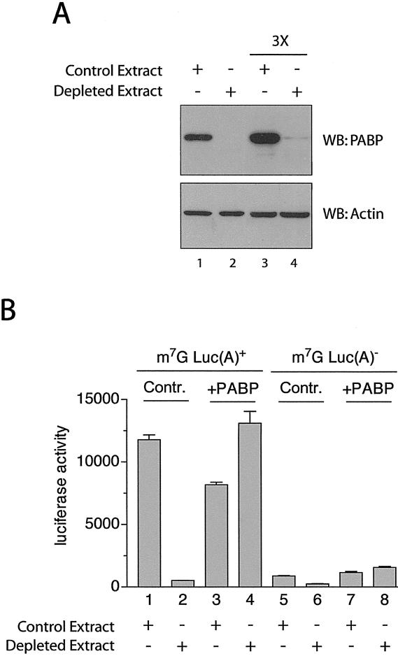
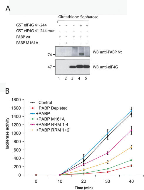
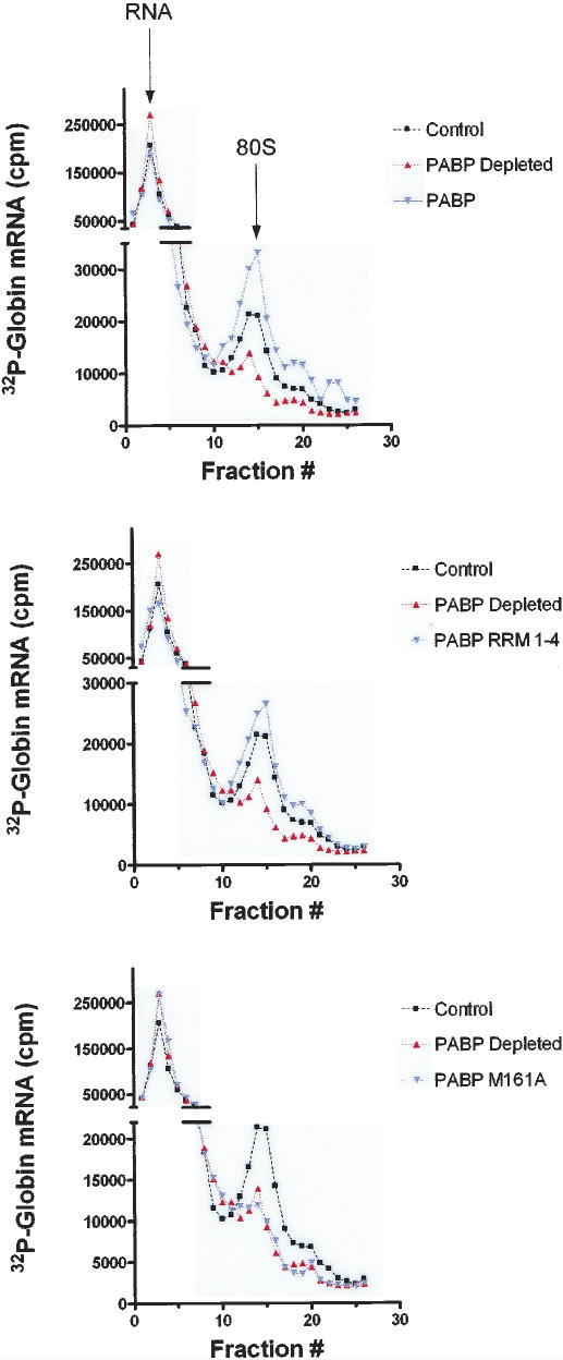
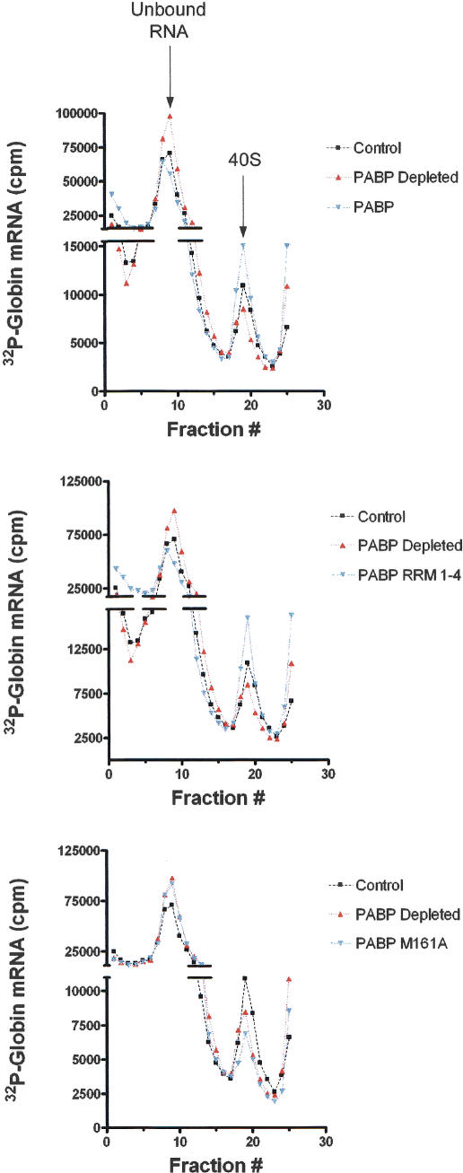
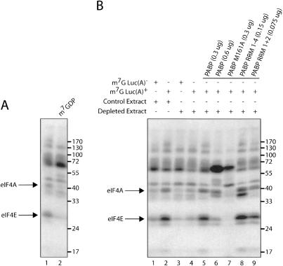
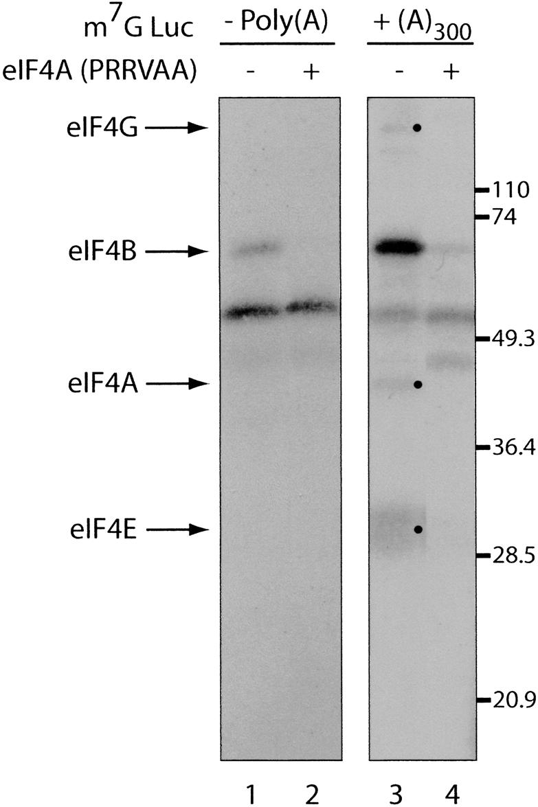
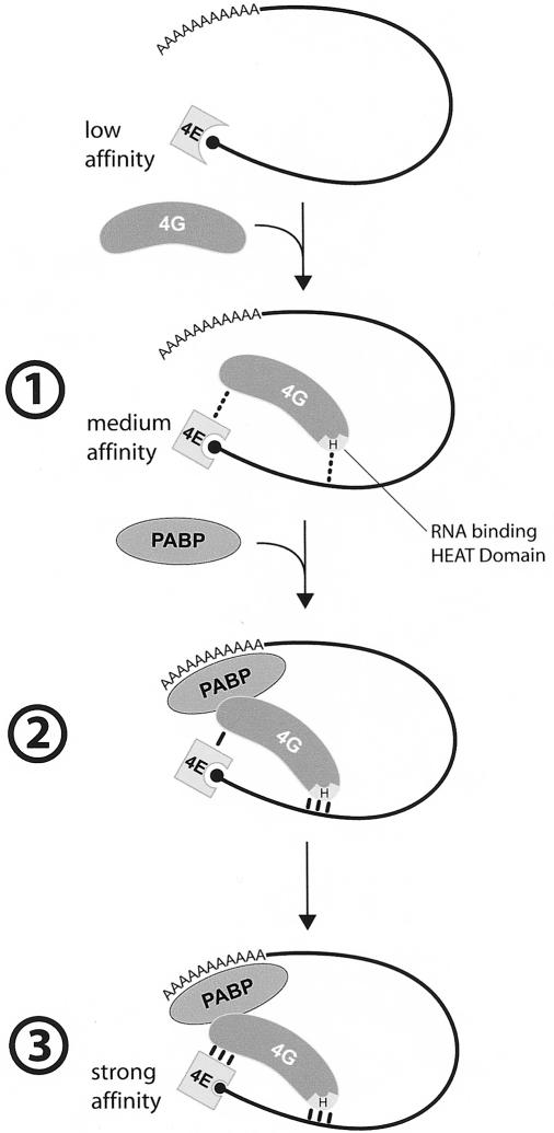
Translation initiation is a multistep process involving several canonical translation factors, which assemble at the 5'-end of the mRNA to promote the recruitment of the ribosome. Although the 3' poly(A) tail of eukaryotic mRNAs and its major bound protein, the poly(A)-binding protein (PABP), have been studied extensively, their mechanism of action in translation is not well understood and is confounded by differences between in vivo and in vitro systems. Here, we provide direct evidence for the involvement of PABP in key steps of the translation initiation pathway. Using a new technique to deplete PABP from mammalian cell extracts, we show that extracts lacking PABP exhibit dramatically reduced rates of translation, reduced efficiency of 48S and 80S ribosome initiation complex formation, and impaired interaction of eIF4E with the mRNA cap structure. Supplementing PABP-depleted extracts with wild-type PABP completely rectified these deficiencies, whereas a mutant of PABP, M161A, which is incapable of interacting with eIF4G, failed to restore translation. In addition, a stronger inhibition (approximately twofold) of 80S as compared to 48S ribosome complex formation (approximately 65% vs. approximately 35%, respectively) by PABP depletion suggests that PABP plays a direct role in 60S subunit joining. PABP can thus be considered a canonical translation initiation factor, integral to initiation complex formation at the 5'-end of mRNA.


Figures

Figure 1.
Figure 1.
Depletion of PABP from Krebs-2 extracts results in diminished translation. (A, top panel) Western blot (SDS-10% PAGE) analysis of extracts treated with GST (Control Extract, lanes 1,3) or GST-Paip2 (Depleted Extract, lanes 2,4) probed using anti-PABP polyclonal antibody. (Bottom panel) Same membrane probed with an anti-β-actin antibody (Sigma) as a loading control. Three times more extract was loaded in lanes 3 and 4 (marked 3×) versus lanes 1 and 2, to reach the antibody's detection limit (lane 4). (B) Capped poly(A)+ (1–4) or capped poly(A) (5–8) luciferase mRNA (2 μg/mL) was translated in Krebs-2 extracts that were either depleted of PABP (Depleted Extract) or mock-depleted (Control Extract). The reactions were supplemented with either recombinant human PABP (10 μg/mL) or control buffer, as indicated. Luciferase activity was measured as described in Materials and Methods and is expressed in relative light units (RLU) × 103.
Figure 2.
Figure 2.
Interaction with eIF4G is critical for PABP to stimulate translation. (A) PABP M161A cannot bind to eIF4G. PABP wild type and PABP M161A were incubated with glutathione-Sepharose beads alone (lanes 1,2), or with beads coupled to GST eIF4G 41–244 (lanes 4,5), washed with binding buffer, and eluted with Laemmli buffer. (Lane 3) GST eIF4G 41–244mut, which cannot bind to PABP, was used as a control. Eluted proteins were resolved on an SDS-10% PAGE. Western blot analysis was performed using anti-PABP antibody (top panel) or anti-eIF4G antibody (bottom panel). The positions of molecular weight markers are indicated on the left. (B) Translation of a capped luciferase poly(A)+ mRNA in PABP-depleted Krebs-2 extract supplemented with different forms of PABP. Luciferase activity was measured at several time points in mock-depleted (control, black line) or depleted extracts supplemented with no protein (PABP Depleted, red line), or supplemented with equimolar amounts of wild-type PABP (blue line), PABP M161A (green line), PABP RRM 1–4 (fuchsia line), or PABP RRM 1 + 2 (orange line). Luciferase activity was measured as described in Materials and Methods and is expressed in RLU × 103, as an average of four experiments.
Figure 3.
Figure 3.
80S ribosome initiation complex formation is deficient in PABP-depleted extracts (representative experiment of four independent experiments). Ribosome initiation complex profiles of control extract (black line, squares), PABP-depleted extract (red line, triangles), and supplemented extract (blue line, inverted triangles) are overlaid for comparison. Extracts were supplemented with equimolar amounts of either wild-type PABP (10 μg/mL; top panel), PABP RRM 1–4 (6 μg/mL; middle panel), or PABP M161A (10 μg/mL; lower panel). Peaks corresponding to unbound RNA and to RNA in complex with 80S ribosomes are indicated with arrows (top panel).
Figure 4.
Figure 4.
40S ribosome initiation complex formation is deficient in PABP-depleted extracts (representative experiment of four independent experiments). 40S initiation complex profiles of control extract (black line, squares), PABP-depleted extract (red line, triangles), and PABP-depleted extract supplemented with PABP (blue line, inverted triangles) are overlaid for comparison. Extracts were supplemented with equimolar amounts of either wild-type PABP (10 μg/mL; top panel), PABP RRM 1–4 (6 μg/mL; middle panel), or PABP M161A (10 μg/mL; lower panel). Peaks corresponding to unbound RNA and to RNA in complex with 40S ribosomes are indicated with arrows (top panel).
Figure 5.
Figure 5.
PABP mediates poly(A)-tail-induced initiation factor binding to the m7G cap. (A) Cap-labeled Luc(A) and Luc(A)+ mRNA were incubated with reticulocyte lysate. Proteins were cross-linked to mRNA by oxidation and subsequent reduction. Bands corresponding to eIF4E and eIF4A are indicated with arrows on the left. The addition of cap analog is indicated above. (B) Cap-labeled Luc(A) and Luc(A)+ mRNA were incubated with Mock-Depleted (control extract) and PABP-Depleted (depleted extract) reticulocyte lysate. Extracts were supplemented with equimolar amounts of recombinant proteins (indicated above). The positions of molecular weight markers are indicated on the right.
Figure 6.
Figure 6.
The poly(A) tail stimulates initiation factor binding to the m7G cap. Cap-labeled Luc(A) (left panel) and Luc(A)+ (right panel) mRNA were incubated with rabbit reticulocyte lysate in the absence (lanes 1,3) or presence (lanes 2,4) of an eIF4A dominant-negative mutant (PRRVAA, 100 μg/mL) (Svitkin et al. 2001b), and subjected to UV cross-linking. Samples were analyzed by SDS-15% PAGE and bands were revealed by autoradiography. Bands corresponding to initiation factors are indicated with arrows on the left and bullets. The positions of molecular weight markers are indicated on the right.
Figure 7.
Figure 7.
Model for PABP enhancement of eIF4E binding to the mRNA 5′-cap. (1) eIF4G enhances eIF4E affinity for the mRNA 5′-cap structure, through the interaction of the eIF4G HEAT (Marcotrigiano et al. 2001) domain (H) with mRNA. (2) PABP enhances eIF4G affinity for the mRNA. (3) eIF4G affinity for eIF4E is increased as a consequence of the increased eIF4G binding to the mRNA.

References

    1. Afonina E., Neumann, M., and Pavlakis, G.N. 1997. Preferential binding of poly(A)-binding protein 1 to an inhibitory RNA element in the human immunodeficiency virus type 1 gag mRNA. J. Biol. Chem. 272: 2307–2311. - PubMed
    1. Bi X. and Goss, D.J. 2000. Wheat germ poly(A)-binding protein increases the ATPase and the RNA helicase activity of translation initiation factors eIF4A, eIF4B, and eIF-iso4F. J. Biol. Chem. 275: 17740–17746. - PubMed
    1. Byrd M.P., Zamora, M., and Lloyd, R.E. 2002. Generation of multiple isoforms of eukaryotic translation initiation factor 4GI by use of alternate translation initiation codons. Mol. Cell. Biol. 22: 4499–4511. - PMC - PubMed
    1. Copeland P.R. and Wormington, M. 2001. The mechanism and regulation of deadenylation: Identification and characterization of Xenopus PARN. RNA 7: 875–886. - PMC - PubMed
    1. Edery I., Humbelin, M., Darveau, A., Lee, K.A., Milburn, S., Hershey, J.W., Trachsel, H., and Sonenberg, N. 1983. Involvement of eukaryotic initiation factor 4A in the cap recognition process. J. Biol. Chem. 258: 11398–11403. - PubMed

Publication types

Substances