. 2005 Jan 1;19(1):164-75.
doi: 10.1101/gad.1253605.

Noncanonical Wnt signaling regulates midline convergence of organ primordia during zebrafish development

Affiliations

Noncanonical Wnt signaling regulates midline convergence of organ primordia during zebrafish development

Takaaki Matsui et al. Genes Dev. .

Abstract

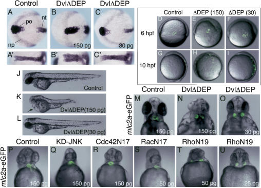
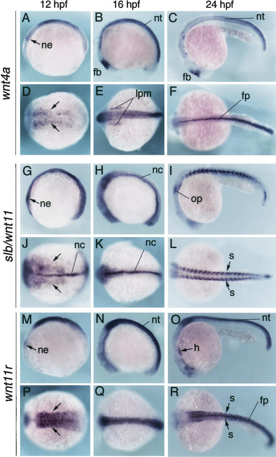
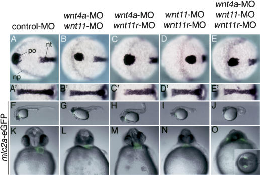
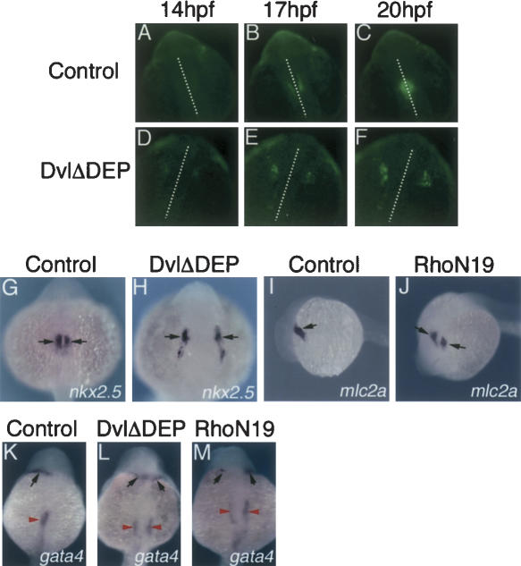
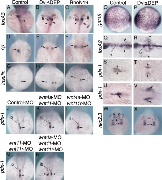
Several components of noncanonical Wnt signaling pathways are involved in the control of convergence and extension (CE) movements during zebrafish and Xenopus gastrulation. However, the complexity of these pathways and the wide patterns of expression and activity displayed by some of their components immediately suggest additional morphogenetic roles beyond the control of CE. Here we show that the key modular intracellular mediator Dishevelled, through a specific activation of RhoA GTPase, controls the process of convergence of endoderm and organ precursors toward the embryonic midline in the zebrafish embryo. We also show that three Wnt noncanonical ligands wnt4a, silberblick/wnt11, and wnt11-related regulate this process by acting in a largely redundant way. The same ligands are also required, nonredundantly, to control specific aspects of CE that involve interaction of Dishevelled with mediators different from that of RhoA GTPase. Overall, our results uncover a late, previously unexpected role of noncanonical Wnt signaling in the control of midline assembly of organ precursors during vertebrate embryo development.


Figures

Figure 1.
Figure 1.
Early and late roles of noncanonical Wnt signaling in zebrafish embryos. (AI) Inhibition of nc-Wnt signaling by DvlΔDEP affects CE movements. (AC) hgg1 (polster, po), dlx3 (anterior edge of the neural plate, np), and ntl (developing notochord, nt) transcripts in 10 hpf embryos injected with 150 pg of mRNA encoding the negative control alkaline phosphatase (AP; Control, A), and 150 (B) or 30 (C) pg of mRNA encoding DvlΔDEP. Animal pole views, anterior to the left (AC); dorsal view, anterior to the left (A–C′). High concentrations of DvlΔDEP led to severe defects in CE movements, while lower concentration of DvlΔDEP altered CE only slightly. (DI) Distribution of transplanted FITC-labeled cells at onset of gastrulation; lateral views, dorsal to the right. (D,G) Wild-type control. (E,H) DvlΔDEP (150 pg). (F,I) DvlΔDEP (30 pg). (DF) Shield (6 hpf). (GI) Tail bud (10 hpf). Dorsal convergence of mesoendodermal cells during zebrafish gastrulation was inhibited by high concentrations of DvlΔDEP (E,H), but not lower concentrations of DvlΔDEP (F,I). (JL) Morphology of 48 hpf embryos injected with Control AP (J), DvlΔDEP (K,L), lateral views, anterior to the left. As consequences of CE defects, high doses of DvlΔDEP led to short A/P axis, microcephaly, and microphthalmia. In addition, DvlΔDEP injection induced pericardial edema (arrow in K). (MO) Effects of DvlΔDEP on zebrafish heart development. Transgenic zebrafish embryos carrying mlc2a–eGFP reporter were injected with Control AP mRNA (M) or DvlΔDEP mRNA (N,O) and allowed to grow for 48 h. (N,O) Injection of DvlΔDEP even in low concentrations led to the formation of two laterally positioned hearts (cardia bifida) in zebrafish embryos. (MO) Ventral views, anterior to the top. (PU) Effect of downstream effectors of Dvl on heart tube assembly. mlc2a-eGFP transgenic zebrafish embryos were injected with mRNA encoding Control AP (150 pg) (P), KD-JNK (150 pg) (Q), Cdc42N17 (150 pg) (R), RacN17 (50 pg) (S), RhoN19 (25–50 pg) (T,U) and allowed to develop until 48 hpf. (T,U) Inhibition of Rho GTPase led to cardia bifida. Ventral views, anterior to the top.
Figure 2.
Figure 2.
Spatial and temporal expression of wnt4a, slb/wnt11, and wnt11r during zebrafish heart morphogenesis. wnt4a (AF), slb/wnt11 (GL), wnt11r (MR) transcripts in zebrafish embryos at 12 hpf (A,D,G,J,M,P), 16 hpf (B,E,H,K,N,Q), and 24 hpf (C,F,I,L,O,R). Lateral (AC,GI,MO) and dorsal (DF,JK,PR) views, anterior to the left. Arrows in panels D, J, and P point at lateral edges of mesoendoderm. (ne) Neural ectoderm; (nt) neural tube; (fb) forebrain; (lpm) lateral plate mesoderm; (fp) floorplate; (nc) notochord; (op) otic placode; (s) somite; (h) heart.
Figure 3.
Figure 3.
Combined, redundant action of wnt4a,slb/wnt11, and wnt11r is required for heart tube assembly. (AE) hgg1 (polster, po), dlx3 (anterior edge of the neural plate, np), and ntl (developing notochord, nt) transcripts in 10 hpf zebrafish embryos injected with Control-MO (7.5 ng) (A), wnt4a-MO (3 ng) plus slb/wnt11-MO (1.5 ng) (B), wnt4a-MO (3 ng) plus wnt11r-MO (3 ng) (C), slb/wnt11-MO (1.5 ng) plus wnt11r-MO (3 ng) (D), or wnt4a-MO (3 ng) plus slb/wnt11-MO (1.5 ng) plus wnt11r-MO (3 ng) (E). Animal pole views, anterior to the left (AE); dorsal view, anterior to the left (A′–E′). Double or triple knockdowns of wnt ligands led to similar defects of CE movements during gastrulation. (FO) mlc2a–eGFP transgenic zebrafish embryos were injected with Control-MO (F,K), wnt4a-MO plus slb/wnt11-MO (G,L), wnt4a-MO plus wnt11r-MO (H,M), slb/wnt11-MO plus wnt11r-MO (I,N), or wnt4a-MO plus slb/wnt11-MO plus wnt11r-MO (J,O), and allowed to develop until 31 hpf. (FJ) Lateral views, anterior to the left. (KO) Ventral views, anterior to the top. (G,J) Extensive areas of cell death were observed in double knockdown of wnt4a and slb/wnt11 or triple knockdowns of wnt4a, slb/wnt11, and wnt11r. (O) Interestingly, only triple knockdowns of wnt4a, slb/wnt11, and wnt11r led to a significant occurence of cardia bifida. The inset in O shows cardia bifida caused by the triple knockdown (dorsal view, anterior to the left). See also Table 2.
Figure 4.
Figure 4.
Noncanonical Wnt/Dvl/RhoA signaling regulates myocardial migration without affecting cell fate of myocardial precursors. (AF) DvlΔDEP injection inhibits myocardial migration to the midline. Transgenic zebrafish embryos carrying a heart-specific carp–eGFP reporter were injected with AP (150 pg) or DvlΔDEP (150 pg) and allowed to develop until 13.5 hpf. Embryos were mounted into 1% agarose and time-lapse images of each embryo were obtained. Time-lapse images of control embryo (AC) and DvlΔDEP-injected embryo (DF) at 14 hpf (A,D), 17 hpf (B,E), and 20 hpf (C,F). White dotted lines indicate embryonic midline. Dorsal views, anterior to the top. (DF) Myocardial precursor cells in DvlΔDEP-injected embryo failed to migrate to the midline. (GM) nkx2.5, mlc2a, gata4 transcripts in control (G,I,K) and DvlΔDEP-injected (H,L) or RhoN19-injected (J,M) embryos at 20 hpf (G,H) and 24 hpf (IM). (G,H,KM) Dorsal views, anterior to the top. (I,J) Lateral views, anterior to the left. nkx2.5 mlc2a- or gata4-expressing myocardial precursors (black arrows) were distributed bilaterally in DvlΔDEP-injected (H,L) or RhoN19-injected (J,M) embryos, but the levels of expression were not changed as compared to those of Control (G,I,K). Note: gata4 is also expressed in endoderm. In DvlΔDEP-injected (L) or RhoN19-injected (M) embryos, gata4-expressing endodermal domains were duplicated (red arrows).
Figure 5.
Figure 5.
Noncanonical Wnt/Dvl/RhoA signaling regulates midline convergence of foregut endoderm precursors without affecting their cell fate. (AI) foxA3 (AC), ceruloplasmin (cp) (DF), and insulin (GI) transcripts in 48 hpf embryos injected with Control AP (A,D,G), DvlΔDEP (B,E,H), or RhoN19 (C,F,I); dorsal views, anterior to the top. Antagonizing nc-Wnt signaling led to the formation of a Y-shaped gut tube (B,C), and the duplication of liver (E,F) and pancreas (H,I) buds. (li) liver buds. (JN) pdx-1 expression in 42 hpf embryos injected with Control-MO (J), wnt4a-MO plus slb/wnt11-MO (K), wnt4a-MO plus wnt11r-MO (L), slb/wnt11-MO plus wnt11r-MO (M), or wnt4a-MO plus slb/wnt11-MO plus wnt11r-MO (N); dorsal views, anterior to the top. Arrows indicate pdx-1-expressing pancreas bud. (ib) intestinal bulb. Only triple knockdown experiments of wnt4, slb/wnt11,and wnt11r resulted in the duplication of pancreas and intestinal bulbs (N). (OX) Expression of gata5 (O,P), foxA2 (Q,R), pdx-1 (SV), and nkx2.3 (W,X) in Control (O,Q, S,U,W) and DvlΔDEP-injected (P,R,T,V,X) embryos. (O,P) Ninety percent epiboly (9 hpf), dorsal views, anterior to the top. Expression levels of gata5 in endodermal precursors were not changed in DvlΔDEP-injected (P) embryos as compared with that of Control (O). (Q,R) Sixteen hours post-fertilization, dorsal views, anterior to the left. foxA2 is expressed in anterior endoderm and ventral neuroectoderm (ne) along the midline. (R) In DvlΔDEP-injected embryos, a large number of foxA2-expressing endodermal cells (arrows) failed to coalesce in the midline. (SV) Pancreas primordia marked by pdx-1 did not coalesce in the midline in embryos injected with DvlΔDEP (T,V). (S,T) Sixteen hours post-fertilization. (U,V) Twenty-four hours post-fertilization. Dorsal views, anterior to the left. (W,X) In DvlΔDEP-injected embryos, posterior nkx2.3-expressing pharyngeal endoderm (double arrows in X) appeared wider along the mediolateral axis, as compared to control (W). Most anterior nkx2.3-expressing pharyngeal endodermal cells (black arrows in W) failed to migrate toward the midline in DvlΔDEP-injected embryos (red asterisks).

References

    1. Alexander J., Rothenberg, M., Henry, G.L., and Stainier, D.Y. 1999. casanova plays an early and essential role in endoderm formation in zebrafish. Dev. Biol. 215: 343–357. - PubMed
    1. Axelrod J.D., Miller, J.R., Shulman, J.M., Moon, R.T., and Perrimon, N. 1998. Differential recruitment of Dishevelled provides signaling specificity in the planar cell polarity and Wingless signaling pathways. Genes & Dev. 12: 2610–2622. - PMC - PubMed
    1. Bakkers J., Kramer, C., Pothof, J., Quaedvlieg, N.E., Spaink, H.P., and Hammerschmidt, M. 2004. Has2 is required upstream of Rac1 to govern dorsal migration of lateral cells during zebrafish gastrulation. Development 131: 525–537. - PubMed
    1. Buckles G.R., Thorpe, C.J., Ramel, M.C., and Lekven, A.C. 2004. Combinatorial Wnt control of zebrafish midbrain-hindbrain boundary formation. Mech. Dev. 121: 437–447. - PubMed
    1. Carachi R. and Azmy, A. 2002. Foregut duplications. Pediatr. Surg. Int. 18: 371–374. - PubMed

Publication types

MeSH terms