. 2005 Jan 11;102(2):315-20.
doi: 10.1073/pnas.0406847102. Epub 2005 Jan 3.

Molecular basis for amyloid fibril formation and stability

Affiliations

Molecular basis for amyloid fibril formation and stability

O Sumner Makin et al. Proc Natl Acad Sci U S A. .

Abstract

The molecular structure of the amyloid fibril has remained elusive because of the difficulty of growing well diffracting crystals. By using a sequence-designed polypeptide, we have produced crystals of an amyloid fiber. These crystals diffract to high resolution (1 A) by electron and x-ray diffraction, enabling us to determine a detailed structure for amyloid. The structure reveals that the polypeptides form fibrous crystals composed of antiparallel beta-sheets in a cross-beta arrangement, characteristic of all amyloid fibers, and allows us to determine the side-chain packing within an amyloid fiber. The antiparallel beta-sheets are zipped together by means of pi-bonding between adjacent phenylalanine rings and salt-bridges between charge pairs (glutamic acid-lysine), thus controlling and stabilizing the structure. These interactions are likely to be important in the formation and stability of other amyloid fibrils.


Figures

Fig. 1.
Fig. 1.
Electron micrographs of fibrous crystals. The AAAK polypeptide assembles after incubation in PBS. Negative-stained transmission electron microscopy shows fibrous crystals in a field at low magnification (a) and the elongated crystals at higher magnification showing the 50-Å striations running parallel to the fiber axis (b).
Fig. 2.
Fig. 2.
Diffraction data from fibrous crystals. (a) X-ray diffraction pattern obtained by using synchrotron radiation from partially aligned, bundled fibers. The fiber axis is horizontal with the incident beam directed orthogonal to the fiber axis. Diffraction spacings were observed to a resolution beyond 2 Å and are shown in Table 2. (b and c) Electron diffraction patterns obtained from the fibrillar crystals with the incident beam parallel to the [0 1 0] zone axis reveal high-resolution, crystalline order before (b) and after (c) contrast enhancement. Discrete diffraction signals were observed out to a resolution of 0.9 Å.
Fig. 4.
Fig. 4.
The structure of the 12-mer peptide colored by residue type (basic, blue; acidic, red; nonpolar, green) or by chain. The structure is generated from a single peptide by imposing the P212121 symmetry, which creates a brick-like arrangement of overlapping peptide molecules (shown in d) and results in a very rigid structure held together with hydrogen-bonding, π–π-bonding, and electrostatic interactions. (a) A single 12-mer peptide labeled with residue names. (b) Four chains (colored blue, green, yellow, and red) showing hydrogen-bonding between the pairs of chains. (c) Two chains colored by residue type, with a higher-magnification image highlighting the intersheet π–π aromatic interactions. (d) Crystal packing with differently colored chains. The higher-magnification image highlights a cluster of phenylalanine side chains showing intersheet interactions and stacking between hydrogen-bonded β-strands. (e) Surface representation of crystal packing elongated along the fiber axis to show the close packing of the structure. (f) A view down the a axis colored by chain.
Fig. 3.
Fig. 3.
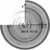
Comparison of observed and calculated x-ray diffraction patterns. The observed x-ray diffraction pattern from the partially aligned fibrous crystals is shown with the top left quadrant showing the calculated diffraction pattern. The calculated pattern is simulated from the model structure. It is clear where the observed and calculated diffraction peaks match, and this shows excellent agreement between diffraction signal positions.

References

    1. Eanes, E. D. & Glenner, G. G. (1968) J. Histochem. Cytochem. 16, 673-677. - PubMed
    1. Geddes, A. J., Parker, K. D., Atkins, E. D. T. & Beighton, E. (1968) J. Mol. Biol. 32, 343-358. - PubMed
    1. Astbury, W., Dickinson, S. & Bailey, K. (1935) Biochem. J. 29, 2351-2360. - PMC - PubMed
    1. Dobson, C. (2001) Phil. Trans. Soc. London Ser. B 356, 133-145. - PMC - PubMed
    1. Hirota-Nakaoka, N., Hasegawa, K., Naiki, H. & Goto, Y. (2003) J. Biochem. (Tokyo) 134, 159-164. - PubMed

Publication types

Associated data