Comparative Study
. 2005 Jan 3;201(1):105-15.
doi: 10.1084/jem.20040624.

Hypoxia-induced neutrophil survival is mediated by HIF-1alpha-dependent NF-kappaB activity

Affiliations
Comparative Study

Hypoxia-induced neutrophil survival is mediated by HIF-1alpha-dependent NF-kappaB activity

Sarah R Walmsley et al. J Exp Med. .

Abstract

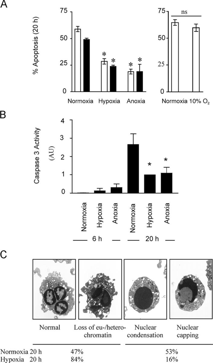
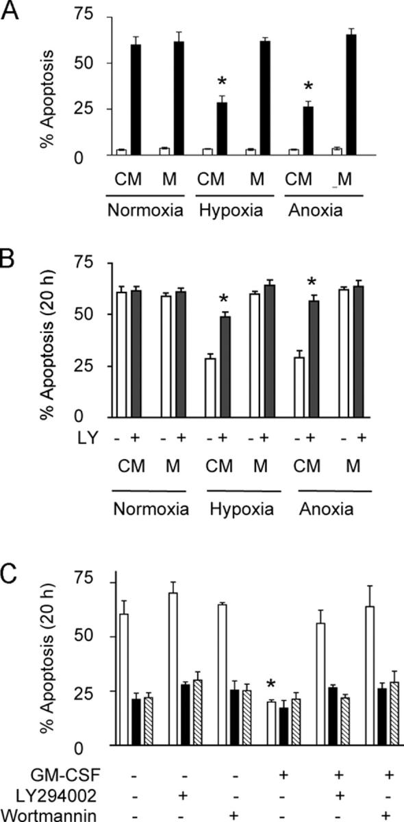
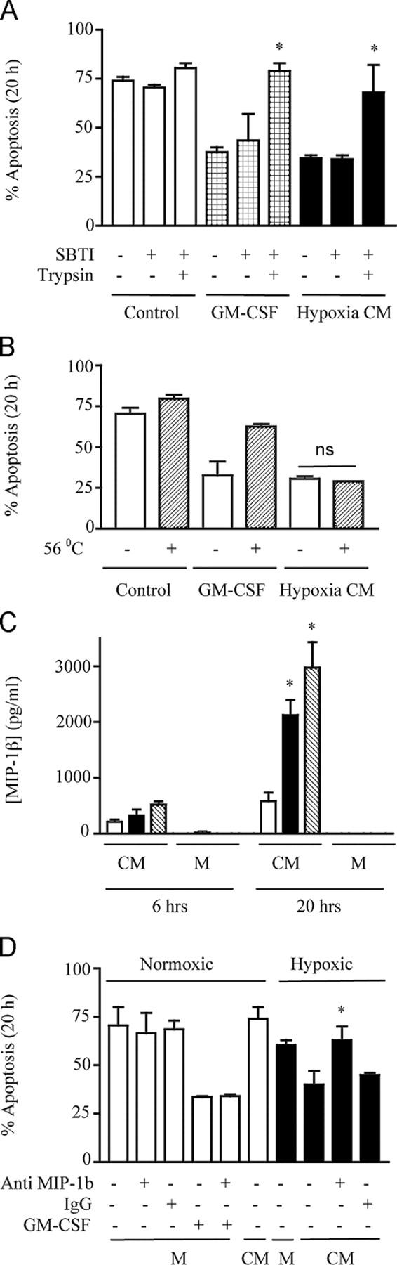
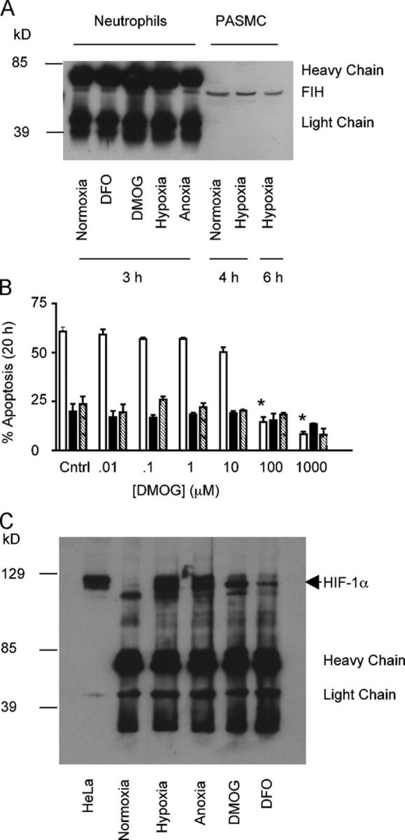
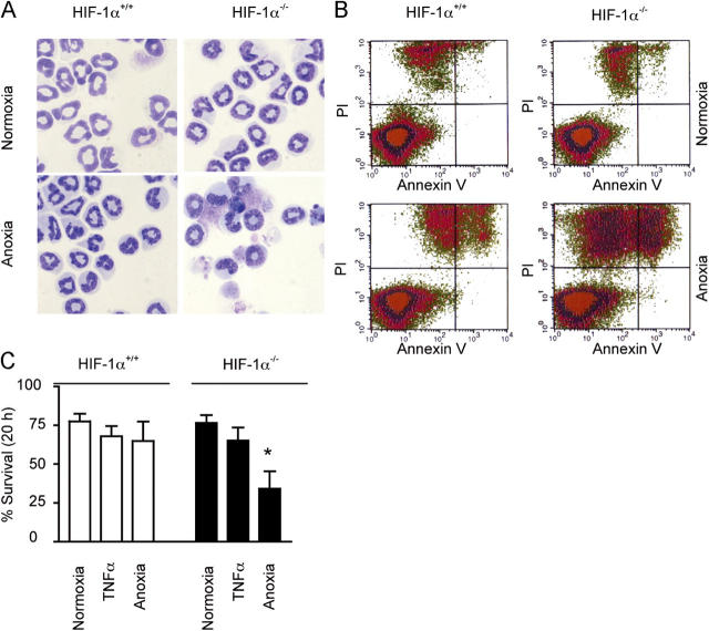
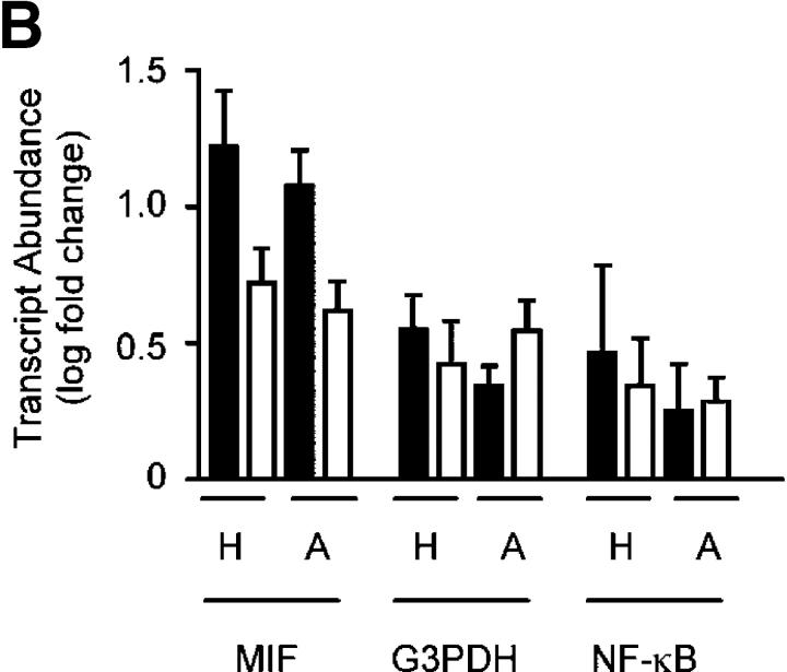
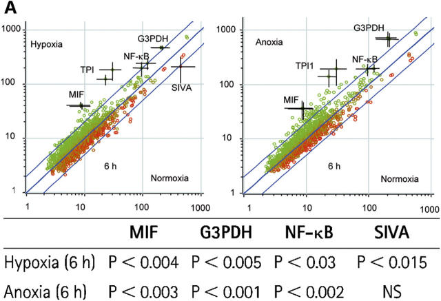
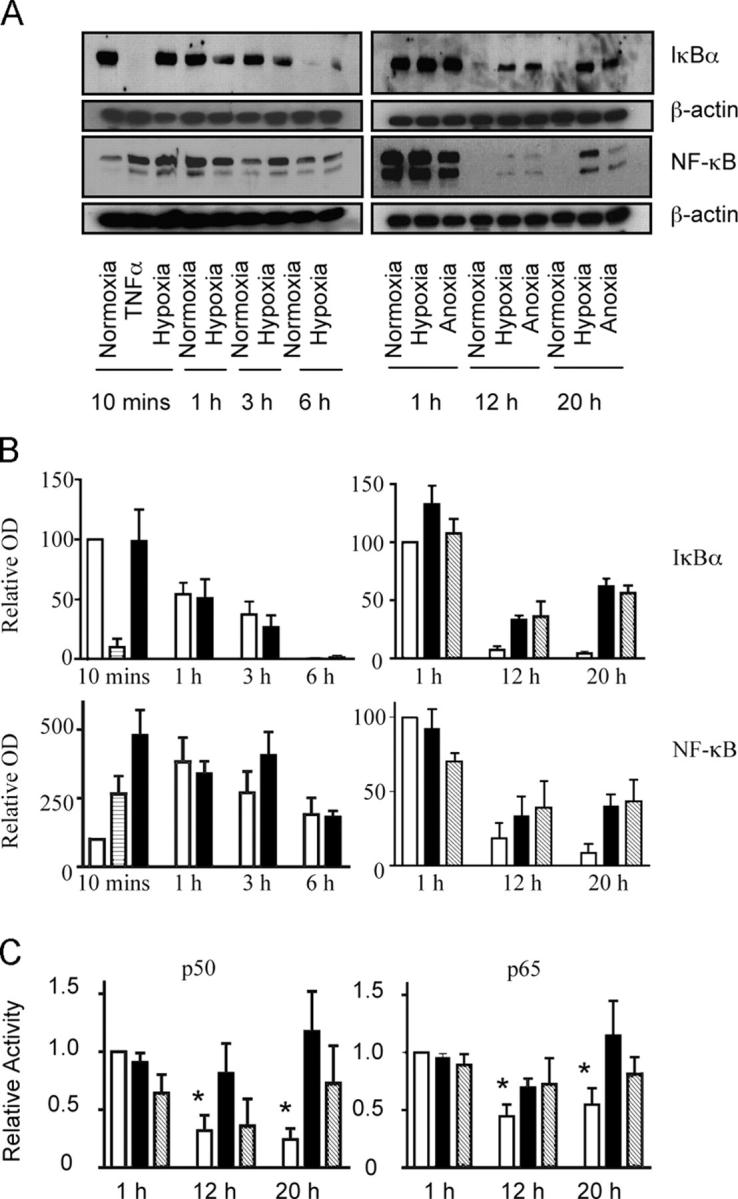
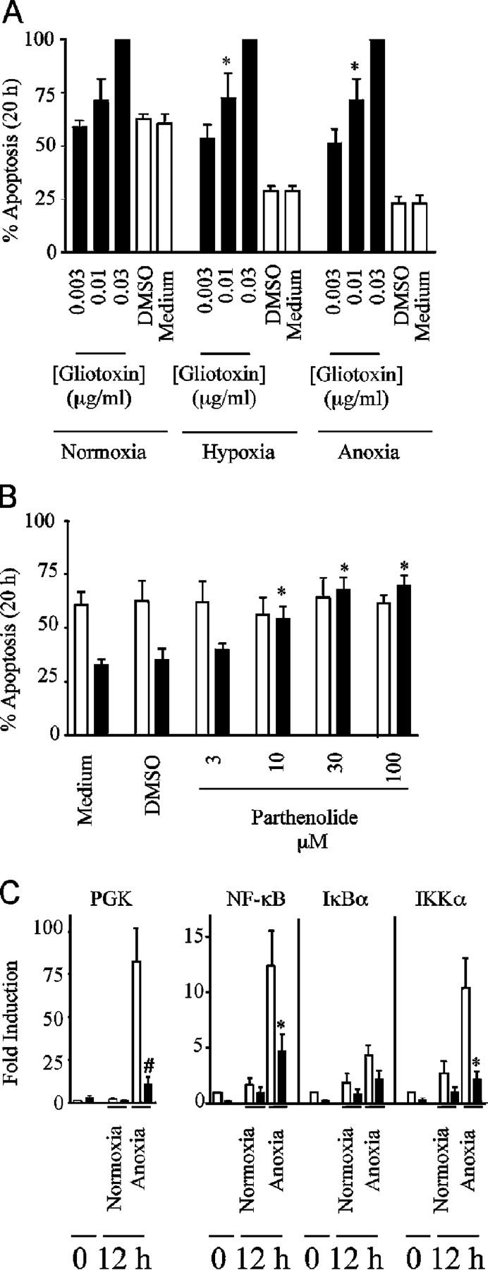
Neutrophils are key effector cells of the innate immune response and are required to migrate and function within adverse microenvironmental conditions. These inflammatory sites are characterized by low levels of oxygen and glucose and high levels of reductive metabolites. A major regulator of neutrophil functional longevity is the ability of these cells to undergo apoptosis. We examined the mechanism by which hypoxia causes an inhibition of neutrophil apoptosis in human and murine neutrophils. We show that neutrophils possess the hypoxia-inducible factor (HIF)-1alpha and factor inhibiting HIF (FIH) hydroxylase oxygen-sensing pathway and using HIF-1alpha-deficient myeloid cells demonstrate that HIF-1alpha is directly involved in regulating neutrophil survival in hypoxia. Gene array, TaqMan PCR, Western blotting, and oligonucleotide binding assays identify NF-kappaB as a novel hypoxia-regulated and HIF-dependent target, with inhibition of NF-kappaB by gliotoxin or parthenolide resulting in the abrogation of hypoxic survival. In addition, we identify macrophage inflammatory protein-1beta as a novel hypoxia-induced neutrophil survival factor.


Figures

Figure 1.
Figure 1.
Hypoxia inhibits constitutive neutrophil apoptosis. (A) Apoptosis. After in vitro culture of human peripheral blood neutrophils for 20 h, cells were assessed for apoptosis by morphology (open bars) or FACS analysis of AV/PI staining (shaded bars). Results represent mean ± SEM (n = 3), *, P < 0.05 compared with normoxic controls. (B) Caspase 3 activity. At the indicated times, caspase 3 activity (arbitrary units) was measured in neutrophil whole cell lysates after culture under different oxygen tensions. Results represent mean ± SEM (n = 4); *, P < 0.05 compared with time-matched normoxic controls. (C) Electron microscopy of neutrophil apoptosis. Electron microscopy appearance of neutrophils aged for 20 h in vitro displaying progressive changes associated with apoptosis; this progression was inhibited in cells incubated under hypoxic or anoxic conditions.
Figure 2.
Figure 2.
Hypoxia induces the release of a PI3-kinase–dependent survival factor. (A) Survival factor secretion. Conditioned medium (CM) obtained from normoxic, hypoxic, or anoxic neutrophils was transferred to freshly isolated cells with subsequent analysis of apoptosis at 6 h (open bars) and 20 h (shaded bars). Results represent mean ± SEM (n = 3); *, P < 0.05 compared with medium (M)-only controls. (B) Survival effect of CM is PI3-kinase dependent. Neutrophils were cultured with CM in the presence of the PI3-kinase inhibitor LY294002 (shaded bars), and apoptosis was assessed by morphology. Results represent mean ± SEM (n = 3); *, P < 0.05 compared with medium (M)-only controls. (C) PI3-kinase– independent hypoxic survival. Neutrophils were cultured in normoxia (open bars), hypoxia (shaded bars), or anoxia (hatched bars) in the presence of PI3-kinase inhibitors LY294002 or wortmannin or the survival cytokine GM-CSF, and apoptosis was assessed morphologically. Results represent mean ± SEM (n = 3); *, P < 0.05 compared with normoxic control.
Figure 3.
Figure 3.
MIP-1β is a novel hypoxia-stimulated granulocyte survival factor. (A) Trypsin sensitivity. Freshly isolated cells were incubated in conditioned medium (CM) obtained from normoxic neutrophils (control) or hypoxic neutrophils (hypoxia CM). These media were either untreated or treated for 2 h with trypsin (1:250 wt/vol), followed by treatment with soya bean trypsin inhibitor (SBTI) or SBTI alone. The effects of trypsin on GM-CSF (100 ng/ml)–mediated neutrophil survival was examined in parallel. Apoptosis was subsequently analyzed at 20 h, and the results represent mean ± SD (n = 2); *, P < 0.05 compared with matched trypsin-untreated conditions. (B) Heat insensitivity. Neutrophils were incubated with CM obtained from normoxic neutrophils (control), hypoxic neutrophils (hypoxia CM), or GM-CSF (100 ng/ml)–supplemented monofeed that was previously heated to 56°C for 45 min. Apoptosis was subsequently analyzed by morphology at 20 h. Results represent mean ± SEM (n = 3). (C) MIP-1β secretion. MIP-1β released into the CM obtained from normoxic (open bar), hypoxic (shaded bar), or anoxic (hatched bar) neutrophils or unconditioned medium (M) was measured by ELISA at 6 and 20 h. Results represent mean ± SEM (n = 3); *, P < 0.05 compared with time-matched normoxic CM. (D) MIP-1β antibody blocks transferable survival. CM obtained from normoxic or hypoxic neutrophils or medium alone (M) was incubated in the presence (+) or absence (−) of 100 μg/ml anti–MIP-1β antibody or 100 μg/ml of total goat IgG isotype control for 30 min at room temperature, before being added to freshly isolated cells. GM-CSF (100 ng/ml) in the presence (+) or absence (−) of anti–MIP-1β or IgG controls were run in parallel, and apoptosis was assessed by 20-h morphology. Results represent mean ± SD (n = 2); *, P < 0.05 compared with matched MIP-1β antibody–untreated conditions.
Figure 4.
Figure 4.
Competitive inhibition of hydroxylase enzymes mimics hypoxic survival. (A) Human neutrophils possess FIH. After culture, under each oxygen tension or in the presence of either the iron chelator DFO or the hydroxylase inhibitor DMOG, whole cell lysates were prepared from peripheral blood neutrophils; immunoprecipitation was performed with an antibody to FIH and separated on SDS-PAGE. Control lysates were also prepared from pulmonary artery smooth muscle cells and run directly on SDS-PAGE without immunoprecipitation. (B) DMOG inhibits human neutrophil apoptosis. Neutrophils were cultured with increasing concentrations of DMOG under normoxia (open bars), hypoxia (shaded bars), and anoxia (hatched bars), and apoptosis was assessed by morphology. Results represent mean ± SEM (n = 3); *, P < 0.05 compared with normoxic control. (C) Hypoxia stabilizes HIF-1α in human neutrophils. Lysates were prepared from neutrophils after culture in hypoxia, anoxia, normoxia or in the presence of DMOG or DFO. Lysates were immunoprecipitated with an antibody to HIF-1α and separated by SDS-PAGE. Both blots are representative of n = 3.
Figure 5.
Figure 5.
HIF-1α regulates neutrophil survival at reduced oxygen tensions. (A) Apoptotic morphology. Murine bone marrow–derived neutrophils were cultured for 20 h under normoxic or anoxic conditions. Apoptosis was assessed by morphology. (B) Annexin V/ Pi. Identical sets of cells were analyzed by FACS for AV/PI staining. (C) Reduced survival in HIF-1α knockout cells under anoxic conditions. Percent survival was calculated after AV/PI staining. Results represent mean ± SEM (n = 3); *, P < 0.05 compared with HIF-1α +/+ control.
Figure 6.
Figure 6.
Hypoxia regulates human neutrophil transcript abundance. (A) Regulation of transcript abundance. Log (base 10) mean signal intensities were calculated and compared from array filters prepared using RNA from human peripheral blood neutrophils cultured under the oxygen tensions shown. Points represent the mean (±SEM bars) of n = 3 experiments; each gene is presented in duplicate. Outer lines represent a twofold change between conditions. All significant transcripts are individually named. The table shows the statistical analysis of the gene array data. Cyber-T tests were performed to calculate the Bayesian p values shown. (B) TaqMan confirmation. Array findings (open bars) were validated for hypoxia (H), and anoxia (A) by TaqMan analysis of duplicate RNA samples (shaded bars) using the nonchanging endogenous control CSF3R (granulocytes). Data represent mean ± SEM of n = 3 experiments.
Figure 6.
Figure 6.
Hypoxia regulates human neutrophil transcript abundance. (A) Regulation of transcript abundance. Log (base 10) mean signal intensities were calculated and compared from array filters prepared using RNA from human peripheral blood neutrophils cultured under the oxygen tensions shown. Points represent the mean (±SEM bars) of n = 3 experiments; each gene is presented in duplicate. Outer lines represent a twofold change between conditions. All significant transcripts are individually named. The table shows the statistical analysis of the gene array data. Cyber-T tests were performed to calculate the Bayesian p values shown. (B) TaqMan confirmation. Array findings (open bars) were validated for hypoxia (H), and anoxia (A) by TaqMan analysis of duplicate RNA samples (shaded bars) using the nonchanging endogenous control CSF3R (granulocytes). Data represent mean ± SEM of n = 3 experiments.
Figure 7.
Figure 7.
Hypoxia stimulates the reexpression of NF-κB protein and maintains activity. (A) Reexpression of IκBα and NF-κB protein with prolonged oxygen deprivation. Human neutrophil lysates were prepared after culture in normoxia ± TNF, hypoxia, or anoxia for 10 min–20 h, and the total protein was measured by Western blot. Blots shown are representative of n = 3–9 experiments. (B) OD quantification of Western blots. Optical densities of IκBα and NF-κB Western blots were quantified for normoxia (open bars), hypoxia (shaded bars), anoxia (hatched bars) and TNF-α (striped bars) using Scion corporation software and normalized to normoxia 10 min (10 min-6 h blots) or normoxia 1 h (1–20 h blots). Data represent mean ± SEM (n = 4). (C) Decreased p50 and p65 activity with prolonged normoxia. p50 or p65 DNA binding activity was measured by ELISA in normoxic (open bars), hypoxic (shaded bars) or anoxic (hatched bars) neutrophil lysates at the time points shown. Data represent mean ± SEM for n = 3 experiments, * P < 0.05 compared with normoxia 1 h.
Figure 8.
Figure 8.
NF-κB activity is required for hypoxic neutrophil survival and dependent on HIF-1α expression. (A) Inhibition of hypoxic survival with gliotoxin. Neutrophils were incubated under the oxygen tensions shown in the presence or absence of gliotoxin for 20 h. Apoptosis was assessed by morphology. Results represent mean ± SEM of n = 3 experiments; *, P < 0.05 compared with oxygen-matched medium only controls. (B) Inhibition of hypoxic survival with parthenolide. Neutrophils were incubated in normoxia (open bars) or hypoxia (shaded bars) in the presence or absence of parthenolide for 20 h. Apoptosis was assessed by morphology. Results represent mean ± SEM of n = 3 experiments; *, P < 0.05 compared with oxygen-matched medium-only controls. (C) Hypoxic induction of PGK, NF-κB, and IKKα message is inhibited in HIF-1α knockout mice. TaqMan analysis of PGK, NF-κB, IκBα, and IKKα transcript abundance relative to β-actin was performed on cDNA isolated from bone marrow–derived murine neutrophils from wild-type (open bars) and HIF-1α knockout mice (shaded bars) after culture in normoxia and anoxia. Data represent mean fold changes ± SD from pooled cDNA from 16 mice in two independent experiments; *, P < 0.05 and #, P < 0.005 compared with matched wild-type controls.

Similar articles

Cited by

References

    1. Nunn, J.F. 1993. Nunn's Applied Respiratory Physiology. Cambridge University Press, Cambridge, UK. 658 pp.
    1. Ellis, G.A., S.E. Edmonds, K. Gaffney, R.B. Williams, and D. Blake. 1994. Synovial tissue oxygenation profile in inflamed and non-inflamed knee joints. Br. J. Rheumatol. 33:172.
    1. Niinikoski, J., T.K. Hunt, and J. Englebert Dunphy. 1972. Oxygen supply in healing tissue. Am. J. Surg. 123:247–252. - PubMed
    1. Levene, P.A., and G.M. Meyer. 1912. The action of leucocytes on glucose. J. Biol. Chem. 11:361–370.
    1. Masson, N., C. William, P. Maxwell, C.W. Pugh, and P.J. Ratcliffe. 2001. Independent function of two destruction domains in hypoxia-inducible factor-α chains activated by prolyl hydroxylation. EMBO J. 20:5197–5206. - PMC - PubMed

Publication types

MeSH terms