Comparative Study
. 2005 Jan 3;201(1):139-48.
doi: 10.1084/jem.20041057.

Synergy of IL-21 and IL-15 in regulating CD8+ T cell expansion and function

Affiliations
Comparative Study

Synergy of IL-21 and IL-15 in regulating CD8+ T cell expansion and function

Rong Zeng et al. J Exp Med. .

Abstract

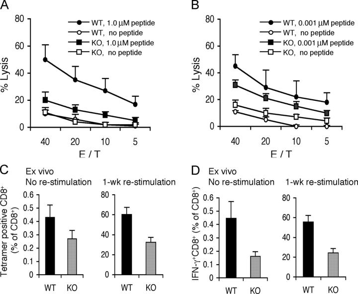
Interleukin (IL)-21 is the most recently recognized of the cytokines that share the common cytokine receptor gamma chain (gamma(c)), which is mutated in humans with X-linked severe combined immunodeficiency. We now report that IL-21 synergistically acts with IL-15 to potently promote the proliferation of both memory (CD44high) and naive (CD44low) phenotype CD8+ T cells and augment interferon-gamma production in vitro. IL-21 also cooperated, albeit more weakly, with IL-7, but not with IL-2. Correspondingly, the expansion and cytotoxicity of CD8+ T cells were impaired in IL-21R-/- mice. Moreover, in vivo administration of IL-21 in combination with IL-15 boosted antigen-specific CD8+ T cell numbers and resulted in a cooperative effect on tumor regression, with apparent cures of large, established B16 melanomas. Thus, our studies reveal that IL-21 potently regulates CD8+ T cell expansion and effector function, primarily in a synergistic context with IL-15.


Figures

Figure 1.
Figure 1.
IL-21 acts synergistically with IL-15 to expand CD8 + T cells. 5 × 10⁶ splenocytes were pooled from two to three wild-type mice and cultured for 7 d in medium containing IL-15, IL-21, or combinations of these cytokines, as indicated. The number of NK cells (A), T cells (B), CD8+ T cells (C), and CD4+ T cells (D) were determined as NK1.1+TCRβ, NK1.1TCRβ+, CD8+CD4, and CD8CD4+ cells, respectively. Results shown are means ± SD from three experiments. (E and F) Cells cultured in medium for 0 d (a) or 7 d (b), or in 100 ng/ml of IL-15, IL-21, or both cytokines (c, d, and e, respectively) were analyzed by flow cytometry for 7 d. The percentages of NK1.1TCRβ+ and NK1.1+TCRβ cells (E) and CD8CD4+ and CD8+CD4 cells (F) are indicated in the quadrant corners.
Figure 2.
Figure 2.
IL-21 acts in concert with IL-15 or IL-7 but not with IL-2 to expand CD8 + T cells. (A and B) 5 × 10⁶ splenocytes pooled from three wild-type mice were cultured for 3, 5, and 7 d in medium containing 100 U/ml IL-2, 100 ng/ml IL-7, 100 ng/ml IL-15, 100 ng/ml IL-21, or combinations of these cytokines, as indicated. CD8+ T (A) and CD4+ T (B) cell subsets were identified as CD8+CD4 and CD8CD4+, respectively. Results shown are means ± SD from three experiments. (C) Representative flow cytometric analysis of cells cultured for 7 d as described in A and B. Percentages of selected cell populations are indicated in the quadrant corners. (D) Cells from A were also stained with annexin V and propidium iodide (PI). Percentages of double negative subpopulations corresponding to viable cells are indicated in the quadrant corners. Data representative of three separate experiments are shown.
Figure 3.
Figure 3.
IL-15 and IL-21 cooperatively enhance effector function of memory-phenotype CD8 + T cells. (A and B) 5 × 10⁶ splenocytes pooled from two to three wild-type mice were cultured for 7 d in medium containing 100 ng/ml of IL-15, IL-21, or combinations of these cytokines, as indicated (b–e). The naive cells (A, subpanel a) were splenocytes from a littermate that were directly analyzed without culture. Cells were analyzed by flow cytometry after staining with (A) CD8-allophycocyanin, CD4-FITC, CD44-CyChrome, and IL-2Rβ-PE or (B) CD8-allophycocyanin, IL-2Rβ-FITC, CD44-CyChrome, and CD62L-PE, respectively. Data representative of three separate experiments are shown. The absolute numbers of gated CD8+ T cells and the percentages of CD44high, CD44intermediate, and CD44low subpopulations are indicated in A. The percentages of CD62Llow and CD62Lhigh subpopulations are indicated in B. (C and D) Freshly isolated splenocytes or cells cultured for 7 d with the indicated cytokines were stimulated with anti-CD3 and anti-CD28 for 0, 1, 2, or 4 h. (C) Representative flow cytometric profiles. (D) The absolute number of IFN-γ–producing CD8+ T cells. Mean values for three similar experiments are shown. The increase in IFN-γ+CD8+ T cells with IL-15 and IL-21 was highly significant.
Figure 4.
Figure 4.
IL-15 and IL-21 cooperatively increase cell cycle progression of both CD44high and CD44low CD8 + T cells. (A) All cell subpopulations were isolated and stained with CFSE as described in Materials and Methods. The “basal” CFSE profile on day 0 is shown as the dashed line. Cells were cultured in complete medium at 2–5 × 10⁵ cells/ml without cytokine or with 100 ng/ml of IL-15 and/or IL-21 for 4 or 7 d, as indicated, and analyzed by flow cytometry. The indicated cell numbers (×10−6) are an average of cell numbers from three independent experiments. In each experiment, the starting cell numbers were normalized to 3.75 × 10⁶ (CD8+ T cells), 0.75 × 10⁶ (CD44highCD8+ T cells), and 2.5 × 10⁶ (CD44lowCD8+ T cells), respectively, which allowed comparison of the different experiments. Flow cytometric results shown are representative of three experiments. (B) Cells in A were also stained with annexin V and PI. The percentages of double negative subpopulations corresponding to viable cells are indicated in the quadrant corners. Data representative of three separate experiments are shown.
Figure 5.
Figure 5.
Antigen-specific CD8 + T cell responses are impaired in IL-21R −/− mice. Five mice in each group were immunized i.p. with 5 × 10⁶ PFU of vPE16. (A and B) On day 5, splenocytes of immunized WT and IL-21R−/− (KO) mice were restimulated with 1.0 μM (A) or 0.001 μM (B) P18-I10 for 1 wk, and lytic activity was measured by a 5-h 51Cr release assay. P815 cells pulsed with 1.0 μM (A) or 0.001 μM (B) P18-I10 were used as target cells. Means ± SEM are shown. (C) The frequency of P18-I10–specific splenic CD8+ T cells was measured by H-2Dd-P18-I10 tetramer staining ex vivo without restimulation in vitro (left) or after 1 wk of restimulation with 1.0 μM P18-I10 (right). Shown is the mean percent ± SEM of tetramer-positive CD8+ T cells in total CD8+ T cells for five mice per group. The difference in the two groups was statistically significant (P < 0.05). (D) The frequency of IFN-γ+CD8+ T cells in spleens was measured by intracellular staining ex vivo without restimulation in vitro (left) or after 1 wk of restimulation with 1.0 μM P18-I10 (right). Cells were stimulated with 1.0 μM P18-I10 for 10 h in the presence of 1 μg/ml brefeldin A. Shown is the mean percent ± SEM of IFN-γ–positive (IFN-γ+) CD8+ T cells in total CD8+ T cells for five mice per group. The difference in the two groups was statistically significant (P < 0.05).
Figure 6.
Figure 6.
In vivo administration of IL-21 in combination with IL-15 yields tumor regression with cures of large, established B16 melanomas. (A) Sublethally irradiated female C57BL/6 mice were implanted subcutaneously with B16 melanomas. 8 d later, mice were treated by adoptively transferring cultured pmel-1 splenocytes (0.5 × 10⁶ Vβ13+ CD8+ T cells) as indicated and, where indicated, vaccinating with rFPVhgp100. In some conditions, IL-15 and IL-21 were administered twice daily for six doses (5 μg each/dose; five to seven mice in each group). (B) Sublethally irradiated female C57BL/6 mice were implanted subcutaneously with B16 melanomas. 10 d later, mice were treated by adoptively transferring cultured pmel-1 splenocytes (106 Vβ13+ CD8+ T cells) and vaccinating with rFPVhgp100. IL-15, IL-21, or both cytokines was administered twice daily for six doses (10 μg each /dose; five to seven mice in each group). A repeat experiment showed similar results. Shown are mean tumor sizes ± SEM. (C) Shown are Vβ13+CD8+ T cell numbers from mice from two experiments (days 21 and 28 from Experiment 1 in B and day 20 from Experiment 2, which was terminated at day 23). *, Mice not receiving cytokine had all died from their tumors, so samples were not available for analysis.
Figure 7.
Figure 7.
Several genes are synergistically regulated by IL-21 and IL-15 in CD8 + T cells. Naive CD8+ T cells were isolated, treated without cytokine or with 100 ng/ml of IL-15 and/or IL-21 for 4 h, and subjected to RNA isolation and Affymetrix Gene Chip analysis as described in Materials and Methods. Results shown are from five independent experiments. Shown in A are the overall expression patterns of genes that are induced or repressed more than twofold by IL-15, IL-21, or both cytokines. Also shown are selected genes that are preferentially regulated by IL-15 alone (B), IL-21 alone (C), or IL-15 plus IL-21 (D).

Similar articles

Cited by

References

    1. Noguchi, M., H. Yi, H.M. Rosenblatt, A.H. Filipovich, S. Adelstein, W.S. Modi, O.W. McBride, and W.J. Leonard. 1993. Interleukin-2 receptor gamma chain mutation results in X-linked severe combined immunodeficiency in humans. Cell. 73:147–157. - PubMed
    1. Leonard, W.J. 2001. Cytokines and immunodeficiency diseases. Nat. Rev. Immunol. 1:200–208. - PubMed
    1. Ozaki, K., K. Kikly, D. Michalovich, P.R. Young, and W.J. Leonard. 2000. Cloning of a type I cytokine receptor most related to the IL-2 receptor beta chain. Proc. Natl. Acad. Sci. USA. 97:11439–11444. - PMC - PubMed
    1. Parrish-Novak, J., S.R. Dillon, A. Nelson, A. Hammond, C. Sprecher, J.A. Gross, J. Johnston, K. Madden, W. Xu, J. West, et al. 2000. Interleukin 21 and its receptor are involved in NK cell expansion and regulation of lymphocyte function. Nature. 408:57–63. - PubMed
    1. Kasaian, M.T., M.J. Whitters, L.L. Carter, L.D. Lowe, J.M. Jussif, B. Deng, K.A. Johnson, J.S. Witek, M. Senices, R.F. Konz, et al. 2002. IL-21 limits NK cell responses and promotes antigen-specific T cell activation: a mediator of the transition from innate to adaptive immunity. Immunity. 16:559–569. - PubMed

Publication types

MeSH terms