. 2005 Jan;115(1):94-101.
doi: 10.1172/JCI23237.

Oncogenic AKAP9-BRAF fusion is a novel mechanism of MAPK pathway activation in thyroid cancer

Affiliations

Oncogenic AKAP9-BRAF fusion is a novel mechanism of MAPK pathway activation in thyroid cancer

Raffaele Ciampi et al. J Clin Invest. 2005 Jan.

Abstract

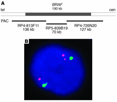
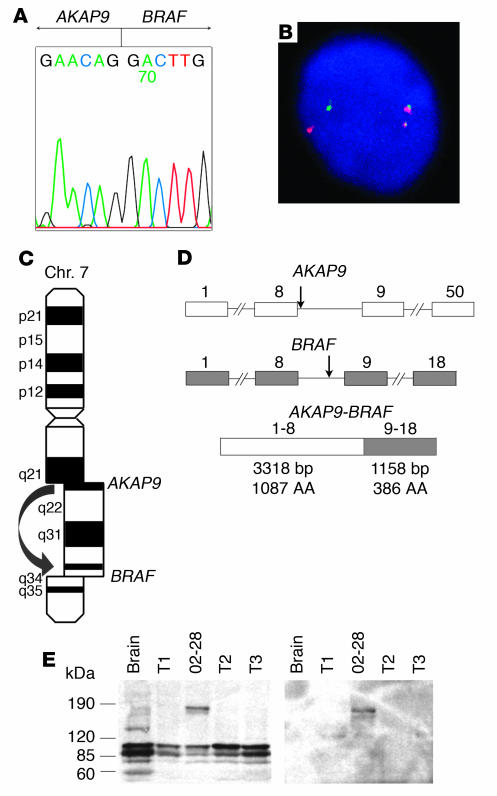
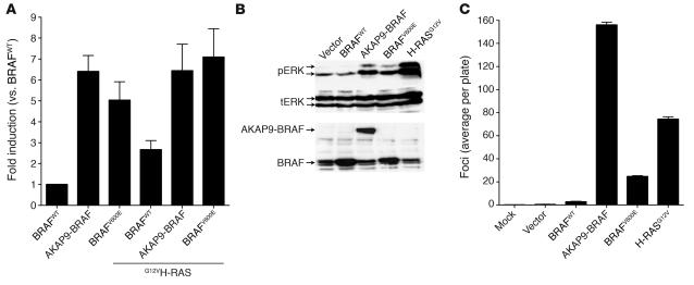
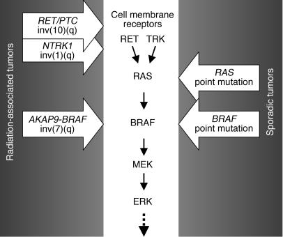
Genes crucial for cancer development can be mutated via various mechanisms, which may reflect the nature of the mutagen. In thyroid papillary carcinomas, mutations of genes coding for effectors along the MAPK pathway are central for transformation. BRAF point mutation is most common in sporadic tumors. By contrast, radiation-induced tumors are associated with paracentric inversions activating the receptor tyrosine kinases RET and NTRK1. We report here a rearrangement of BRAF via paracentric inversion of chromosome 7q resulting in an in-frame fusion between exons 1-8 of the AKAP9 gene and exons 9-18 of BRAF. The fusion protein contains the protein kinase domain and lacks the autoinhibitory N-terminal portion of BRAF. It has elevated kinase activity and transforms NIH3T3 cells, which provides evidence, for the first time to our knowledge, of in vivo activation of an intracellular effector along the MAPK pathway by recombination. The AKAP9-BRAF fusion was preferentially found in radiation-induced papillary carcinomas developing after a short latency, whereas BRAF point mutations were absent in this group. These data indicate that in thyroid cancer, radiation activates components of the MAPK pathway primarily through chromosomal paracentric inversions, whereas in sporadic forms of the disease, effectors along the same pathway are activated predominantly by point mutations.


Figures

Figure 1
Figure 1
Identification of the BRAF gene rearrangement. (A) Genomic region on 7q containing the BRAF gene and position of PAC clones used as a probe for FISH. (B) Interphase nucleus from the index tumor showing split of 1 BRAF signal (red) and preservation of 2 chromosome 7 centromeric signals (green), which indicates the rearrangement of the BRAF gene.
Figure 2
Figure 2
BRAF is recombined with the AKAP9 gene and results in expression of a fusion protein. (A) Sequence of the 5′ RACE product showing a fusion point between AKAP9 and BRAF. (B) Confirmation of the reciprocal fusion by FISH with probes corresponding to the BRAF (red) and AKAP9 (green) genes. (C) The fusion is a result of paracentric chromosomal inversion inv(7)(q21–22q34). Chr., chromosome. (D) Genomic structure of the BRAF and AKAP9 genes showing the location of breakpoints (arrows) and the organization of the chimeric cDNA. Exons are represented by boxes and introns by lines. Numbers above indicate exon numbers. (E) Western blot analysis using BRAF antibody (left) and AKAP9 antibody (right), showing an approximately 170-kDa protein, corresponding to the predicted molecular weight of the fusion protein in the index case (number 02-28). Three other papillary carcinomas (T1–T3) are shown for comparison. Wild-type AKAP9 is 453 kDa in size and was not detected in this Western blot.
Figure 3
Figure 3
Subcellular localization of AKAP9 and AKAP9-BRAF detected by immunohistochemistry with an AKAP9 antibody. (A) In a papillary thyroid carcinoma negative for the fusion, the protein is predominantly targeted to centrosomes, seen as a single perinuclear dot. There is also weak diffuse cytoplasmic staining. (B) The tumor expressing AKAP9-BRAF reveals diffuse cytoplasmic distribution of the chimeric protein and the perinuclear dot is no longer visible. The shift in localization involves only tumor cells but not adjacent vascular endothelial cells, which preserve the dotted pattern of staining (arrow). Magnification, ×200.
Figure 4
Figure 4
The AKAP9-BRAF chimeric protein has increased BRAF kinase activity and results in NIH3T3 cell transformation. (A) In vitro kinase activity of myc-tagged BRAFWT, AKAP9-BRAF, and BRAFV600E in the absence or presence of H-RASG12V in COS7 cells. BRAF kinase activity was measured in myc-IgG immunoprecipitates using MEK1 substrate phosphorylation as a read-out. Each sample was assayed in triplicate, and bars represent the standard deviations from the mean. Similar results were obtained in 2 independent transfections. (B) Western blots of lysate from cells stably transfected with pEFP, pEFP-BRAFWT, pEFP–AKAP9-BRAF, pEFP-BRAFV600E, or pBabe puro–H-RASG12V probed with antibodies to phospho-ERK1/2 (pERK), total ERK (tERK), or C-terminus of BRAF. Similar results were obtained in 3 additional independent experiments. (C) Foci formation of NIH3T3 cells transfected with pEFP-BRAFWT, pEFP–AKAP9-BRAF, pEFP-BRAFV600E, or pBabe puro–H-RASG12V. Bars represent the average number of foci per plate from 6 plates. Similar results were obtained in 2 independent experiments.
Figure 5
Figure 5
Analysis of post-Chernobyl tumors that developed 5–6 years after exposure. (A) Southern blot of RT-PCR products amplified with primers Ex8A and Ex10B and hybridized with probe PR showing 2 positive cases (C32 and C27). Other tumor samples are negative (C13, C23, C24, C18, and C31). NC, negative control; PC, positive control (index case). (B) Immunohistochemical analysis of tumor C32 with AKAP9 antibody showing a shift in subcellular localization of the protein from a single dot/centrosomal pattern observed in the entrapped non-neoplastic follicles (arrows) to diffuse cytoplasmic staining in tumor cells. Magnification, ×200.
Figure 6
Figure 6
Detection of V600E BRAF mutation cDNA from post-Chernobyl tumors that developed 9–12 years after exposure using LightCycler real-time PCR followed by fluorescence melting curve analysis. (A) Real-time amplification of cDNA samples. (B) Detection of point mutations by the postamplification fluorescence melting curve analysis based on a distinct melting temperature of duplexes formed between the WT probe and either WT (62.8°C) or mutant (57.0°C) sequences. The mutations in both samples are heterozygous, since both mutant and WT peaks are detectable.
Figure 7
Figure 7
Activation of MAPK pathway in papillary thyroid carcinogenesis involves predominantly point mutations in sporadic tumors or chromosomal rearrangements in radiation-associated tumors.

Comment in

References

    1. Kimura ET, et al. High prevalence of BRAF mutations in thyroid cancer: genetic evidence for constitutive activation of the RET/PTC-RAS-BRAF signaling pathway in papillary thyroid carcinoma. Cancer Res. 2003;63:1454–1457. - PubMed
    1. Soares P, et al. BRAF mutations and RET/PTC rearrangements are alternative events in the etiopathogenesis of PTC. Oncogene. 2003;22:4578–4580. - PubMed
    1. Frattini M, et al. Alternative mutations of BRAF, RET and NTRK1 are associated with similar but distinct gene expression patterns in papillary thyroid cancer. Oncogene. 2004;23:7436–7440. - PubMed
    1. Takahashi M, Ritz J, Cooper GM. Activation of a novel human transforming gene, ret, by DNA rearrangement. Cell. 1985;42:581–588. - PubMed
    1. Takahashi M. Structure and expression of the ret transforming gene. IARC Sci. Publ. 1988;1988:189–197. - PubMed

Publication types

MeSH terms