

Dopaminergic neurons generated from monkey embryonic stem cells function in a Parkinson primate model
- PMID: 15630449
- PMCID: PMC539189
- DOI: 10.1172/JCI21137
Dopaminergic neurons generated from monkey embryonic stem cells function in a Parkinson primate model
Abstract
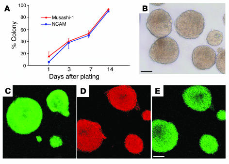
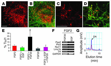
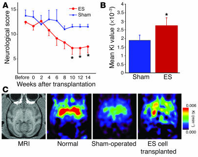
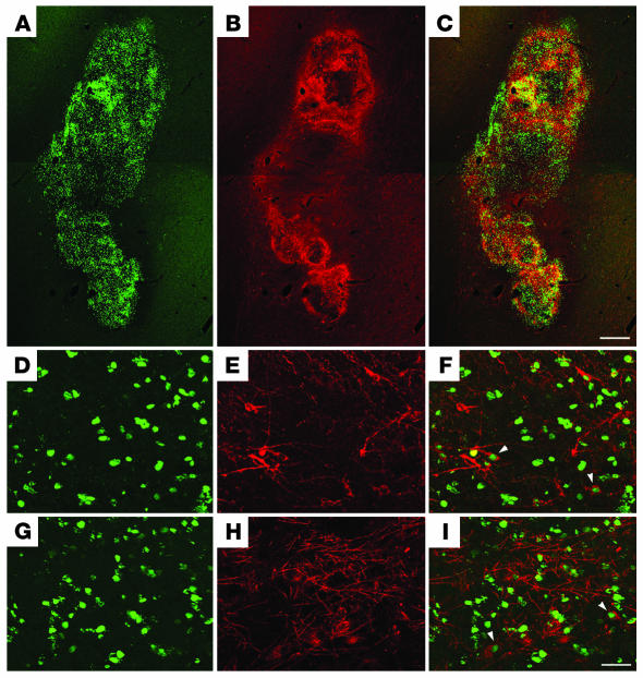
Parkinson disease (PD) is a neurodegenerative disorder characterized by loss of midbrain dopaminergic (DA) neurons. ES cells are currently the most promising donor cell source for cell-replacement therapy in PD. We previously described a strong neuralizing activity present on the surface of stromal cells, named stromal cell-derived inducing activity (SDIA). In this study, we generated neurospheres composed of neural progenitors from monkey ES cells, which are capable of producing large numbers of DA neurons. We demonstrated that FGF20, preferentially expressed in the substantia nigra, acts synergistically with FGF2 to increase the number of DA neurons in ES cell-derived neurospheres. We also analyzed the effect of transplantation of DA neurons generated from monkey ES cells into 1-methyl-4-phenyl-1,2,3,6-tetrahydropyridine-treated (MPTP-treated) monkeys, a primate model for PD. Behavioral studies and functional imaging revealed that the transplanted cells functioned as DA neurons and attenuated MPTP-induced neurological symptoms.
Figures

Comment in
-
The promise of stem cells in Parkinson disease.J Clin Invest. 2005 Jan;115(1):23-5. doi: 10.1172/JCI24012. J Clin Invest. 2005. PMID: 15630437 Free PMC article.
References
-
- Bergman H, Deuschl G. Patho-physiology of Parkinson’s disease: from clinical neurology to basic neuroscience and back. Mov. Disord. 2002;17(Suppl. 3):S28–S40. - PubMed
-
- Dostrovsky JO, Hutchison WD, Lozano AM. The globus pallidus, deep brain stimulation, and Parkinson’s disease. Neuroscientist. 2002;8:284–290. - PubMed
-
- Miyasaki JM, Martin W, Suchowersky O, Weiner WJ, Lang AE. Practice parameter: initiation of treatment for Parkinson’s disease: an evidence-based review: report of the Quality Standards Subcommittee of the American Academy of Neurology. Neurology. 2002;58:11–17. - PubMed
-
- Freed CR, et al. Transplantation of embryonic dopamine neurons for severe Parkinson’s disease. N. Engl. J. Med. 2001;344:710–719. - PubMed
-
- Hagell P, Brundin P. Cell survival and clinical outcome following intrastriatal transplantation in Parkinson disease. J. Neuropathol. Exp. Neurol. 2001;60:741–752. - PubMed
Publication types
MeSH terms
Substances
LinkOut - more resources
Full Text Sources
Other Literature Sources
Medical
