

doi: 10.1172/JCI19189.
Revascularization of ischemic tissues by PDGF-CC via effects on endothelial cells and their progenitors
Affiliations
- PMID: 15630451
- PMCID: PMC535797
- DOI: 10.1172/JCI19189
Item in Clipboard
Revascularization of ischemic tissues by PDGF-CC via effects on endothelial cells and their progenitors
J Clin Invest.
2005 Jan.
Abstract
The angiogenic mechanism and therapeutic potential of PDGF-CC, a recently discovered member of the VEGF/PDGF superfamily, remain incompletely characterized. Here we report that PDGF-CC mobilized endothelial progenitor cells in ischemic conditions; induced differentiation of bone marrow cells into ECs; and stimulated migration of ECs. Furthermore, PDGF-CC induced the differentiation of bone marrow cells into smooth muscle cells and stimulated their growth during vessel sprouting. Moreover, delivery of PDGF-CC enhanced postischemic revascularization of the heart and limb. Modulating the activity of PDGF-CC may provide novel opportunities for treating ischemic diseases.
Figures
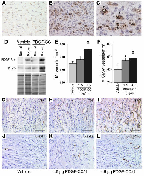
Therapeutic revascularization with PDGF-CC in ischemic heart. (A–C) Immunostaining, revealing low PDGF-Rα expression in a subset of microvessels in an uninjured myocardium (A) and strongly increased PDGF-Rα expression in sprouting vessels in the border region of a myocardial infarct, from where vessels revascularize the infarct (B and C). C shows a detail of PDGF-Rα expression in microvessels (arrows). (D) Upper and middle panels: immunoprecipitation and subsequent Western blotting for PDGF-Rα (upper) and pTyr (middle) showed that PDGF-Rα was upregulated in the ischemic myocardial regions bordering the infarct where vessels start to grow. Note also that PDGF-Rα was activated more in the borders than the normal (nonischemic) regions and maximally after PDGF-CC treatment. Lower panel: Coomassie staining revealing comparable loading. (E and F) PDGF-CC protein treatment increased TM+ (E) and SMA+ (F) vessel density in the infarcted areas in a dose-dependent manner. *P < 0.05 vs. vehicle. (G–I) TM immunostaining of myocardial vessels, revealing increased vessel densities after PDGF-CC treatment. (J–L) α-SMA immunostaining of myocardial vessels, revealing increased vessel densities after PDGF-CC treatment. Scale bars: 50 μm in A, B, G–L and 20 μm in C.
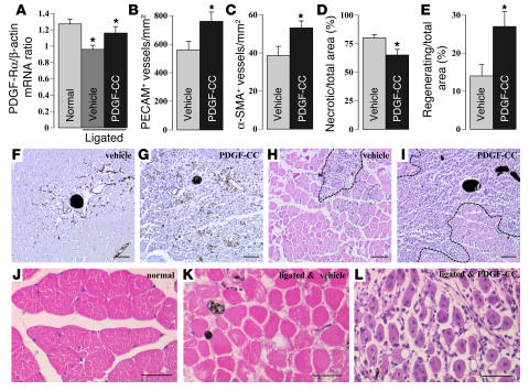
Therapeutic revascularization with PDGF-CC in ischemic limbs. (A) RNAse protection analysis, showing that PDGF-Rα expression in the gastrocnemius muscle was decreased at 2 days after femoral artery ligation but restored to normal levels after PDGF-CC treatment. The ratio of the PDGF-Rα levels (arbitrary units), normalized for β-actin levels, is shown. (B and C) PDGF-CC protein treatment increased the PECAM capillary (B) and α-SMA+ arteriolar (C) density in the ischemic gastrocnemius muscle. (D and E) PDGF-CC protein treatment decreased muscle necrosis (D) and increased muscle regeneration (E) in the gastrocnemius muscle at 7 days after femoral artery ligation. Areas are percentage of total muscle area. *P < 0.05 (A–E). (F and G) Compared with vehicle (F), PDGF-CC protein treatment increased the density of PECAM vessels in the regenerating areas of the ischemic gastrocnemius muscle (G). No signs of edema, hemorrhage. or fibrosis were observed. (H and I) H&E staining, showing larger areas of regenerating myocytes (small cells with central nuclei) after PDGF-CC treatment (I) than after treatment with vehicle (H). The regions containing regenerating myocytes are surrounded by a dashed black line in both panels. (J–L) Higher magnification of H&E-stained sections of a normal gastrocnemius muscle (J); ischemic muscle, treated with vehicle, containing numerous necrotic ghost myocytes, and few blood vessels (K); ischemic muscle, treated with PDGF-CC, containing numerous regenerating myocytes with a central nucleus and numerous blood vessels (L). Values are mean ± SEM of at least 15 mice. The lumen of the arterioles is filled with dark bismuth gelatin in F–L. Scale bars: 50 μm.
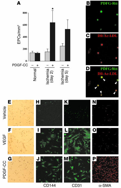
Effects of PDGF-CC on endothelial progenitors. (A) PDGF-CC treatment increased EPC mobilization from day 2 to day 5 after hind limb ischemia but did not affect EPC mobilization in normal conditions. *P < 0.05. Values are mean ± SEM of 10 mice. (B–D) Double labeling of EPCs for PDGF-Rα (green in B) and Dil-ac-LDL (red in C), showing coexpression (yellow in D). (E–P) After 2 weeks of stimulation, both PDGF-CC and VEGF induced the expression of EC surface markers CD144 (VE-cadherin) and CD31 (PECAM), while vehicle-treated cells remained negative. Only PDGF-CC induced prominent α-SMA expression, while cells treated with VEGF or vehicle displayed background levels of α-SMA expression. Unstained cells in E–G show that both VEGF and PDGF-CC promoted stem/progenitor cell adherence.
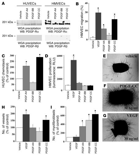
Effect of PDGF-CC on EC migration and outgrowth of microvessels. (A) WGA precipitation and subsequent immunoblotting of extracts of control and ligand-stimulated HUVECs and HMVECs, revealing detectable expression of PDGF-Rα but not of PDGF-Rβ. By immunoblotting, PDGF-Rα was tyrosine phosphorylated in both cell lines (data not shown). (B and C) In the scrape wound assay (B), PDGF-CC stimulated HMVEC migration with a potency similar to that of VEGF, while PDGF-AA and -BB had an intermediate or no effect, respectively. In the Boyden chamber assay (C), both PDGF-AA and -CC induced HUVEC chemotaxis, while PDGF-BB was inactive. (D) None of the PDGFs affected HMVEC proliferation, while VEGF potently promoted cell proliferation. *P < 0.05. Values are mean ± SEM. RLU, relative luminescence units. (E–G) Micrographs of aortic rings, displaying microvascular sprouts and perivascular cells. Compared to vehicle (E), PDGF-CC enhanced the outgrowth of both microvascular sprouts and fibroblast-like cells (F), while VEGF stimulated microvascular outgrowth (G). (H and I) In the aortic ring assay, PDGF-CC and VEGF induced microvessel outgrowth, while PDGF-AA and -BB were inactive (H). Each PDGF form stimulated myofibroblast outgrowth from the aortic ring, but PDGF-CC was more potent than PDGF-BB and PDGF-AA, whereas VEGF was inactive (I). P < 0.05 (H and I). WB, Western blot.
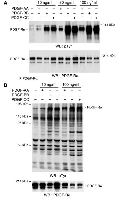
Tyrosine phosphorylation of PDGF-Rα and downstream proteins in response to PDGF-CC. (A) Immunoprecipitation of equal amounts of PAEC/Rα extracts for PDGF-Rα and subsequent immunoblotting for pTyr, revealing a higher degree of PDGF-Rα tyrosine phosphorylation after 10-minute stimulation with PDGF-BB or -CC than with PDGF-AA. The lower panel shows an immunoblot of PDGF-Rα, displaying the amount of receptor present in each sample. (B) Immunoblotting of equal amounts of PAEC/Rα extracts for pTyr after stimulation with PDGF-AA, -BB, or -CC for 10 minutes, revealing that a similar set of proteins (judged on the basis of their molecular weight) was phosphorylated but that, in general, the phosphorylation signals were stronger in response to PDGF-BB and -CC than PDGF-AA. The lower panel shows an immunoblot of PDGF-Rα, displaying the amount of receptor present in each sample. For both A and B, though equal amounts of cell lysate were immunoprecipitated (A) and/or loaded (A and B), PDGF-Rα levels differed among the various lanes, presumably reflecting effects on receptor internalization and degradation. PDGF-Rα is present as a doublet due to differences in glycosylation.
References
-
- Li X, et al. PDGF-C is a new protease-activated ligand for the PDGF alpha-receptor. Nat. Cell Biol. 2000;2:302–309. - PubMed
-
- Bergsten E, et al. PDGF-D is a specific, protease-activated ligand for the PDGF beta- receptor. Nat. Cell Biol. 2001;3:512–516. - PubMed
-
- LaRochelle WJ, et al. PDGF-D, a new protease-activated growth factor. Nat. Cell Biol. 2001;3:517–521. - PubMed
-
- Heldin CH, Westermark B. Mechanism of action and in vivo role of platelet-derived growth factor. Physiol. Rev. 1999;79:1283–1316. - PubMed
-
- Gilbertson DG, et al. Platelet-derived growth factor C (PDGF-C) a novel growth factor that binds to PDGF alpha and beta receptor. J. Biol. Chem. 2001;276:27406–27414. - PubMed
Publication types
MeSH terms
Substances
LinkOut - more resources
Full Text Sources
Other Literature Sources
Medical
