. 2005 Jan;115(1):177-86.
doi: 10.1172/JCI21867.

Possible involvement of pregnane X receptor-enhanced CYP24 expression in drug-induced osteomalacia

Affiliations

Possible involvement of pregnane X receptor-enhanced CYP24 expression in drug-induced osteomalacia

Jean Marc Pascussi et al. J Clin Invest. 2005 Jan.

Abstract

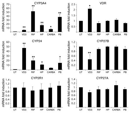
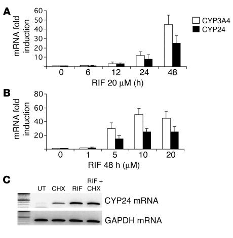
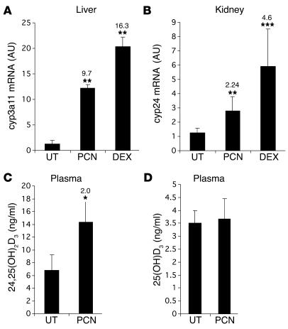
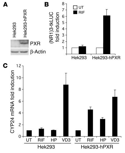
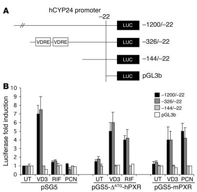
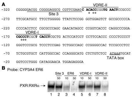
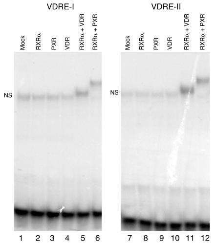
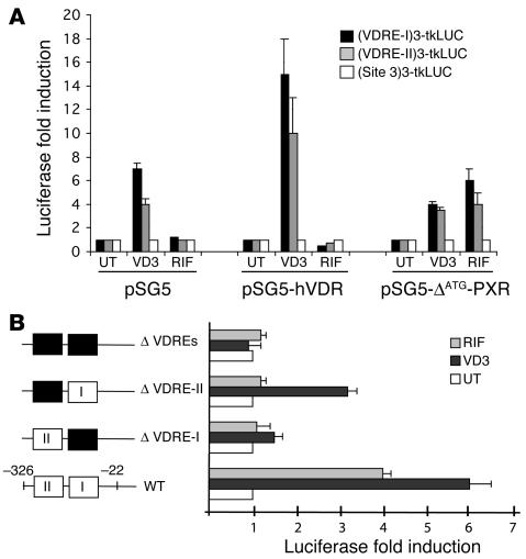
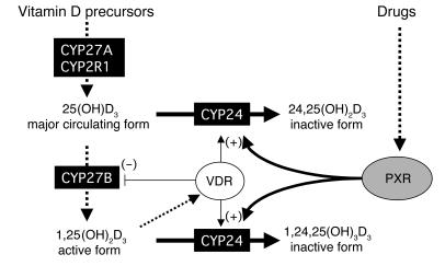
Vitamin D controls calcium homeostasis and the development and maintenance of bones through vitamin D receptor activation. Prolonged therapy with rifampicin or phenobarbital has been shown to cause vitamin D deficiency or osteomalacia, particularly in patients with marginal vitamin D stores. However, the molecular mechanism of this process is unknown. Here we show that these drugs lead to the upregulation of 25-hydroxyvitamin D(3)-24-hydroxylase (CYP24) gene expression through the activation of the nuclear receptor pregnane X receptor (PXR; NR1I2). CYP24 is a mitochondrial enzyme responsible for inactivating vitamin D metabolites. CYP24 mRNA is upregulated in vivo in mice by pregnenolone 16alpha-carbonitrile and dexamethasone, 2 murine PXR agonists, and in vitro in human hepatocytes by rifampicin and hyperforin, 2 human PXR agonists. Moreover, rifampicin increased 24-hydroxylase activity in these cells, while, in vivo in mice, pregnenolone 16alpha-carbonitrile increased the plasma concentration of 24,25-dihydroxyvitamin D(3). Transfection of PXR in human embryonic kidney cells resulted in rifampicin-mediated induction of CYP24 mRNA. Analysis of the human CYP24 promoter showed that PXR transactivates the sequence between -326 and -142. We demonstrated that PXR binds to and transactivates the 2 proximal vitamin D-responsive elements of the human CYP24 promoter. These data suggest that xenobiotics and drugs can modulate CYP24 gene expression and alter vitamin D(3) hormonal activity and calcium homeostasis through the activation of PXR.


Figures

Figure 1
Figure 1
Effect of 1α,25(OH)2D3, rifampicin, hyperforin, carbamazepine, and phenobarbital on CYP24, CYP2R1, CYP27B, CYP27A, VDR, and CYP3A4 mRNAs in human hepatocytes. Human hepatocytes were cultured for 48 hours in the absence (UT) or presence of the indicated compounds: 50 nM 1α,25(OH)2D3 (VD3), 20 μM rifampicin (RIF), 2 μM hyperforin (HP), 20 μM carbamazepine (CARBA), or 500 μM phenobarbital (PB). Total RNA was isolated using TRIZOL reagent. One microgram of total RNA was reverse-transcribed, and CYP3A4, CYP24, CYP2R1, CYP27B, CYP27A, VDR, and GAPDH mRNAs were quantified by real-time RT-PCR analysis using the LightCycler apparatus (Roche Diagnostics Corp.). Data presented are means ± SE (from 5 different cultures from 5 different liver donors) of the ratio of mRNA levels in treated cells to corresponding levels in untreated cells, normalized with respect to GAPDH mRNA levels, which themselves exhibited no significant variation. Statistically significant inductions compared with those in untreated cells are marked with asterisks: *P < 0.05 and **P < 0.01.
Figure 2
Figure 2
Effects of rifampicin on CYP24 gene transcription in human hepatocytes. (A) Time course of induction of CYP24 and CYP3A4 by rifampicin (RIF). Hepatocytes were cultured for the indicated times with or without 20 μM rifampicin. Total RNA was reverse-transcribed and analyzed by quantitative RT-PCR using primers for CYP3A4 (white bars), CYP24 (black bars), and GAPDH. Data presented are means ± SE (from 3 different cultures from 3 different liver donors) of the ratio of mRNA levels in treated cells to corresponding levels in untreated cells, normalized with respect to GAPDH mRNA levels. (B) Dose-dependent induction of CYP24 and CYP3A4 by rifampicin. Hepatocytes were cultured for 48 hours in the absence or presence of increasing concentrations of rifampicin, from 1 μM to 20 μM. Total RNA was extracted and analyzed by quantitative RT-PCR for CYP24 (black bars), CYP3A4 (white bars), and GAPDH mRNA content. Data presented are means ± SE (from 3 different cultures from 3 different liver donors) of the ratio of mRNA levels in treated cells to corresponding levels in untreated cells, normalized with respect to GAPDH mRNA levels. (C) Rifampicin has direct transcriptional effects on CYP24. Hepatocytes from liver donor number 220 were untreated (UT) or pretreated with 10 μg/ml CHX for 1 hour before addition of 20 μM rifampicin. Total RNA was harvested 24 hours later, reverse-transcribed, and analyzed by semiquantitative RT-PCR for CYP24 (45 cycles) and GAPDH (25 cycles) mRNA content.
Figure 3
Figure 3
In vivo modulation of CYP24 by PXR agonists. (A and B) Effect of pregnenolone 16α-carbonitrile and dexamethasone on cyp24 mRNA abundance. Mice (n = 5) were injected i.p. for 6 consecutive days with dexamethasone (DEX; 10 mg/kg/d), pregnenolone 16α-carbonitrile (PCN; 100 mg/kg), or corn oil (UT). Total RNA from liver or kidney was prepared, and 1 μg was reverse-transcribed. The relative levels of cyp3a11 and cyp24 mRNAs were determined in duplicate for each mouse by real-time PCR using cyp3a11-specific (A) and cyp24-specific (B) primers. Cyclophilin mRNA levels were used as a reference standard. Data are means ± SE of the ratio of mRNA levels in treated mice to corresponding levels in untreated mice, normalized with respect to cyclophilin mRNA levels. (C and D) Effect of pregnenolone 16α-carbonitrile on vitamin D3 metabolites in mouse plasma. Mice (n = 5) were injected i.p. for 6 consecutive days with pregnenolone 16α-carbonitrile (100 mg/kg) or corn oil and plasma samples. Pooled mouse plasma (1–2 mice, 300 μl, n = 3) or 50-μl plasma samples (n = 5) were analyzed for 24,25(OH)2D3 (C) or 25(OH)D3 metabolites (D), respectively, as described in Methods. Statistically significant expressions compared with untreated mice are marked with asterisks: *P < 0.05, **P < 0.01, and ***P < 0.005. Fold change relative to control mice is indicated.
Figure 4
Figure 4
Rifampicin induces the CYP24 gene in a Hek293 cell line stably transfected with PXR. (A) Expression of human PXR in parental and pIRES-hPXR/neo–transfected Hek293 cells. Whole-cell extracts were prepared from parental Hek293 cells or pIRES-hPXR/neo–transfected cells (Hek293-hPXR). Proteins were separated on 10% SDS-PAGE, and PXR and β-actin expression was monitored by Western blotting (Santa Cruz Biotechnology Inc.). (B) Rifampicin activates an NR1-LUC reporter gene in Hek293-hPXR cells. Parental Hek293 or Hek293-hPXR cell lines were transiently transfected with CYP2B6 NR1-LUC reporter gene together with pRSV-β-gal transfection control plasmid. After transfection, cells were treated with 0.1% DMSO (UT; white bars) or 10 μM rifampicin (RIF; black bars). Values represent β-gal–corrected luciferase activities normalized to the corresponding level in untreated Hek293 cells and are the average of duplicates ± SE. These were replicated in independent experiments. (C) Rifampicin and hyperforin induce the CYP24 gene in Hek293-hPXR cells. Parental Hek293 or Hek293-hPXR cell lines were untreated (UT) or treated with 50 nM 1α,25(OH)2D3 (VD3), 10 μM rifampicin (RIF), or 2 μM hyperforin (HP) in triplicate. Forty-eight hours later, total RNA was extracted and analyzed by quantitative RT-PCR for CYP24 and GAPDH mRNA content. Values represent the average ± SE.
Figure 5
Figure 5
PXR transactivates the proximal human CYP24 promoter. (A) VDREs present in the human CYP24 (hCYP24) gene. Schematic representation of the human CYP24 constructs used in this work. (B) Ligand-activated PXR transactivates the –326 to –143 promoter region of human CYP24. Hek293 cells were transiently transfected with human PXR (pSG5-ØATG-hPXR), mouse PXR (pSG5-mPXR), or control expression vector (pSG5) in the presence of reporter vectors driven by the indicated human CYP24 promoter sequences, together with pRSV-β-gal transfection control plasmid. Cells were treated with vehicle (0.1% DMSO; UT), 50 nM 1α,25(OH)2D3 (VD3), 10 μM rifampicin (RIF), or 10 μM pregnenolone 16α-carbonitrile (PCN) for 24 hours. Cells were then harvested and analyzed for both luciferase and β-gal activities. Values represent β-gal–corrected luciferase activities normalized to the corresponding level in untreated Hek293 cells and are the average of duplicates ± SE. These were replicated in independent experiments.
Figure 6
Figure 6
CYP2A4 VDREs compete with the CYP3A4 ER6 probe for binding to the PXR:RXRα heterodimer in electrophoretic mobility shift assay. (A) PXR-responsive region of CYP24 promoter sequence. VDRE sequences (VDRE-II, –294 to –274, and VDRE-I, –174 to –151) are overlined, and half-sites are indicated in boldface letters. The region between –316 and –291 (site 3), which contains sequences that are homologous to various direct repeats, is underlined. Circles under nucleotides denote those nucleotides that were changed in mutation constructs. The TATA box is indicated. (B) CYP24 VDRE-II and VDRE-I motifs compete with the CYP3A4 ER6 probe for binding to the PXR:RXRα heterodimer. Radiolabeled CYP3A4 ER6 oligonucleotide (50,000 cpm) was incubated in the presence of PXR and RXRα proteins prepared by in vitro translation using a transcription-translation coupled system (lane 1). In parallel experiments, incubation was performed in the presence of a 50-fold molar excess of the CYP24 site 3 region (lane 2), or a 10- to 50-fold molar excess of unlabeled CYP3A4 ER6 (lanes 3 and 4), CYP24 VDRE-I (lanes 5 and 6), and VDRE-II (lanes 7 and 8).
Figure 7
Figure 7
PXR and VDR bind as heterodimers with RXRα to the VDRE-I and VDRE-II motifs of the human CYP24 promoter in electrophoretic mobility shift assay. Radiolabeled CYP24 VDRE-I (lanes 1–6) and VDRE-II (lanes 7–12) oligonucleotides (50,000 cpm) were incubated in the absence or presence of PXR, RXRα, or VDR protein, alone or in association as indicated, before being loaded onto the gel. Mock: control TNT lysate. NS, nonspecific band.
Figure 8
Figure 8
Functional and mutational analysis of human CYP24 VDREs. (A) Thymidine kinase promoter luciferase reporter vectors (pGL3tkLUC) driven by 3 repeats of oligonucleotides from the human CYP24 promoter (site 3, VDRE-I, and VDRE-II) were cotransfected with control expression plasmid (pSG5), human VDR (pSG5-hVDR), or human PXR (pSG5-ØATG-hPXR) into Hek293 cells, together with pRSV-β-gal transfection control plasmid. Cells were treated with vehicle (0.1% DMSO; UT), 50 nM 1α,25(OH)2D3 (VD3), or 10 μM rifampicin (RIF) for 24 hours. Cells were then harvested and analyzed for both luciferase and β-gal activities. Values represent β-gal–corrected luciferase activities normalized to the corresponding level in untreated Hek293 cells and are the average of duplicates ± SE. They were replicated in independent experiments. (B) Hek293 cells were transiently cotransfected with pSG5-ØATG-hPXR expression vector along with various human CYP24 promoter sequence–containing reporter constructs, together with pRSV-β-gal transfection control plasmid. Cells were treated with vehicle, 50 nM 1α,25(OH)2D3, or 10 μM rifampicin for 24 hours. Reporter constructs were as follows: –316 to –22 wild-type sequence in pGL3b (WT) or point mutants of VDRE-I (–168CCC mutated to GTT; ØVDRE-I), VDRE-II (–289CACC to AAAA; ØVDRE-II), or both VDREs (ØVDREs). Values represent β-gal–corrected luciferase activities normalized to the corresponding level in untreated Hek293 cells and are the average of duplicates ± SE. They were replicated in independent experiments.
Figure 9
Figure 9
Proposed model for PXR-mediated drug-induced osteopenia or osteomalacia. While 1α,25(OH)2D3 maintains vitamin D homeostasis via downregulation of vitamin D3 biosynthesis enzymes (CYP27B) and upregulation of the vitamin D–inactivating enzyme (CYP24), drugs that activate PXR may be responsible for the acceleration of vitamin D catabolism through the upregulation of CYP24, leading to vitamin D deficiency and eventually to osteopenia or osteomalacia.

Comment in

References

    1. Cheng JB, Motola DL, Mangelsdorf DJ, Russell DW. De-orphanization of cytochrome P450 2R1. J. Biol. Chem. 2003;278:38084–38093. - PMC - PubMed
    1. Wikvall K. Cytochrome P450 enzymes in the bioactivation of vitamin D to its hormonal form. Int. J. Mol. Med. 2001;7:201–209. - PubMed
    1. Brown AJ, Dusso A, Slatopolsky E. Vitamin D. Am. J. Physiol. 1999;277:F157–F175. - PubMed
    1. Carlberg C, Polly P. Gene regulation by vitamin D3. Crit. Rev. Eukaryot. Gene Expr. 1998;8:19–42. - PubMed
    1. Kato S. The function of vitamin D receptor in vitamin D action. J. Biochem. 2000;127:717–722. - PubMed

Publication types

MeSH terms