. 2005 Jan;3(1):e5.
doi: 10.1371/journal.pbio.0030005. Epub 2004 Dec 28.

The structure of a rigorously conserved RNA element within the SARS virus genome

Affiliations

The structure of a rigorously conserved RNA element within the SARS virus genome

Michael P Robertson et al. PLoS Biol. 2005 Jan.

Abstract

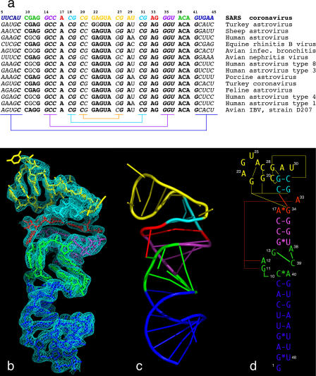
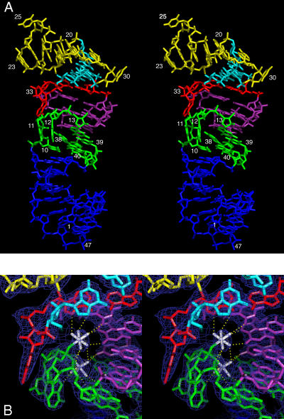
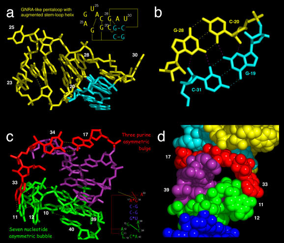
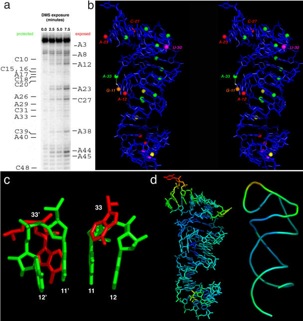
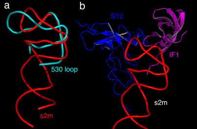
We have solved the three-dimensional crystal structure of the stem-loop II motif (s2m) RNA element of the SARS virus genome to 2.7-A resolution. SARS and related coronaviruses and astroviruses all possess a motif at the 3' end of their RNA genomes, called the s2m, whose pathogenic importance is inferred from its rigorous sequence conservation in an otherwise rapidly mutable RNA genome. We find that this extreme conservation is clearly explained by the requirement to form a highly structured RNA whose unique tertiary structure includes a sharp 90 degrees kink of the helix axis and several novel longer-range tertiary interactions. The tertiary base interactions create a tunnel that runs perpendicular to the main helical axis whose interior is negatively charged and binds two magnesium ions. These unusual features likely form interaction surfaces with conserved host cell components or other reactive sites required for virus function. Based on its conservation in viral pathogen genomes and its absence in the human genome, we suggest that these unusual structural features in the s2m RNA element are attractive targets for the design of anti-viral therapeutic agents. Structural genomics has sought to deduce protein function based on three-dimensional homology. Here we have extended this approach to RNA by proposing potential functions for a rigorously conserved set of RNA tertiary structural interactions that occur within the SARS RNA genome itself. Based on tertiary structural comparisons, we propose the s2m RNA binds one or more proteins possessing an oligomer-binding-like fold, and we suggest a possible mechanism for SARS viral RNA hijacking of host protein synthesis, both based upon observed s2m RNA macromolecular mimicry of a relevant ribosomal RNA fold.


Figures

Figure 1
Figure 1. The Primary, Secondary, and Tertiary Structures of the SARS s2m RNA
(A) Phylogenetic comparisons of s2m RNA sequences from various coronavirus and astrovirus species. The SARS RNA sequence is color-coded to match the color scheme used throughout. Conserved sequences are highlighted as bold letters, and co-varying sequences involved in conventional RNA helical base-pairing are indicated in italics. Sequence complements are indicated using color-coded brackets. (B) The 2.7-Å experimental SIRAS platinum-phased and solvent-flattened electron density map contoured at 1.25 root mean square deviation. The map allowed unambiguous tracing of the RNA molecule because the density was unambiguous for all backbone atoms and all nucleotide bases except U(25), U(30), and U(48). (C) A corresponding ribbon diagram highlighting the unusual fold. (D) Schematic representation of the s2m RNA secondary structure, with tertiary structural interactions indicated as long-range contacts. The schematic diagram is designed to approximate the representation of the fold. The GNRA-like pentaloop structure is shown in yellow, A-form RNA helices are shown in blue and purple, the three-purine asymmetric bulge is in red, and the seven-nucleotide bubble is in green. Long-range tertiary contacts are indicated by thin red and yellow lines.
Figure 2
Figure 2. Stereo Representations of the SARS s2m RNA Structure
(A) The overall SARS s2m RNA three-dimensional structure and (B) a detailed view of tertiary contacts the and [Mg(H2O)5]2+ binding sites in the context of the experimentally phased electron density map (dark blue). The [Mg(H2O)5]2+ complex ions, depicted as white octahedra, bind to the pro-R and pro-S phosphate oxygen atoms of A(12). An extensive network of potential hydrogen bonds between the metal-coordinated water molecules and the RNA is shown as yellow dotted lines.
Figure 3
Figure 3. Tertiary Structural Interactions in the SARS s2m RNA
(A) Close-up of the pentaloop structure together with the augmenting helix, shown in yellow, and the perpendicular junction formed with the A-form stem, shown in cyan. The pink hydrogen bonds indicate base-quartet hydrogen bonding, as shown in (B). The 90° kink thus formed is facilitated by a very sharp bend in the backbone involving unpaired residues 29 and 30. (B) Formation of the junction of two perpendicular helices is facilitated by a base quartet composed of two G–C pairs. (C) The unusual pairing between A(17) and G(34) facilitates formation of a long-range tertiary contact between A(33) of the three-purine asymmetric bulge and G(11) and A(12) of the seven-nucleotide asymmetric bubble. A(38) forms a base triple with C(39) and G(13), forcing G(11) and A(12) out of the main helix. (D) Space-filling representation of the region shown in (C), but rotated approximately 180°. A tunnel is created by the tertiary contacts between A(33) of the purine asymmetric bulge (red), G(11) and A(12) of the seven-nucleotide bubble (green), and the helical region between them (purple). The non-bridging phosphate oxygens of G(11) and A(12) line the surface of the cavity, creating a negatively charged region into which Mg2+ ions are observed to bind.
Figure 4
Figure 4. Chemical Probing of the SARS s2m RNA in Solution
(A) An autoradiogram of DMS modification of the s2m RNA in solution. (B) Mapping the results of DMS, kethoxal, and CMCT modifications onto a stereo representation of the RNA structure. Red spheres represent strongly reactive N1 positions of adenosines and N3 positions of cytidine residues in the presence of DMS, and yellow spheres represent weaker reaction. Green spheres represent positions that appear to be protected from DMS. The orange sphere represents reaction with kethoxal at the N1 position of G(11), and magenta spheres represent CMCT reactions with uridines. (C) The most extensive crystal packing interaction involves stacking of G(11) upon its symmetry mate, G(11)′. (D) Temperature factors mapped onto all non-hydrogen atoms (left) and the phosphate backbone (right) of the s2m RNA crystal structure. U(25) is the most disordered residue in the structure and has the highest temperature factor. Density of the base of U(25) is not apparent even after refinement. Most of the rest of the structure is rather well ordered.
Figure 5
Figure 5. SARS Virus RNA Macromolecular Mimicry
(A) The SARS s2m RNA structure (red) is superimposed upon the 530 loop of 16S rRNA (cyan), revealing the similar stem-loop folds. (B) The IF-1 (magenta) and S12 protein (blue) that bind to the 16S rRNA 530 loop (now hidden) are shown relative to the same s2m RNA superposition, suggesting that their eukaryotic homologs might plausibly bind to the s2m RNA.

References

    1. Marra MA, Jones SJ, Astell CR, Holt RA, Brooks-Wilson A, et al. The genome sequence of the SARS-associated coronavirus. Science. 2003;300:1399–1404. - PubMed
    1. Jonassen CM, Jonassen TO, Grinde B. A common RNA motif in the 3′ end of the genomes of astroviruses, avian infectious bronchitis virus and an equine rhinovirus. J Gen Virol. 1998;79:715–718. - PubMed
    1. Zarembinski TI, Hung LW, Mueller-Dieckmann HJ, Kim KK, Yokota H, et al. Structure-based assignment of the biochemical function of a hypothetical protein: A test case of structural genomics. Proc Natl Acad Sci U S A. 1998;95:15189–15193. - PMC - PubMed
    1. Doudna JA. Structural genomics of RNA. Nat Struct Biol. 2000;7(Suppl):954–956. - PubMed
    1. Rota PA, Oberste MS, Monroe SS, Nix WA, Campagnoli R, et al. Characterization of a novel coronavirus associated with severe acute respiratory syndrome. Science. 2003;300:1394–1399. - PubMed

Publication types