. 2005 Feb;3(2):e31.
doi: 10.1371/journal.pbio.0030031. Epub 2004 Dec 28.

Cobalamin-independent methionine synthase (MetE): a face-to-face double barrel that evolved by gene duplication

Affiliations

Cobalamin-independent methionine synthase (MetE): a face-to-face double barrel that evolved by gene duplication

Robert Pejchal et al. PLoS Biol. 2005 Feb.

Abstract

Cobalamin-independent methionine synthase (MetE) catalyzes the transfer of a methyl group from methyltetrahydrofolate to L-homocysteine (Hcy) without using an intermediate methyl carrier. Although MetE displays no detectable sequence homology with cobalamin-dependent methionine synthase (MetH), both enzymes require zinc for activation and binding of Hcy. Crystallographic analyses of MetE from T. maritima reveal an unusual dual-barrel structure in which the active site lies between the tops of the two (betaalpha)(8) barrels. The fold of the N-terminal barrel confirms that it has evolved from the C-terminal polypeptide by gene duplication; comparisons of the barrels provide an intriguing example of homologous domain evolution in which binding sites are obliterated. The C-terminal barrel incorporates the zinc ion that binds and activates Hcy. The zinc-binding site in MetE is distinguished from the (Cys)(3)Zn site in the related enzymes, MetH and betaine-homocysteine methyltransferase, by its position in the barrel and by the metal ligands, which are histidine, cysteine, glutamate, and cysteine in the resting form of MetE. Hcy associates at the face of the metal opposite glutamate, which moves away from the zinc in the binary E.Hcy complex. The folate substrate is not intimately associated with the N-terminal barrel; instead, elements from both barrels contribute binding determinants in a binary complex in which the folate substrate is incorrectly oriented for methyl transfer. Atypical locations of the Hcy and folate sites in the C-terminal barrel presumably permit direct interaction of the substrates in a ternary complex. Structures of the binary substrate complexes imply that rearrangement of folate, perhaps accompanied by domain rearrangement, must occur before formation of a ternary complex that is competent for methyl transfer.


Figures

Figure 1
Figure 1. Multiple Alignment of MetE from T. maritima (METE_THEMA), E. coli (METE_ECOLI), Saccharomyces cerevisiae (METE_YEAST), and A. thaliana (METE_ARATH)
Conservation in the N-terminal domain is indicated in aqua while conservation in the C-terminal domain is shown in yellow. Zinc ligands His618, Cys620, Glu642, and Cys704 are highlighted in green. The conserved repeat at β4 is marked by asterisks. Main barrel elements are designated β(1–8)F and α(1–8)F and β(1–8)H and α(1–8)H for the N- and C-terminal barrels, respectively. Extension elements are labeled alphabetically and numbered based on the β strand that they follow. For example, α1AF follows β1F and precedes α1F.
Figure 2
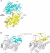
Figure 2. The Fold of MetE and Similarities between the Two Barrels
(A) MetE folds into two (βα)8 barrels. The N-terminal barrel (aqua) is joined to the C-terminal barrel (yellow) by a 35-residue inter-domain linker (gray) that spans 65 Å. Except for the α1 helix (at the right), the linker residues are in extended conformations. This view is along the approximate 2-fold axis that relates the two barrels. The drawing is based on coordinates for zinc-replete MetE in complex with CH3-H4folate (Table 1). The zinc ligands, zinc, and CH3-H4folate are shown in ball-and-stick representation. This figure and all subsequent figures were prepared using RIBBONS [43]. See Figure 1 for the nomenclature used to describe secondary structures. (B) A side-by-side view of the barrels of MetE, arranged to show the similarities of the β–α loop extensions. Major extensions that follow the first four β strands of the barrels are shown in cyan and gold for the N-terminal and C-terminal barrels, respectively. The drawing is based on coordinates for the zinc-replete binary complex with folate (not shown). The active site is located in the C-terminal barrel (on the right) between the extra-barrel β hairpin of the β2–α2 loop and the C-termini of the barrel strands. Zinc is gray and the zinc ligands, His618, Cys620, Glu642, and Cys704, are shown in ball-and-stick mode.
Figure 3
Figure 3. The Resting-State Zinc Is Coordinated in a Tetrahedral Fashion by Four Protein Residues
Figure 4
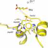
Figure 4. The Geometry at the Zinc Ion in Complexes with Hcy
(A) Interactions of Hcy in the MetE·Hcy binary complex. The amino group of Hcy is bound by hydrogen bonds to Asp577, Glu462, and the backbone carbonyl of Ile409; the Hcy carboxyl group interacts with the backbone amide and side chain hydroxyl of Ser411. The Hcy sulfur is coordinated to zinc via a long (3.15 Å) bond, which is eclipsed in this view. (B) Superposition of the MetE resting state (gray) and the Hcy binary complex (yellow). Upon Hcy binding, zinc and His618 move away from Glu642 and closer to Hcy, and the zinc site adopts trigonal bipyramidal geometry with three strong equatorial ligands (Zn–NHis618, 2.07 Å; Zn–SCys620, 2.23 Å; Zn–SCys704, 2.24 Å) and two distant axial ligands (Zn–OGlu642, 2.90 Å; Zn–SHcy, 3.13 Å). Zinc moves 0.75 Å toward the substrate in the Hcy complex. Full inversion at zinc, upon tight binding of Hcy to MetE, would displace the metal ion approximately 1.5 Å. (C) The MetH·Hcy complex. The zinc configuration in substrate-free MetH is opposite to that found in MetE; binding of Hcy occurs without inversion in MetH and in BHMT. (D) Difference electron density for the MetE·Hcy complex, showing the geometry at the metal-binding site. The map was computed after simulating annealing and refinement of a model omitting the zinc and its five neighbors. The long Zn–Hcy and Zn–Glu642 interactions are indicated with dashes. Contour levels are 2σ (green) and 6σ (orange).
Figure 5
Figure 5. Interactions of CH3-H4folate with MetE
(A) A stereoview of T. maritima MetE showing the substrate and metal-binding sites. This is a composite picture in which Hcy from the MetE·Hcy complex has been positioned by superposition on the structure of the MetE·CH3-H4folate binary complex. The substrates and metal ligands are displayed in ball-and-stick mode; Hcy is in green. (B) A stereoview of the zinc site and bound CH3-H4folate. Folate is bound by conserved residues in the N-terminal barrel (aqua) and the C-terminal barrel (yellow) with the N5-CH3 facing away from the zinc, at a distance of almost 14 Å. The pterin interacts with the long hairpins of both barrels and with the extra-barrel helices α3AH and α4AH. The groove that binds the glutamate tail of the substrate is bordered by α1AF, by the β hairpin (β2BH and β2CH) and α1AH of the C-terminal domain, and by the conserved DMV sequence that begins α2AH.
Figure 6
Figure 6. Superposition of the Hcy Binary Complex (Yellow) and Substrate-Free (Gray) Enzymes Showing Local Changes in the DMV Region
Met468 moves toward the Hcy substrate, rearranging the start of helix α2AH, and a new hydrogen bond is formed between the carbonyls of Met468 and Thr531. This position of Met468 stabilizes a rotamer of Trp539 that favors folate binding.
Figure 7
Figure 7. Superposition of MetE and MetH Zn+2/Hcy Barrels
In MetH (gray) the sulfur of Hcy is positioned close to the center of the barrel for interaction with methylcobalamin. In MetE (yellow) the α1–β1 and α8–β8 connectors make large incursions across the top of the barrel, displacing Hcy to the other side of the barrel.
Figure 8
Figure 8. Disruption of Hcy and Zinc Binding in the N-Terminal Barrel
Overhaul of the Zn+2/Hcy site in the N-terminal domain (aqua) following gene duplication is accomplished through mutation and small backbone displacements. The competent Zn/Hcy site from the C-terminal barrel is at the left; the remodeled site from the N-terminal barrel is at the right. Important substitutions that disable Hcy and zinc binding are Trp146, Phe230, Tyr232, and Asp253 (see text).

References

    1. Ludwig ML, Matthews RG. Structure-based perspectives on B12-dependent enzymes. Annu Rev Biochem. 1997;66:269–313. - PubMed
    1. Evans JC, Huddler DP, Hilgers MT, Romanchuk G, Matthews RG, et al. Structures of the N-terminal modules imply large domain motions during catalysis by methionine synthase. Proc Natl Acad Sci U S A. 2004;101:3729–3736. - PMC - PubMed
    1. Peariso K, Zhou ZS, Smith AE, Matthews RG, Penner-Hahn JE. Characterization of the zinc sites in cobalamin-independent and cobalamin-dependent methionine synthase using zinc and selenium x-ray absorption spectroscopy. Biochemistry. 2001;40:987–993. - PubMed
    1. Matthews RG, Smith AE, Zhou ZS, Taurog RE, Bandarian V, et al. Cobalamin-dependent and cobalamin-independent methionine synthases: Are there two solutions to the same chemical problem? Helvetica Chimica Acta. 2003;86:3939–3954.
    1. Hightower KE, Fierke CA. Zinc-catalyzed sulfur alkylation: Insights from protein farnesyltransferase. Curr Opin Chem Biol. 1999;3:176–181. - PubMed

Publication types

MeSH terms

Substances

LinkOut - more resources