

An unusual fold for potassium channel blockers: NMR structure of three toxins from the scorpion Opisthacanthus madagascariensis
- PMID: 15631621
- PMCID: PMC1186715
- DOI: 10.1042/BJ20041705
An unusual fold for potassium channel blockers: NMR structure of three toxins from the scorpion Opisthacanthus madagascariensis
Abstract
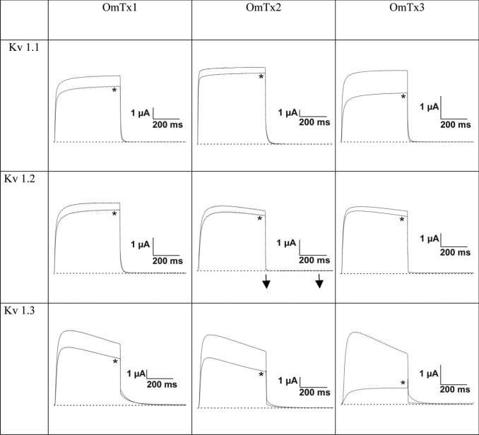
The Om-toxins are short peptides (23-27 amino acids) purified from the venom of the scorpion Opisthacanthus madagascariensis. Their pharmacological targets are thought to be potassium channels. Like Csalpha/beta (cystine-stabilized alpha/beta) toxins, the Om-toxins alter the electrophysiological properties of these channels; however, they do not share any sequence similarity with other scorpion toxins. We herein demonstrate by electrophysiological experiments that Om-toxins decrease the amplitude of the K+ current of the rat channels Kv1.1 and Kv1.2, as well as human Kv1.3. We also determine the solution structure of three of the toxins by use of two-dimensional proton NMR techniques followed by distance geometry and molecular dynamics. The structures of these three peptides display an uncommon fold for ion-channel blockers, Csalpha/alpha (cystine-stabilized alpha-helix-loop-helix), i.e. two alpha-helices connected by a loop and stabilized by two disulphide bridges. We compare the structures obtained and the dipole moments resulting from the electrostatic anisotropy of these peptides with those of the only other toxin known to share the same fold, namely kappa-hefutoxin1.
Figures


Similar articles
-
Solution structure of IsTX. A male scorpion toxin from Opisthacanthus madagascariensis (Ischnuridae).Eur J Biochem. 2004 Oct;271(19):3855-64. doi: 10.1111/j.1432-1033.2004.04322.x. Eur J Biochem. 2004. PMID: 15373831
-
The new kappa-KTx 2.5 from the scorpion Opisthacanthus cayaporum.Peptides. 2011 Jul;32(7):1509-17. doi: 10.1016/j.peptides.2011.05.017. Epub 2011 May 23. Peptides. 2011. PMID: 21624408
-
Synthesis, 1H NMR structure, and activity of a three-disulfide-bridged maurotoxin analog designed to restore the consensus motif of scorpion toxins.J Biol Chem. 2000 May 5;275(18):13605-12. doi: 10.1074/jbc.275.18.13605. J Biol Chem. 2000. PMID: 10788477
-
Potassium channel blockers from the venom of the Brazilian scorpion Tityus serrulatus ().Toxicon. 2016 Sep 1;119:253-65. doi: 10.1016/j.toxicon.2016.06.016. Epub 2016 Jun 25. Toxicon. 2016. PMID: 27349167 Review.
-
Scorpion toxins specific for Na+-channels.Eur J Biochem. 1999 Sep;264(2):287-300. doi: 10.1046/j.1432-1327.1999.00625.x. Eur J Biochem. 1999. PMID: 10491073 Review.
Cited by
-
Arachnids of medical importance in Brazil: main active compounds present in scorpion and spider venoms and tick saliva.J Venom Anim Toxins Incl Trop Dis. 2015 Aug 13;21:24. doi: 10.1186/s40409-015-0028-5. eCollection 2015. J Venom Anim Toxins Incl Trop Dis. 2015. PMID: 26273285 Free PMC article.
-
Solution structure and functional analysis of HelaTx1: the first toxin member of the κ-KTx5 subfamily.BMB Rep. 2020 May;53(5):260-265. doi: 10.5483/BMBRep.2020.53.5.256. BMB Rep. 2020. PMID: 32172732 Free PMC article.
-
Molecular Dynamics Simulation Reveals Specific Interaction Sites between Scorpion Toxins and Kv1.2 Channel: Implications for Design of Highly Selective Drugs.Toxins (Basel). 2017 Nov 1;9(11):354. doi: 10.3390/toxins9110354. Toxins (Basel). 2017. PMID: 29104247 Free PMC article.
-
A Deeper Examination of Thorellius atrox Scorpion Venom Components with Omic Techonologies.Toxins (Basel). 2017 Dec 12;9(12):399. doi: 10.3390/toxins9120399. Toxins (Basel). 2017. PMID: 29231872 Free PMC article.
-
New tricks of an old pattern: structural versatility of scorpion toxins with common cysteine spacing.J Biol Chem. 2012 Apr 6;287(15):12321-30. doi: 10.1074/jbc.M111.329607. Epub 2012 Jan 10. J Biol Chem. 2012. PMID: 22238341 Free PMC article.
References
-
- Bontems F., Roumestand C., Gilquin B., Toma F. Refined structure of charybdotoxin: common motifs in scorpion toxins and insect defensins. Science. 1991;254:1521–1523. - PubMed
-
- Blanc E., Romi-Lebrun R., Bornet O., Nakajima T., Darbon H. Solution structure of two new toxins from the venom of the Chinese scorpion Buthus martensi Karsch blockers of potassium channels. Biochemistry. 1998;37:12412–12418. - PubMed
-
- Fajloun Z., Ferrat G., Carlier E., Fathallah M., Lecomte C., Sandoz G., di Luccio E., Mabrouk K., Legros C., Darbon H., et al. Synthesis, 1H NMR structure, and activity of a three-disulfide-bridged maurotoxin analog designed to restore the consensus motif of scorpion toxins. J. Biol. Chem. 2000;275:13605–13612. - PubMed
-
- Renisio J., Romi-Lebrun R., Blanc E., Bornet O., Nakajima T., Darbon H. Solution structure of BmKTX, a K+ blocker toxin from the Chinese scorpion Buthus martensi. Proteins. 2000;38:70–78. - PubMed
Publication types
MeSH terms
Substances
Associated data
- Actions
- Actions
- Actions
LinkOut - more resources
Full Text Sources