. 2005 Jan 3;168(1):29-33.
doi: 10.1083/jcb.200409067.

Glycogen synthase kinase-3 is an endogenous inhibitor of Snail transcription: implications for the epithelial-mesenchymal transition

Affiliations

Glycogen synthase kinase-3 is an endogenous inhibitor of Snail transcription: implications for the epithelial-mesenchymal transition

Robin E Bachelder et al. J Cell Biol. .

Abstract

We report that the activity of glycogen synthase kinase-3 (GSK-3) is necessary for the maintenance of the epithelial architecture. Pharmacological inhibition of its activity or reducing its expression using small interfering RNAs in normal breast and skin epithelial cells results in a reduction of E-cadherin expression and a more mesenchymal morphology, both of which are features associated with an epithelial-mesenchymal transition (EMT). Importantly, GSK-3 inhibition also stimulates the transcription of Snail, a repressor of E-cadherin and an inducer of the EMT. We identify NFkappaB as a transcription factor inhibited by GSK-3 in epithelial cells that is relevant for Snail expression. These findings indicate that epithelial cells must sustain activation of a specific kinase to impede a mesenchymal transition.


Figures

Figure 1.
Figure 1.
GSK-3 maintains the epithelial phenotype. (A) MCF10A cells were incubated with either DMSO or 25 μM SB415286 (Biosource International) in 0.5% FBS-containing medium. Cell morphology was assessed after 72 h by phase contrast microscopy. Expression of Cyclin D1, Stat-1, E-cadherin, and vimentin was assessed by immunoblotting. (B) HaCaT cells were incubated with DMSO or SB415286, and E-cadherin, Stat-1, and cyclin D1 expression was assessed as indicated in A. Similar results were obtained in four independent experiments. (C) E-cadherin and GAPDH mRNA levels were determined after 48 h of drug treatment by RT-PCR. (D) MCF10A cells were cotransfected with GSK-3α– and GSK-3β–specific inhibitory RNA pools (α+β siRNA) or a nonspecific control pool (Scr). RNA was extracted from these cells 48 h after transfection, and E-cadherin and GAPDH mRNA levels were determined by RT-PCR. GSK-3 and Stat-1 expression was assessed by immunoblotting.
Figure 2.
Figure 2.
GSK-3 inhibits Snail transcription in epithelial cells. (A) MCF10A and HaCaT cells were either incubated with 25 μM of the GSK-3 inhibitor SB415286 or with DMSO in 0.5% FBS-containing medium. Alternatively, these cells were transfected with GSK-3α– and GSK-3β–specific inhibitory RNA pools (α+β siRNA) or a nonspecific control pool (Scr). Snail and GAPDH mRNA levels were assessed by reverse transcription, followed by PCR. GSK-3 and Stat-1 expression was assessed by immunoblotting. (B) HaCaT cells were cotransfected with a Snail promoter–driven and a control β-galactosidase reporter construct. These transfectants were then incubated with DMSO or SB415286, as described in A, for 24 h. Data are reported as relative luciferase activity (normalized to β-galactosidase activity), ± SD, and are representative of three independent experiments. (C) MCF10A and HeLa cells were treated with SB415286 as described in A, and Snail expression was assessed by immunofluorescence.
Figure 3.
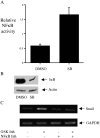
Figure 3.
GSK-3 inhibits NFκB, an activator of Snail transcription. (A) HaCaT cells were transfected transiently with a luciferase construct driven by NFκB binding sites in addition to a control renilla luciferase reporter gene. These transfectants were then incubated with DMSO or SB415286, as described for Fig. 2, for 15 h. Data are reported as relative luciferase activity (normalized to renilla luciferase activity), ± SD, and are representative of two independent experiments. (B) MCF10A cells were incubated with DMSO or SB415286 as described for Fig. 2. Equivalent amounts of total cellular protein were extracted from these cells, subjected to SDS-PAGE, and immunoblotted with an IκB- or actin-specific antibody. Results are representative of those obtained in two independent experiments. (C) MCF10A cells were incubated for 2 h with an NFκB-specific inhibitor (SN50; Calbiochem) or control peptide (SN50M; Calbiochem ). These cells were then incubated with DMSO or 25 μM SB415286. RNA was isolated after 15 h, and the levels of Snail and GAPDH were determined by RT-PCR. Similar results were obtained in three independent experiments.

References

    1. Barbera, M.J., I. Puig, D. Dominguez, S. Julien-Grille, S. Guaita-Esteruelas, S. Peiro, J. Baulida, C. Franci, S. Dedhar, L. Larue, and A. Garcia De Herreros. 2004. Regulation of Snail transcription during epithelial to mesenchymal transition of tumor cells. Oncogene. 23:7345–7354. - PubMed
    1. Batlle, E., E. Sancho, C. Franci, D. Dominguez, M. Monfar, J. Baulida, and A. Garcia De Herreros. 2000. The transcription factor snail is a repressor of E-cadherin gene expression in epithelial tumour cells. Nat. Cell Biol. 2:84–89. - PubMed
    1. Beals, C.R., C.M. Sheridan, C.W. Turck, P. Gardner, and G.R. Crabtree. 1997. Nuclear export of NF-ATc enhanced by glycogen synthase kinase-3. Science. 275:1930–1934. - PubMed
    1. Blanco, M.J., G. Moreno-Bueno, D. Sarrio, A. Locascio, A. Cano, J. Palacios, and M.A. Nieto. 2002. Correlation of Snail expression with histological grade and lymph node status in breast carcinomas. Oncogene. 21:3241–3246. - PubMed
    1. Bringuier, P.P., R. Umbas, H.E. Schaafsma, H.F. Karthaus, F.M. Debruyne, and J.A. Schalken. 1993. Decreased E-cadherin immunoreactivity correlates with poor survival in patients with bladder tumors. Cancer Res. 53:3241–3245. - PubMed

Publication types

MeSH terms