. 2005 Jan 3;168(1):67-77.
doi: 10.1083/jcb.200408188.

Identification of cytoplasmic residues of Sec61p involved in ribosome binding and cotranslational translocation

Affiliations

Identification of cytoplasmic residues of Sec61p involved in ribosome binding and cotranslational translocation

Zhiliang Cheng et al. J Cell Biol. .

Abstract

The cytoplasmic surface of Sec61p is the binding site for the ribosome and has been proposed to interact with the signal recognition particle receptor during targeting of the ribosome nascent chain complex to the translocation channel. Point mutations in cytoplasmic loops six (L6) and eight (L8) of yeast Sec61p cause reductions in growth rates and defects in the translocation of nascent polypeptides that use the cotranslational translocation pathway. Sec61 heterotrimers isolated from the L8 sec61 mutants have a greatly reduced affinity for 80S ribosomes. Cytoplasmic accumulation of protein precursors demonstrates that the initial contact between the large ribosomal subunit and the Sec61 complex is important for efficient insertion of a nascent polypeptide into the translocation pore. In contrast, point mutations in L6 of Sec61p inhibit cotranslational translocation without significantly reducing the ribosome-binding activity, indicating that the L6 and L8 sec61 mutants affect different steps in the cotranslational translocation pathway.


Figures

Figure 1.
Figure 1.
Point mutations in L6 of Sec61p. (A) Secondary structure of L6 (M. jannaschii SecY) and sequence alignment between eukaryotic and M. jannaschii L6 segments. Identities are boxed and asterisks indicate residues subjected to mutagenesis. (B) Yeast strains RGY401 (ssh1Δ) and RGY402 (SSH1) that had been transformed with plasmids expressing wild-type or mutant (R275*, R275S, R275L, or R275G) alleles of Sec61p were streaked on 5-fluoroorotic acid plates and allowed to grow for 2 d at 30°C. Sec61R275* has a termination codon at position 275. (C and D) Growth rates of L6 sec61 mutants were compared by serial dilution analysis (C) as described in Materials and methods and used to assign the L6 sec61ssh1Δ mutants to growth phenotype categories (D).
Figure 2.
Figure 2.
Point mutations in L8 of Sec61p. (A) Secondary structure of L8 (M. jannaschii SecY) and sequence alignment between eukaryotic and M. jannaschii L8 segments. The locations of α-helical segments in L8 of M. jannaschii SecY are indicated below the alignment. Identities are boxed and asterisks indicate residues subjected to mutagenesis. The L8 region of M. jannaschii SecY contains a two-residue insertion (KS) relative to eukaryotic Sec61 sequences. (B) Yeast strains RGY401 (ssh1Δ) and RGY402 (SSH1) that had been transformed with plasmids expressing wild-type SEC61, or mutant alleles (R406*, R406E, L6L8EE, or K396D) of Sec61p were streaked onto 5-fluoroorotic acid plates and allowed to grow for 2 d at 30°C. (C and D) Serial dilution experiments were performed as described in Fig. 1 C's legend and used to assign the L8 sec61ssh1Δ mutants to growth phenotype categories (D).
Figure 3.
Figure 3.
Translocation defects in sec61 mutants are suppressed by expression of Ssh1p. (A) Wild-type yeast (RGY402; closed squares) and ssh1Δ mutants expressing wild-type Sec61p (circles), sec61R275E (open squares), or sec61R406E (triangles) were grown to mid-log phase at 30°C in SEG media. The cultures were diluted into YPD media at 0 h and allowed to grow for 8–12 h at 30°C. (B and D) Wild-type and mutant yeast cultures were pulse labeled for 7 min at 30°C after 4 h of growth in SD media at 30°C. One sample of wild-type cells was treated with tunicamycin (wt + TM) for 30 min before pulse labeling. DPAPB-HA (B) and CPY (D) immunoprecipitates were resolved by SDS-PAGE. The ER (p1), Golgi (p2), and precursor (ppCPY) forms of CPY and the glycosylated (D) and nonglycosylated (p-D) forms of DPAPB-HA are labeled. In D, white lines indicate that intervening lanes of Endo H digestion products have been removed for clarity. Translocation of CPY or integration of DPAPB-HA was quantified with a BioRad FX Molecular Imager. (C) Wild-type yeast (RGY402; closed squares) and ssh1Δ mutants expressing wild-type Sec61p (circles), sec61R275E (triangles), or sec61R406E (open squares) were pulse labeled to evaluate integration of DPAPB-HA as described in B, after 1, 2, 4, 8, or 24 h of growth in SD media. As needed, cell cultures were diluted with fresh SD media to maintain an A600 of <0.8. (E) Pulse-labeled sec61L6DDD spheroplasts were osmotically lysed and centrifuged at 500 g to remove unbroken cells. Spheroplast lysates were incubated on ice with trypsin (100 μg/ml) as indicated. The lane designated 15-TX contained trypsin plus Triton X-100. Trypsin was inactivated with PMSF before immunoprecipitation.
Figure 4.
Figure 4.
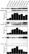
Differential effect of Sec61p mutations on SRP-dependent and SRP-independent translocation pathways. Integration of DPAPB-HA and translocation of CPY and Gas1p were evaluated by pulse labeling of wild-type and mutant yeast strains that were grown for 4 h in SD media at 30°C. Pulse labeling and immunoprecipitation of proteins were conducted as described in Fig. 3's legend.
Figure 5.
Figure 5.
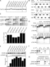
Transport pathways affected by Sec61 mutations. All experiments were conducted after 4 h of growth in SD media at 30°C. (A) Equal amounts of total protein (25 μg) were resolved by SDS-PAGE for protein immunoblot analysis using a COOH-terminal–specific antibody to Sec61p. (B) Total cell extracts were prepared for SDS-PAGE with or without prior digestion by Endo H. Deglycosylated mature CPY (dgm) is resolved from vacuolar CPY (m) and nontranslocated prepro-CPY (p). The asterisk designates an incomplete Endo H digestion product. (C) Degradation of CPY*HA in L6 and L8 sec61 mutants. Cell extracts prepared at 30-min intervals after cycloheximide addition were resolved by SDS-PAGE. Nontranslocated ppCPY*HA and translocated p1CPY*HA were detected using anti-HA antibodies. Protease digestion experiments confirmed that p1CPY*HA, but not ppCPY*HA, was in a membrane-enclosed compartment (not depicted). The apparent half-life of p1CPY*HA, determined according to a first-order decay process, is plotted below representative time courses. (D) Yeast cultures were pulse labeled for 7 min and chased for 10, 20, or 30 min. The nontranslocated precursor (p-Gas1), the translocated ER form (Gas1), and the mature form (m-Gas1) of Gas1p are labeled. (E) Protein immunoblot detection of p-DPAPB-HA and mature DPAPB-HA in total cell extracts resolved by SDS-PAGE. Protein immunoblots (C and E) were quantified by densitometry. (F) Differential centrifugation of spheroplast lysates prepared from the sec61L6DDDssh1Δ mutant. Total lysates (T) and supernatant (S) and pellet (P) fractions were obtained after centrifugation at 500 g, 13,000 g, and 100,000 g. (G) The P13 fraction (T) was resuspended in buffer A (50 mM Hepes, pH 7.5, 150 mM KOAc, 5 mM Mg(OAc)2, and 1 mM DTT) adjusted to 250 mM sucrose and applied to a sucrose step gradient in buffer A with 1.6-M and 2-M sucrose layers. After centrifugation for 1 h at 100,000 g, the gradient was resolved into the following fractions: (1) 0.25 sample load plus 0.25/1.6-M interface, (2) 1.6-M sucrose layer plus 1.6/2-M interface, (3) 2-M sucrose layer, and (4) pellet. The P13 fraction (T) was solubilized in 3% digitonin and 500 mM KOAc and centrifuged at 100,000 g for 1 h to obtain supernatant (S) and pellet (P) fractions.
Figure 6.
Figure 6.
Binding of ribosomes to yeast PK-RM and Sec61 proteoliposomes. (A–C) Scatchard plots of ribosome binding to PK-RM (A and B) or Sec61p proteoliposomes (C) isolated from wild type (SEC61ssh1Δ) or from L6 (A and C) and L8 (B and C) sec61ssh1Δ mutants. (D) Sec61 heterotrimers (150–300 fmol) purified from wild type and from selected L6 and L8 mutants were incubated in the presence or absence of 900 fmol of yeast ribosomes before centrifugation to obtain supernatant (S) and pellet (P) fractions. After SDS-PAGE, Sbh1p was detected using anti-FLAG antibodies.
Figure 7.
Figure 7.
Point mutations in L6 and L8 define a contact surface for cytoplasmic ligands of the Sec61 complex. (A) A ribbon diagram of SecYEG complex showing the three subunits (SecY, green; SecE, cyan; and SecG, magenta) as viewed from within the plane of the membrane. The L6 (blue) and L8 (white) regions in SecY are highlighted. The SecY residue that aligns with a Sec61 residue subjected to mutagenesis is designated by a colored side chain; mutagenesis of red, but not yellow, side chains caused growth defects. (B) An expanded view of A showing that the critical residues in Sec61p are located at the tips of L6 and L8. (C) A top view of the SecYEG complex. The subunits, loops, and mutagenized residues are colored as in A. The dimerization interface for the SecYEG complex is formed by the transmembrane span of SecE (cyan chain). The asterisk designates the proposed translocation pore in SecYEG that is plugged by the short TM2α helix. (D) An expanded top view of the L6 and L8 regions. SecE is hidden to simplify the image. The figure was created with MacPyMOL software using SEC YEG structure (PDB 1RHZ).

References

    1. Arnold, C.E., and K.D. Wittrup. 1994. The stress response to loss of signal recognition particle function in Saccharomyces cerevisiae. J. Biol. Chem. 269:30412–30418. - PubMed
    1. Baxter, B.K., P. James, T. Evans, and E.A. Craig. 1996. SSI1 encodes a novel Hsp70 of the Saccharomyces cerevisiae endoplasmic reticulum. Mol. Cell. Biol. 16:6444–6456. - PMC - PubMed
    1. Beckmann, R., D. Bubeck, R. Grassucci, P. Penczek, A. Verschoor, G. Blobel, and J. Frank. 1997. Alignment of conduits for the nascent polypeptide chain in the ribosome-Sec61 complex. Science. 278:2123–2126. - PubMed
    1. Beckmann, R., C.M. Spahn, N. Eswar, J. Helmers, P.A. Penczek, A. Sali, J. Frank, and G. Blobel. 2001. Architecture of the protein-conducting channel associated with the translating 80S ribosome. Cell. 107:361–372. - PubMed
    1. Corsi, A.K., and R. Schekman. 1996. Mechanism of polypeptide translocation into the endoplasmic reticulum. J. Biol. Chem. 271:30299–30302. - PubMed

Publication types

MeSH terms