. 2005 Jan;166(1):61-71.
doi: 10.1016/S0002-9440(10)62232-8.

Matrix metalloproteinase-9 knockout confers resistance to corneal epithelial barrier disruption in experimental dry eye

Affiliations

Matrix metalloproteinase-9 knockout confers resistance to corneal epithelial barrier disruption in experimental dry eye

Stephen C Pflugfelder et al. Am J Pathol. 2005 Jan.

Abstract

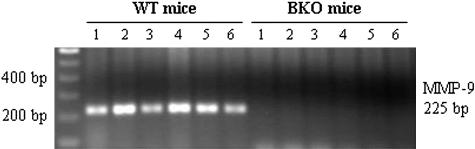
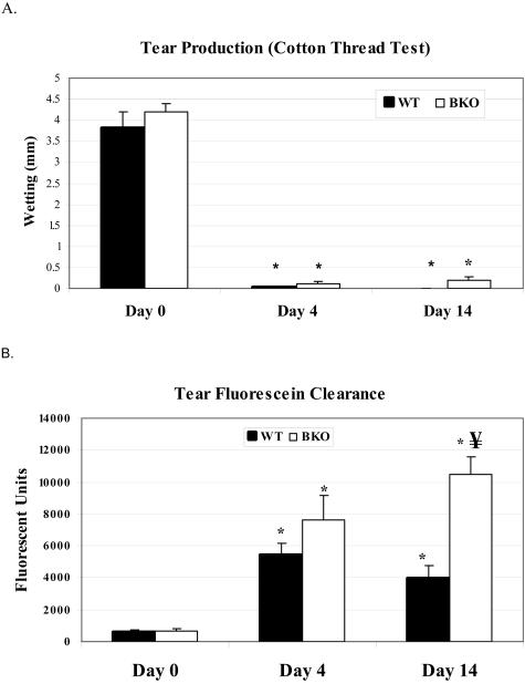
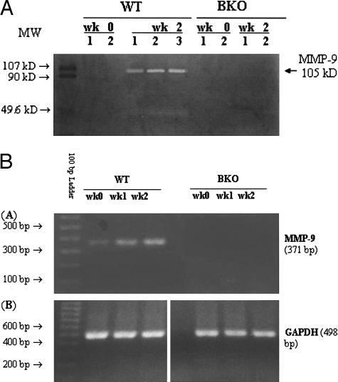
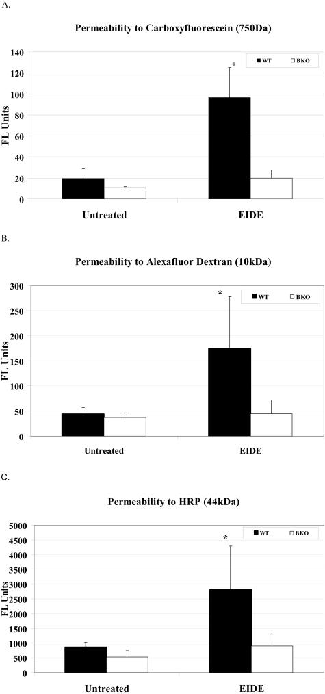
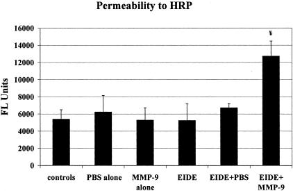
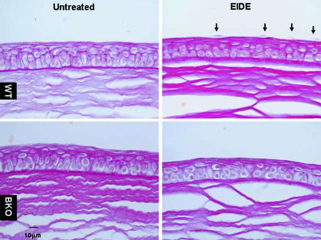
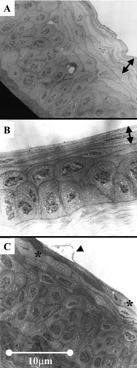
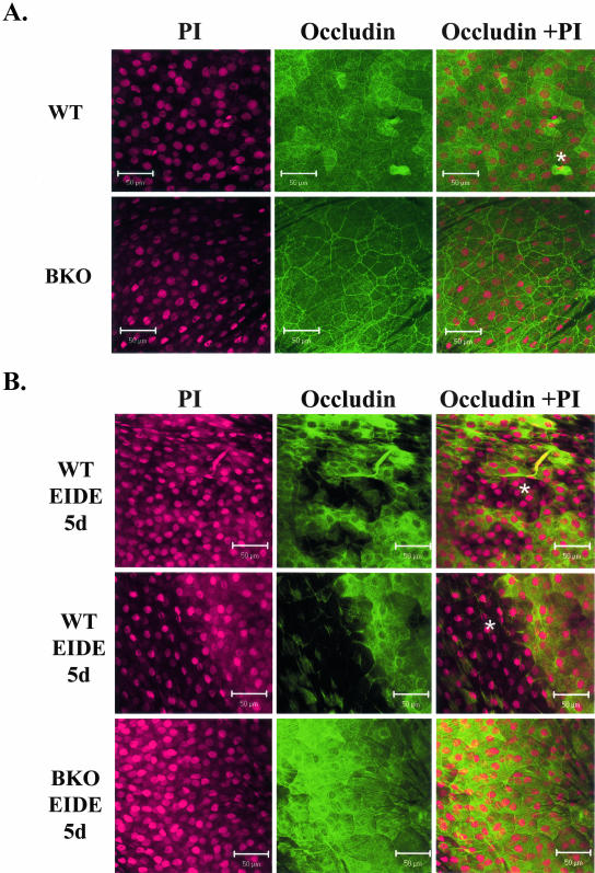
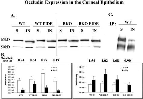
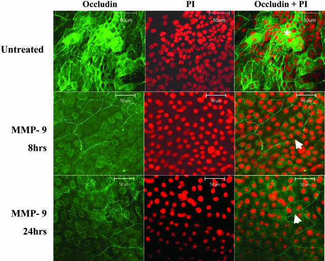
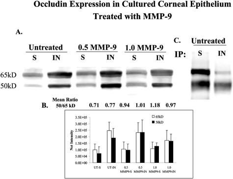
Altered corneal epithelial barrier function is the cause for ocular irritation and visual morbidity in dry eye disease. Increased matrix metalloproteinase (MMP)-9 activity has been observed in the tear fluid of dry eye patients. To determine the pathogenic role of MMP-9 in the corneal epithelial disease of dry eye, the effects of experimentally induced dry eye on corneal epithelial morphology and barrier function were compared in MMP-9 knockout mice and their wild-type littermates. Dry eye was created through cholinergic blockade and exposure to a desiccating environment. The tear fluid MMP-9 concentration increased in response to dryness in wild-type mice. Corneal epithelial permeability to three different-sized molecules increased in dry eye wild-type mice, but not in MMP-9 knockout mice. Topical administration of active MMP-9 to dry eye MMP-9 knockout mice significantly increased corneal epithelial permeability. Compared to MMP-9 knockout mice, wild-type mice showed greater desquamation of differentiated apical corneal epithelial cells that expressed the tight junction protein occludin in response to dryness. This was accompanied by an increase in lower sized (50 kd) occludin in the corneal epithelia of wild-type mice. These findings could be replicated in cultured human corneal epithelial cells that were treated with active MMP-9. These studies indicate that increased MMP-9 activity on the ocular surface in response to dryness disrupts corneal epithelial barrier function. This appears to be because of accelerated loss of tight junction bearing superficial corneal epithelial cells, perhaps by proteolytic cleavage of occludin.


Figures

Figure 1
Figure 1
Genotype determination by PCR using mouse tail genomic DNA and a specific primer pair designed from the MMP-9 gene exon 2 sequence, which was knocked out in BKO mice. The PCR products were analyzed by 1.5% agarose gel electrophoresis. A 225-bp band was generated from all six WT mice (left, lanes 1 to 6) tested but not from six BKO mice (right, lanes 1 to 6).
Figure 2
Figure 2
A: Aqueous tear production measured in mm with a cotton thread in mice before (day 0) and after 4 and 14 days of EIDE. *, P < 0.001 compared to baseline. B: Measurement of clearance of fluorescein dye from the tear fluid 15 minutes after instillation of 1 μl of 2% sodium fluorescein in mice before (day 0) and 4 and 14 days after EIDE. *, P < 0.001 compared to baseline; ¥, P < 0.001 compared to WT at day 14. Values are mean ± SD from 14 eyes of seven mice.
Figure 3
Figure 3
A: Gelatin zymography of pooled tear fluid washings obtained from WT and BKO mice before (wk 0) and after 2 weeks (wk 2) of EIDE. Mouse MMP-9 has a molecular weight of 107 kd. B: Expression of MMP-9 and GAPDH RNA in the corneal epithelium of WT and BKO mice by RT-PCR before (wk 0) and after 1 (wk 1) and 2 (wk 2) weeks of EIDE.
Figure 4
Figure 4
Corneal permeability to carboxyfluorescein (A, FL units = units of fluorescein emission at 530 nm), AFD (B, FL units = units of fluorescein emission at 530 nm), and HRP (C, FL units = units of fluorescein emission from activated peroxidase substrate Amplex Red at 590 nm) in untreated control and after 2 weeks of EIDE in WT and BKO mice Values are mean ± SD for four eyes. A: *, P = 0.002 WT with EIDE versus untreated control WT and BKO with EIDE; B: *, P = 0.048 WT with EIDE versus untreated control WT and BKO with EIDE; C: *, P < 0.03 WT with EIDE versus untreated control WT and treated BKO.
Figure 5
Figure 5
Corneal permeability to HRP in untreated controls and mice treated 10 hours per day for 2 days with PBS, MMP-9 (1 μl of 1 μg/ml), EIDE, EIDE plus PBS, EIDE plus MMP-9 (1 μl of 1 μg/ml). Drops were administered every 2 hours. Values are mean ± SD for 3 eyes. ¥, Between group difference was 0.002 ANOVA. Post-hoc analysis showed significant differences between EIDE + MMP-9 and controls (P < 0.05), EIDE (P < 0.01), and EIDE + PBS (P < 0.05). FL, fluorescent.
Figure 6
Figure 6
PAS-stained sections of representative central corneas from untreated WT and BKO mice before (left, top, and bottom, respectively) and after EIDE for 14 days (right, top, and bottom, respectively). Detaching apical cells (arrows) were in the corneas of WT mice with EIDE (top, right), but not in BKO mice with EIDE (bottom, right).
Figure 7
Figure 7
Transmission electron micrograph of untreated WT mouse cornea (A), cornea of BKO mouse with EIDE for 14 days (B) (arrows in A and B indicate layers of thinned apical epithelium with barely detectable cell nuclei), and cornea of a WT mouse with EIDE for 14 days (C). Arrow indicates detaching apical epithelial cell. Superficial cells with cleaved nuclei are marked with asterisks.
Figure 8
Figure 8
Confocal microscopy of whole mount corneas stained with polyclonal occludin antisera. A: Apical corneal epithelium of control WT (top) and BKO (bottom) mice. Detached epithelium is noted with asterisk. B: Apical corneal epithelium in WT mice with EIDE for 5 days (top and middle). Areas of epithelial detachment are noted with asterisk. The middle figure shows the border of a large central circular area of detachment (∼1 mm diameter). A representative cornea from a BKO mouse with EIDE for 5 days is shown on the bottom. Cells were counterstained with propidium iodide (PI) (left), and polyclonal occludin antisera (middle). The merged image is on the right.
Figure 9
Figure 9
A: Occludin Western blot of triton soluble (S) and insoluble (IN) fractions of corneal epithelial lysates from control WT and BKO mice and WT and BKO mice with EIDE for 5 days (WT 5 day and BKO 5 day, respectively). B: Graphs of mean 65- and 50-kd band densities from triton soluble (S) and insoluble (IN) corneal epithelial fractions from three blots. The mean 50:65-kd ratio for each sample is provided above the bar graphs. C: Immunoprecipitation of triton soluble (S) and insoluble (IN) fractions of corneal epithelial lysates from control WT mice using polyclonal occludin antisera followed by immunoblotting with monoclonal occludin antibody.
Figure 10
Figure 10
Confocal microscopy of control untreated primary cultured human corneal epithelium in media (top), primary cultured human corneal epithelium treated with MMP-9 (1 μg/ml) for 8 hours (MMP-9 8 hours, middle), and primary cultured human corneal epithelium treated with MMP-9 (1 μg/ml) for 24 hours (MMP-9 24 hours, bottom). In each group, cells were stained with polyclonal occludin antisera (left), counterstained with propidium iodide (PI, middle), and the merged image is on the right. A honeycomb pattern of occludin staining was observed in areas of stratification (asterisk) in control cultures; areas of discontinuity of occludin staining in MMP-9-treated cultures are indicated by arrows.
Figure 11
Figure 11
A: Occludin Western blot of triton soluble (S) and insoluble (IN) fractions of lysates from untreated confluent primary human corneal epithelial cultures and cultures treated with 0.5 and 1.0 μg/ml of activated MMP-9 (0.5 MMP-9 and 1.0 MMP-9, respectively) for 24 hours. B: Graph of mean 65- and 50-kd band densities from triton soluble (S) and insoluble (IN) corneal epithelial fractions from three blots. The mean 50:65-kd ratio for each sample is provided above the bar graphs. C: Immunoprecipitation of triton soluble (S) and insoluble (IN) fractions of corneal epithelial lysates from control human corneal epithelial cultures using polyclonal occludin antisera followed by immunoblotting with monoclonal occludin antibody.

References

    1. Murillo-Lopez F, Pflugfelder SC. Dry eye. Krachmer J, Mannis M, Holland E, editors. St. Louis: Mosby; The Cornea. 1996:pp 663–686.
    1. Yokoi N, Kinoshita S. Clinical evaluation of corneal epithelial barrier function with the slit-lamp fluorophotometer. Cornea. 1995;14:485–489. - PubMed
    1. Gobbels M, Spitznas M. Corneal epithelial permeability of dry eyes before and after treatment with artificial tears. Ophthalmology. 1992;99:873–878. - PubMed
    1. Pflugfelder SC, Tseng SC, Sanabria O, Kell H, Garcia CG, Felix C, Feuer W, Reis BL. Evaluation of subjective assessments and objective diagnostic tests for diagnosing tear-film disorders known to cause ocular irritation. Cornea. 1998;17:38–56. - PubMed
    1. Rolando M, Iester M, Macri A, Calabria G. Low spatial-contrast sensitivity in dry eyes. Cornea. 1998;17:376–379. - PubMed

Publication types

LinkOut - more resources