

Nitric oxide contributes to resistance of the Brown Norway rat to experimental autoimmune encephalomyelitis
- PMID: 15632008
- PMCID: PMC1602296
- DOI: 10.1016/S0002-9440(10)62240-7
Nitric oxide contributes to resistance of the Brown Norway rat to experimental autoimmune encephalomyelitis
Abstract
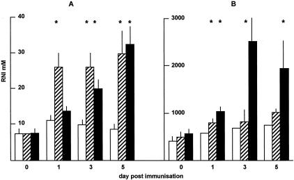
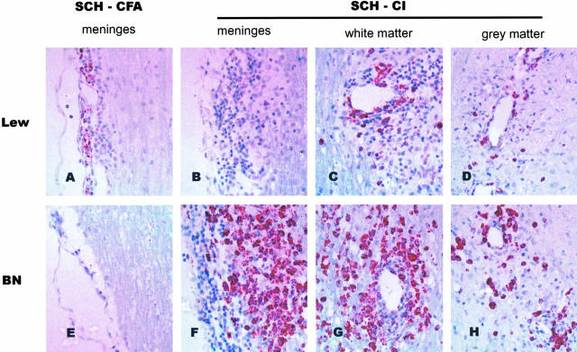
The Brown Norway (BN) rat is reported to be resistant to the induction of experimental autoimmune encephalomyelitis (EAE) and a number of mechanisms have been suggested to explain this resistance. In work reported here we provide evidence that such resistance in the BN rat can be accounted for, at least in part, by their ability to produce higher levels of nitric oxide (NO) than susceptible strains of rats. Spleen cells from the BN rat make significantly more NO following in vitro stimulation than do cells from the Lewis or PVG rat and following in vivo immunization using complete Freund's adjuvant (CFA) the BN rat makes substantially more NO than either susceptible strain. If carbonyl iron is used as adjuvant in vivo there is no increase in NO levels in the BN rat and they are rendered highly susceptible to EAE. Immunizing with CFA simultaneously with neuroantigen and carbonyl iron drives up NO levels and the resistance is restored. EAE produced using carbonyl iron is characterized by extensive macrophage/microglia presence in the central nervous system lesions of the BN rat yet the cytokine profile in the lymph nodes does not differ from that in the EAE Lewis rats.
Figures


Similar articles
-
Limiting-dilution analysis of the frequency of myelin basic protein-reactive T cells in Lewis, PVG/c and BN rats. Implication for susceptibility to autoimmune encephalomyelitis.Immunology. 1990 Feb;69(2):215-21. Immunology. 1990. PMID: 1689693 Free PMC article.
-
The innate immune response to adjuvants dictates the adaptive immune response to autoantigens.J Neuropathol Exp Neurol. 2008 Jun;67(6):543-54. doi: 10.1097/NEN.0b013e31817713cc. J Neuropathol Exp Neurol. 2008. PMID: 18520773
-
Allergic encephalomyelitis in the reputedly resistant Brown Norway strain of rats.J Immunol. 1975 Feb;114(2 Pt 1):597-601. J Immunol. 1975. PMID: 1120901
-
Nitric oxide in interstitial nephritis and other autoimmune diseases.Semin Nephrol. 1999 May;19(3):288-95. Semin Nephrol. 1999. PMID: 10226335 Review.
-
Nitric oxide and experimental autoimmune encephalomyelitis review.J Neuroimmunol. 2025 Jul 15;404:578586. doi: 10.1016/j.jneuroim.2025.578586. Epub 2025 Apr 2. J Neuroimmunol. 2025. PMID: 40220601 Review.
Cited by
-
Down-regulation of Myelin Gene Expression in Human Oligodendrocytes by Nitric Oxide: Implications for Demyelination in Multiple Sclerosis.J Clin Cell Immunol. 2013 Jul 31;4:10.4172/2155-9899.1000157. doi: 10.4172/2155-9899.1000157. J Clin Cell Immunol. 2013. PMID: 24273691 Free PMC article.
-
GM-CSF-neuroantigen fusion proteins reverse experimental autoimmune encephalomyelitis and mediate tolerogenic activity in adjuvant-primed environments: association with inflammation-dependent, inhibitory antigen presentation.J Immunol. 2014 Sep 1;193(5):2317-29. doi: 10.4049/jimmunol.1303223. Epub 2014 Jul 21. J Immunol. 2014. PMID: 25049359 Free PMC article.
-
Myelin-phagocytosing macrophages modulate autoreactive T cell proliferation.J Neuroinflammation. 2011 Jul 25;8:85. doi: 10.1186/1742-2094-8-85. J Neuroinflammation. 2011. PMID: 21781347 Free PMC article.
-
End-point effector stress mediators in neuroimmune interactions: their role in immune system homeostasis and autoimmune pathology.Immunol Res. 2012 Apr;52(1-2):64-80. doi: 10.1007/s12026-012-8275-9. Immunol Res. 2012. PMID: 22396175 Review.
-
Do Natural T Regulatory Cells become Activated to Antigen Specific T Regulatory Cells in Transplantation and in Autoimmunity?Front Immunol. 2013 Aug 2;4:208. doi: 10.3389/fimmu.2013.00208. eCollection 2013. Front Immunol. 2013. PMID: 23935597 Free PMC article.
References
-
- Marlatta MA. Nitric oxide: biosynthesis and biological significance. Trends Biochem Sci. 1989;14:488–492. - PubMed
-
- Moncada S, Palmer RM, Higgs EA. Nitric oxide: physiology, pathophysiology, and pharmacology. Pharmacol Rev. 1991;43:109–142. - PubMed
-
- Lowenstein CJ, Snyder SH. Nitric oxide, a novel biologic mesenger. Cell. 1992;70:705–707. - PubMed
-
- Nathan CF, Hibbs JB. Role of nitric oxide synthesis in macrophage antimicrobial activity. Curr Opin Immunol. 1991;3:65–70. - PubMed
-
- Willenborg DO, Staykova MA, Cowden WB. Our shifting understanding of the role of nitric oxide in autoimmune encephalomyelitis: a review. J Neuroimmunol. 1999;100:21–35. - PubMed
Publication types
MeSH terms
Substances
LinkOut - more resources
Full Text Sources
