. 2005 Jan;166(1):173-83.
doi: 10.1016/S0002-9440(10)62242-0.

Human herpesvirus 6 activates lytic cycle replication of Kaposi's sarcoma-associated herpesvirus

Affiliations

Human herpesvirus 6 activates lytic cycle replication of Kaposi's sarcoma-associated herpesvirus

Chun Lu et al. Am J Pathol. 2005 Jan.

Erratum in

  • Correction.
    [No authors listed] [No authors listed] Am J Pathol. 2021 May;191(5):965. doi: 10.1016/j.ajpath.2021.02.012. Am J Pathol. 2021. PMID: 33894887 Free PMC article. No abstract available.

Abstract

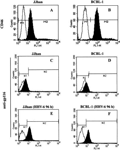
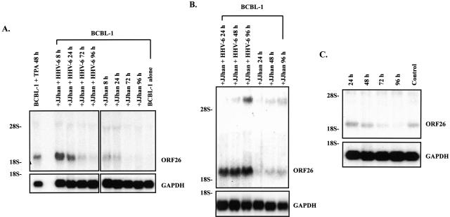
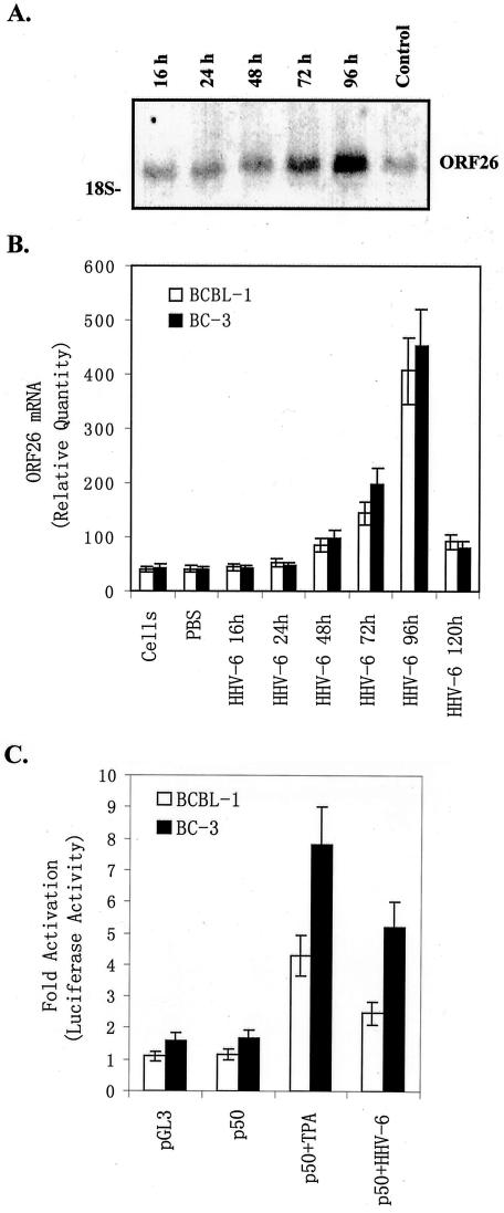
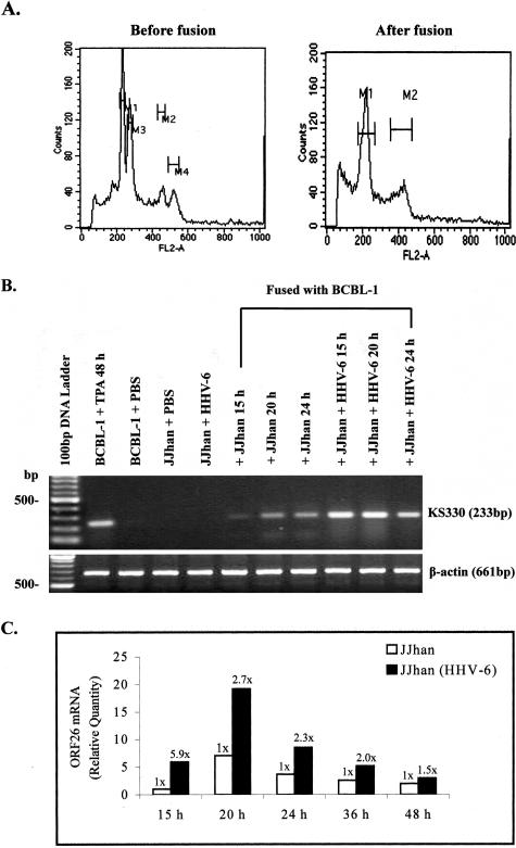
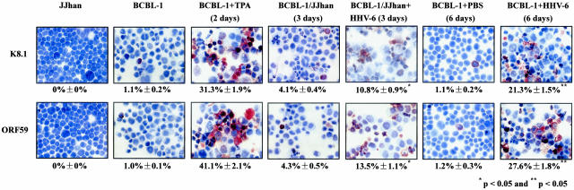
Kaposi's sarcoma-associated herpesvirus (KSHV) or human herpesvirus 8 (HHV-8) is a gamma-herpesvirus consistently identified in Kaposi's sarcoma (KS), primary effusion lymphoma, and multicentric Castleman's disease. KSHV infection appears to be necessary, but not be sufficient for development of KS without other co-factors. However, factors that facilitate KSHV to cause KS have not been well defined. Because patients with KS are often immunosuppressed and susceptible to many infectious agents including human herpesvirus 6 (HHV-6), we investigated the potential of HHV-6 to influence the replication of KSHV. By co-culturing HHV-6-infected T cells with KSHV-latent BCBL-1 cell line, infecting BCBL-1 cells with HHV-6 virions, and generating heterokaryons between HHV-6-infected T cells and BCBL-1 cells, we showed that HHV-6 played a critical role in induction of KSHV replication, as determined by production of lytic phase mRNA transcripts and viral proteins. We confirmed and extended the results by using a luciferase reporter assay in which KSHV ORF50 promoter, the first promoter activated during KSHV replication, drove the luciferase expression. Besides HHV-6, we also found that cytokines such as interferon-gamma partially contributed to induction of KSHV replication in the co-culture system. These findings suggest that HHV-6 may participate in KS pathogenesis by promoting KSHV replication and increasing KSHV viral load.


Figures

Figure 1
Figure 1
Expression of CD46 and HHV-6 gp116 antigens in JJhan and BCBL-1 cells. JJhan (A, C) and BCBL-1 (B, D) cells or HHV-6-infected JJhan (E) and HHV-6-infected BCBL-1 (F) cells were stained with anti-CD46 (top) or with anti-HHV-6 gp116 (middle and bottom) antibodies (fluorescein isothiocyanate-labeled monoclonal antibodies). Fluorescein isothiocyanate-labeled IgG was used as an isotype control antibody. The expressions of CD46 and HHV-6 gp116 were analyzed by FACS. The black shading represents anti-CD46 or anti-gp116-specific antibodies and the white shading represents the isotype control antibody.
Figure 2
Figure 2
Northern blot analysis for ORF26 mRNA in BCBL-1 cells. A: ORF26 mRNA expressed in BCBL-1 cells after co-culture with HHV-6-infected JJhan cells by direct cell-cell contact. Total RNA isolated from BCBL-1 cells treated with TPA for 48 hours (positive control, BCBL-1 + TPA 48 hours), BCBL-1 cells co-cultured with HHV-6-infected JJhan cells (JJhan + HHV-6), 8, 24, 72, and 96 hours, respectively (+JJhan + HHV-6 8 hours, 24 hours, 72 hours, 96 hours), BCBL-1 cells co-cultured with uninfected JJhan cells, 8, 24, 72, and 96 hours, respectively (+JJhan 8 hours, 24 hours, 72 hours, 96 hours), BCBL-1 cells alone (negative control, BCBL-1 alone) were transferred to nylon membrane and ORF26 mRNA expression was detected by Northern blot. The same membrane was stripped and reprobed for GAPDH to demonstrate equal loading of the RNA. B: ORF26 mRNA expressed in BCBL-1 cells cultured for 24 to 96 hours in 50% conditioned medium. ORF26 mRNA expression in BCBL-1 cells cultured in 50% conditioned medium from HHV-6-infected JJhan cells for 24, 48, and 96 hours (+JJhan + HHV-6 24 hours, 48 hours, 96 hours) and BCBL-1 cells cultured in 50% conditioned medium from uninfected JJhan cells for 24, 48, and 96 hours (+JJhan 24 hours, 48 hours, 96 hours) was detected by Northern blot. C: ORF26 mRNA expressed in BCBL-1 cells cultured for 24 to 96 hours in KSHV-depleted conditioned medium. ORF26 mRNA expression in BCBL-1 cells cultured in KSHV-depleted conditioned medium for 24, 48, 72, and 96 hours (24 hours, 48 hours, 72 hours, 96 hours) and normal BCBL-1 cells (control) was detected by Northern blot. All results shown are a representative experiment of at least three independent experiments with similar results.
Figure 3
Figure 3
Infection of BCBL-1 cells with HHV-6 induces lytic cycle replication of KSHV. A: ORF26 mRNA expressed in BCBL-1 cells infected with pelleted HHV-6. ORF26 mRNA expression in BCBL-1 cells infected with pelleted HHV-6 for 16, 24, 48, 72, and 96 hours (16 hours, 24 hours, 48 hours, 72 hours, 96 hours), and uninfected BCBL-1 (control) cells was detected by Northern blot. A representative experiment is shown; at least three independent experiments were performed with similar results. B: Real-time quantitative PCR for ORF26 mRNA in BCBL-1 and BC-3 cells after infection with pelleted HHV-6. ORF26 mRNA expression in BCBL-1 (white bar) and BC-3 (black bar) cells infected with pelleted HHV-6 for 16, 24, 48, 72, 96, and 120 hours was quantitated by real-time quantitative PCR. Results shown were the statistic of three independent experiments performed in triplicate. C: Infection of HHV-6 promotes induction of KSHV ORF50 promoter activity. BCBL-1 (white bar) and BC-3 (black bar) cells were transfected with pGL-3 basic control or p50 plasmids. p50-transfected cells were either untreated (p50), treated with TPA (p50 + TPA), or infected with HHV-6 (p50 + HHV-6). Luciferase activities were measured as induction (n-fold). Results from a representative experiment performed in triplicate are shown; five independent experiments were performed with similar results.
Figure 4
Figure 4
Expression of ORF26 mRNA in BCBL-1 cells fused with HHV-6-infected JJhan cells. A: Detection of fused cells in the heterokaryon assay by flow cytometry. BCBL-1 cells mixed with HHV-6-infected JJhan cells in the equal number (before fusion) and BCBL-1 cells 20 hours after fusing with HHV-6-infected JJhan cells (after fusion) were fixed, stained with propidium iodide, and analyzed for DNA contents of tetraploid cells (M2 and M4 before fusion, M2 after fusion) by flow cytometry. B: RT-PCR analysis of ORF26 mRNA expression in heterokaryons. ORF26 mRNA expression in BCBL-1 cells fusing with JJhan cells for 15, 20, 24 hours (+JJhan 15 hours, 20 hours, 24 hours) or fusing with HHV-6-infected JJhan cells for 15, 20, 24 hours (+JJhan + HHV-6 15 hours, 20 hours, 24 hours) was detected by RT-PCR. As controls, ORF26 mRNA expression in TPA-treated BCBL-1 (BCBL-1 + TPA 48 hours, positive control) cells, PBS-treated BCBL-1 cells (BCBL-1 + PBS), PBS-treated JJhan cells (JJhan + PBS), or HHV-6-infected JJhan cells (JJhan + HHV-6) were also detected. C: Real-time quantitative PCR analysis for ORF26 mRNA in BCBL-1 cells fused for 15 to 48 hours with HHV-6-infected JJhan cells. ORF26 mRNA expression in BCBL-1 fusing with JJhan cells (white bar, JJhan) or BCBL-1 fusing with HHV-6-infected JJhan cells [black bar, JJhan(HHV-6)] for 15 to 48 hours was quantitated by real-time quantitative PCR. Relative quantities of ORF26 expression are represented on the y axis. *, P < 0.05. Results were the statistic of four independent experiments performed in triplicate.
Figure 5
Figure 5
Immunohistochemisty staining of BCBL-1 cells co-cultured with HHV-6-infected JJhan cells and infected with pelleted HHV-6. KSHV lytic proteins ORF K8.1 (top) and ORF59 (bottom) expression in JJhan cells, BCBL-1 cells, TPA-treated BCBL-1 cells (BCBL-1 + TPA), PBS-treated BCBL-1 cells (BCBL-1 + PBS), BCBL-1 cell co-cultured with JJhan cells (BCBL-1/JJhan), BCBL-1 cell co-cultured with HHV-6-infected JJhan cells (BCBL-1/JJhan + HHV-6), or HHV-6 infected BCBL-1 cells (BCBL-1 + HHV-6) were detected by immunohistochemistry with ORF K8.1 and ORF59 monoclonal antibodies. * and **, Statistically significant increases in ORF K8.1 and ORF59 expression compared to that of BCBL-1 cells co-cultured with normal JJhan cells for 3 days and cultured in PBS buffer for 6 days, respectively. Original magnifications, ×60.
Figure 6
Figure 6
IFN-γ partially contributes to induction of KSHV replication. A: Expression of IFN-γ in BCBL-1 cells co-cultured for 8 to 120 hours with HHV-6-infected JJhan cells and cultured in 50% conditioned medium. Supernatants from BCBL-1 cells co-cultured with JJhan (JJhan + BCBL-1), HHV-6-infected JJhan [JJhan(HHV-6) + BCBL-1] or BCBL-1 cells cultured in conditioned medium from JJhan cell culture (SuJJhan + BCBL-1) and HHV-6-infected JJhan cell culture [SuJJhan(HHV-6) + BCBL-1] for various times indicated were collected for detection of IFN-γ by ELISA. Results were the statistic of three independent experiments with duplicate. B: Real-time quantitative PCR analysis for ORF26 mRNA expression in blocking assay by mAb against IFN-γ. Real-time quantitative PCR was used to detected relative quantities of ORF26 mRNA in normal BCBL-1 cells (BCBL-1 alone), IFN-γ-treated BCBL-1 cells (IFN-γ + BCBL-1), BCBL-1 cells co-cultured with JJhan cell (JJhan + BCBL-1), HHV-6-infected JJhan cells [JJhan(HHV-6) + BCBL-1], or HHV-6-infected JJhan cells plus mAb treatment [JJhan(HHV-6) + BCBL-1 + mAb] for various times indicated. The ORF26 mRNA expression in BCBL-1 cells cultured in conditioned medium from JJhan cell (SuJJhan + BCBL-1), HHV-6-infected JJhan cells [SuJJhan(HHV-6) + BCBL-1], or HHV-6-infected JJhan cells plus mAb treatment [SuJJhan(HHV-6) + BCBL-1 + mAb] for various times indicated was also detected by real-time quantitative PCR. The statistic of three independent experiments performed in triplicate was shown. C: Northern blot analysis for ORF26 mRNA expression in blocking assay by mAb against IFN-γ. Northern blot analysis was used to detect ORF26 mRNA in BCBL-1 cells co-cultured with HHV-6-infected JJhan cells at 24 and 48 hours or cultured in conditioned medium at 48 and 72 hours before or after treatment with mAb against IFN-γ, respectively. Lane 1 (from left), BCBL-1 co-cultured with HHV-6-infected JJhan cells (BCBL-1 + JJhan + HHV-6), 24 hours; lane 2, BCBL-1 co-cultured with HHV-6-infected JJhan cells with addition of mAb against IFN-γ (BCBL-1 + JJhan + HHV-6 + mAb), 24 hours; lane 3, BCBL-1 + JJhan + HHV-6, 48 hours; lane 4, BCBL-1 + JJhan + HHV-6 + mAb, 48 hours; lane 5, BCBL-1 cells cultured in conditioned medium from HHV-6-infected JJhan cells with addition of mAb against IFN-γ (BCBL-1 + C.M. + mAb), 72 hours; lane 6, BCBL-1 + C.M., 72 hours; lane 7, BCBL-1 + C.M. + mAb, 48 hours; lane 8, BCBL-1 + C.M., 48 hours. A representative experiment is shown; three independent experiments were run and gave similar results.

Similar articles

Cited by

References

    1. Chang Y, Cesarman E, Pessin MS, Lee F, Culpepper J, Knowles DM, Moore PS. Identification of herpesvirus-like DNA sequences in AIDS-associated Kaposi’s sarcoma. Science. 1994;266:1865–1869. - PubMed
    1. Moore PS, Gao SJ, Dominguez G, Cesarman E, Lungu O, Knowles DM, Garber R, Pellett PE, McGeoch DJ, Chang Y. Primary characterization of a herpesvirus agent associated with Kaposi’s sarcoma. J Virol. 1996;70:549–558. - PMC - PubMed
    1. Katano H, Sata T. Human herpesvirus 8: virology, epidemiology and related diseases. Jpn J Infect Dis. 2000;53:137–155. - PubMed
    1. Schulz TF. Kaposi’s sarcoma-associated herpesvirus (human herpesvirus-8). J Gen Virol. 1998;79:1573–1591. - PubMed
    1. Moore PS, Chang Y. Detection of herpesvirus-like DNA sequences in Kaposi’s sarcoma in patients with and without HIV infection. N Engl J Med. 1995;332:1181–1185. - PubMed

Publication types