. 2005 Jan;166(1):231-40.
doi: 10.1016/S0002-9440(10)62247-X.

Melanocyte-specific proteins are aberrantly trafficked in melanocytes of Hermansky-Pudlak syndrome-type 3

Affiliations

Melanocyte-specific proteins are aberrantly trafficked in melanocytes of Hermansky-Pudlak syndrome-type 3

Raymond E Boissy et al. Am J Pathol. 2005 Jan.

Abstract

Hermansky-Pudlak Syndrome-type 3 (HPS-3) is a relatively mild subtype of HPS with minimal cutaneous and ocular depigmentation. The HPS-3 gene encodes a novel protein of unknown function with a predicted molecular weight of 114 kd. To assess the role of the HPS3 protein in melanization, cultured melanocytes developed from HPS-3 patients were evaluated biochemically and histologically for activity and localization of melanocyte-specific proteins. Endogenous tyrosinase activity of HPS-3 melanocytes was substantial, but tyrosinase activity and melanin synthesis was suppressed in intact melanocytes. However, the level of suppression, as well as extent to which up-regulation by isobutylmethylxanthine and cholera toxin was muted, was less that in HPS-1 melanocytes. Ultrastructurally, HPS-3 melanocytes contained morphologically normal melanosomes, predominantly of stage I and II with minimal stage III and few stage IV melanosomes. Dihydroxyphenylalanine (DOPA) histochemistry demonstrated an increase in melanization of melanosomes. Unique to HPS-3 melanocytes were numerous DOPA-positive 50-nm vesicles and tubular elements present throughout the cell body and dendrites. Tyrosinase, tyrosinase-related protein-1 (Tyrp1), dopachrome tautomerase (Dct), and LAMP1 and 3 localization in HPS-3 melanocytes, as evaluated by immunocytochemistry and confocal microscopy, demonstrated a fine, floccular distribution in contrast to the coarse, granular distribution characteristic of control melanocytes. The localization profile of other proteins expressed by melanocytes (ie, Silver/Pmel17, Melan-A/MART-1, LAMP2, Rab 27, transferrin, c-kit, adaptin-3, and the HPS1 protein) appeared normal. These results suggest that a specific subset of melanocyte proteins are aberrantly trafficked throughout the HPS-3 melanocyte and may be responsible for the reduction in melanin synthesis.


Figures

Figure 1
Figure 1
Melanocytes cultured from patients with HPS-3 exhibit suppressed tyrosinase activity and mild hypomelanosis. Cultures of melanocytes derived from a Caucasian individual (NHML), an African American individual (NHMD), an individual with oculocutaneous albinism type 1 (OCA1), and three individuals with either HPS-1 or HPS-3 were tested in triplicate for (a) tyrosinase activity in cell lysates [shown separately are each HPS-1 (bars from left to right represent patients 3, 1, and 2, respectively) and HPS-3 (bars from left to right represent patients 1, 2, and 3, respectively) melanocyte line], (b) melanin content in cell lysates (data from each HPS-1 and 3 melanocyte line is grouped by genotype, as for c to d also), (c) tyrosinase activity in intact cells, and (d) percent increase in melanin after treatment of cultured melanocytes with isobutyl methylxanthine and cholera toxin for 10 days as described in Materials and Methods. Data are expressed as means ± SEM. *, P < 0.05 for each line compared to the NHML lines.
Figure 2
Figure 2
HPS-3 melanocytes exhibit morphologically normal melanosomes with diminished melanin synthesis. As demonstrated by electron microscopy, the Golgi (G) area (a) and dendritic (D) area (c) of normal human melanocytes (a and c represent NHMD and NHML, respectively) contain melanosomes ranging from the early (I) to the mature (IV) stage. In contrast, the Golgi (G) area (b) and dendritic (D) area (d) of melanocytes of patients with HPS-3 (b and d represent patients 1 and 2, respectively) contain melanosomes representing stages I to III only, with no stage IV melanosomes apparent. Insets demonstrate that the melanofilament arrangement within the stage II melanosomes of the HPS-3 melanocytes appears normal. N = nucleus. Bars: a–d, 2 μm; insets, 1 μm.
Figure 3
Figure 3
After DOPA histochemistry, HPS-3 melanocytes exhibit an extensive distribution of DOPA-positive vesicles and tubules. As demonstrated by electron microscopy, normal human melanocytes (NHML) (a) exhibit DOPA-positive vesicles confined to the Golgi (G) area and notably absent from the lateral side of the nucleus (arrowhead). In contrast, HPS-3 melanocytes (b and c represent patients 2 and 1, respectively) exhibit DOPA-positive vesicles (black arrows) and tubules (white arrows) throughout the cell body peripheral to the Golgi (G) area as well as in the lateral side of the nucleus (arrowhead). Some of the DOPA-positive tubular profiles appear to be continuous with melanosomes (white arrows with asterisks in c and inset to c). In addition, DOPA-positive vesicles (black arrows) and tubules (white arrows) are absent from the dendrites of (d) normal human melanocytes (NHMD) but (e) prevalent in the dendrites of HPS-3 melanocytes (patient 2). Bars: a–e, 2 μm; inset to c, 0.3 μm.
Figure 4
Figure 4
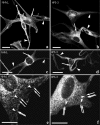
Tyrosinase distribution in HPS-3 melanocytes appears floccular and less granular than in normal human melanocytes (NHM). As demonstrated by immunocytochemical staining of tyrosinase, normal human melanocytes (a and c represent NHMD) exhibit a granular staining pattern that is dense in the cell body (arrow) and sparse in the dendrites (arrowheads). In contrast, HPS-3 melanocytes (b and d represent patients 1 and 2, respectively) exhibit a finer, more floccular staining pattern, also more intense in the cell body (arrows) than in the dendrites (arrowheads). Higher magnification of NHM (e represents NHMD) demonstrates that the granular pattern characteristic of NHM represents immunolabeling predominantly of 0.5 to 1.0 μm melanosomes (arrows) with lack of immunolabeling between melanosomes (arrowheads). In contrast, higher magnification of HPS-3 (f represents patient 1) demonstrates that the floccular staining characteristic of HPS-3 melanocytes represents additional staining (arrowheads) between the immunolabeled melanosomes (arrows). This finer immunolabeling profile represents the numerous DOPA-positive intermelanosomal vesicles and tubules observed with electron microscopy (Figure 3). Bars: a–d, 20 μm; e–f, 8 μm.
Figure 5
Figure 5
Tyrosinase-related protein-1 (Tyrp1) in HPS-3 melanocytes also appears floccular and less granular and without uniform co-localization with tyrosinase. As demonstrated by dual immunocytochemical staining, normal human melanocytes (NHM) derived from light skin (a–c) exhibit a granular staining pattern relatively dense in the cell body (arrow) and sparser in the dendrites (arrowheads) for both tyrosinase (a, red) and Tyrp1 (b, green). Tyrosinase and Tyrp1 exhibit marked co-localization in the merged image (c). In contrast, HPS-3 melanocytes derived from patient 1 (d–f) exhibit a finer, more floccular staining pattern also more intense in the cell body (arrows) than the dendrites (arrowheads) for both tyrosinase (d, red) and Tyrp1 (e, green). Merged images demonstrate that tyrosinase and Tyrp1 exhibit less co-localization in HPS-3 melanocytes (f) than observed in NHM (c). Similar results were obtained using HPS-3-#2 melanocytes (not shown) Bar, 15 μm.
Figure 6
Figure 6
HPS-3 melanocytes exhibit a staining pattern distinct from normal human melanocytes (NHML) for Dct and LAMP 1 and 3 but not for the HPS1 protein or adaptin-3. Normal and HPS-3 melanocytes were immunolabeled for Dct, LAMP1, LAMP2, HPS1 protein, and adaptin-3 as described in Materials and Methods. Dct (b versus a), LAMP1 (d versus c), and LAMP3 (f versus e) exhibit a finer, more floccular staining pattern in HPS-3 melanocytes, as opposed to the granular pattern in NHM. Bar, 10 μm.

Similar articles

Cited by

References

    1. Hermansky F, Pudlak P. Albinism associated with hemorrhagic diathesis and unusual pigmented reticular cells in the bone marrow: report of two cases with histochemical studies. Blood. 1959;14:162–169. - PubMed
    1. Huizing M, Boissy RE, Gahl WA. Hermansky-Pudlak syndrome: vesicle formation from yeast to man. Pigment Cell Res. 2002;15:405–419. - PubMed
    1. King RA, Hearing VJ, Creel DJ, Oetting WS. Albinism. Scriver CR, Beaudet AL, Valle DL, Sly WS, editors. New York: McGraw-Hill; The Metabolic and Molecular Bases of Inherited Disease. 2001:pp 5587–5627.
    1. Witkop CJJ, Krumwiede M, Sedano H, White JG. Reliability of absent platelet dense bodies as a diagnostic criterion for Hermansky-Pudlak syndrome. Am J Hematol. 1987;26:305–311. - PubMed
    1. Gahl WA, Brantly M, Kaiser-Kupfer MI, Iwata F, Hazelwood S, Shotelersuk V, Duffy LF, Kuehl EM, Troendle I, Bernardini I. Genetic defects and clinical characteristics of patients with a form of oculocutaneous albinism (Hermansky-Pudlak syndrome). N Engl J Med. 1998;338:1258–1264. - PubMed

Publication types