. 2005 Jan;17(1):132-48.
doi: 10.1105/tpc.104.026351.

Receptor salvage from the prevacuolar compartment is essential for efficient vacuolar protein targeting

Affiliations

Receptor salvage from the prevacuolar compartment is essential for efficient vacuolar protein targeting

Luis L P daSilva et al. Plant Cell. 2005 Jan.

Abstract

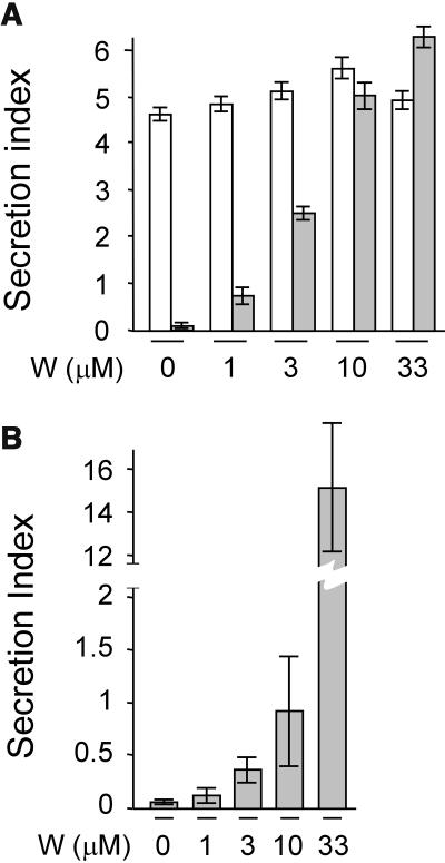
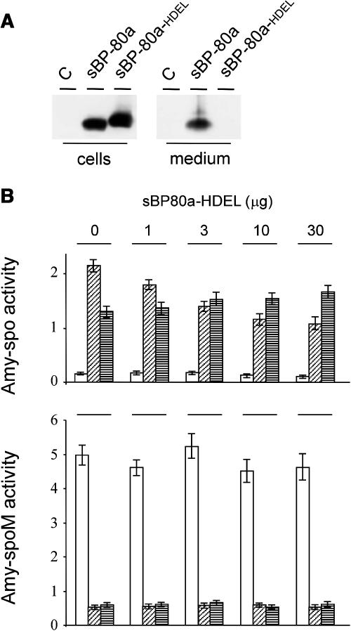
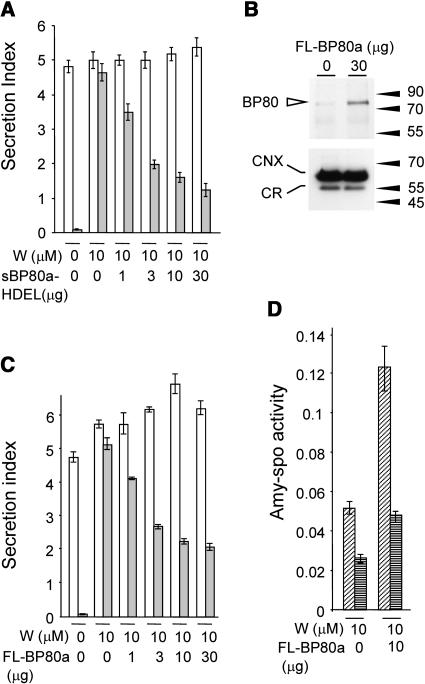
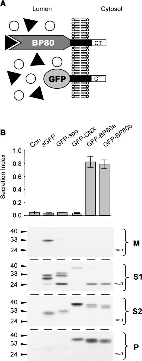
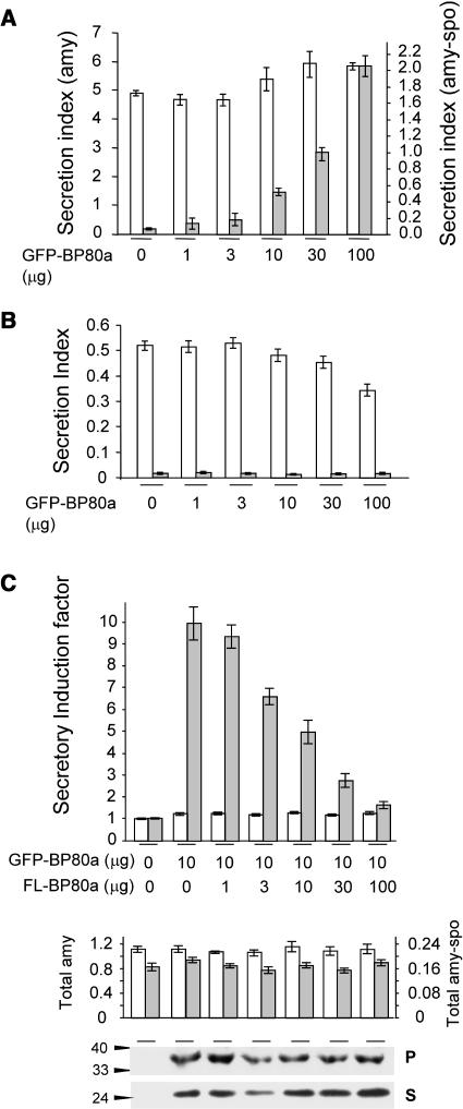
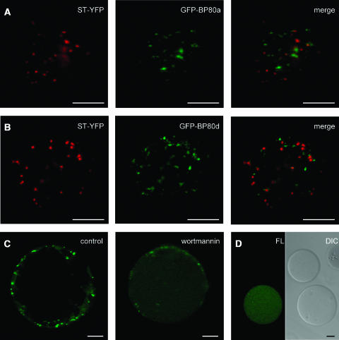
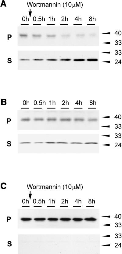
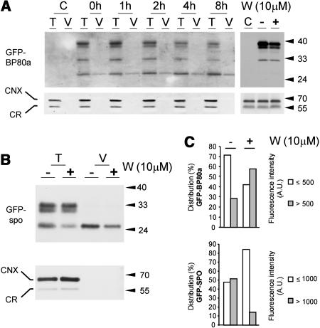
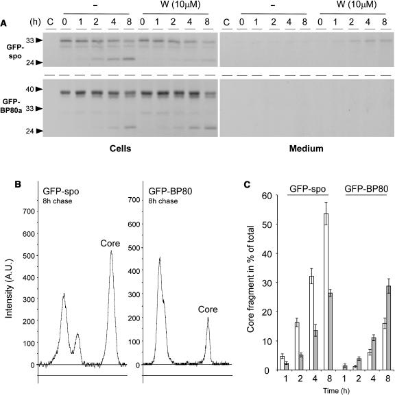
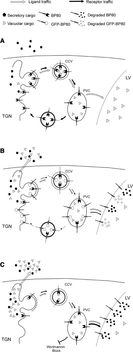
We have characterized the requirements to inhibit the function of the plant vacuolar sorting receptor BP80 in vivo and gained insight into the crucial role of receptor recycling between the prevacuolar compartment and the Golgi apparatus. The drug wortmannin interferes with the BP80-mediated route to the vacuole and induces hypersecretion of a soluble BP80-ligand. Wortmannin does not prevent receptor-ligand binding itself but causes BP80 levels to be limiting. Consequently, overexpression of BP80 partially restores vacuolar cargo transport. To simulate receptor traffic, we tested a truncated BP80 derivative in which the entire lumenal domain of BP80 has been replaced by the green fluorescent protein (GFP). The resulting chimeric protein (GFP-BP80) accumulates in the prevacuolar compartment as expected, but a soluble GFP fragment can also be detected in purified vacuoles. Interestingly, GFP-BP80 coexpression interferes with the correct sorting of a BP80-ligand and causes hypersecretion that is reversible by expressing a 10-fold excess of full-length BP80. This suggests that GFP-BP80 competes with endogenous BP80 mainly at the retrograde transport route that rescues receptors from the prevacuolar compartment. Treatment with wortmannin causes further leakage of GFP-BP80 from the prevacuolar compartment to the vacuoles, whereas BP80-ligands are secreted. We propose that recycling of the vacuolar sorting receptor from the prevacuolar compartment to the Golgi apparatus is an essential process that is saturable and wortmannin sensitive.


Figures

Figure 1.
Figure 1.
Wortmannin Strongly Inhibits the Sequence-Specific Vacuolar Sorting Route. (A) The concentration-dependent influence of the drug wortmannin on the secretion index of amy (white bars) and amy-spo (gray bars) in tobacco mesophyl protoplasts. Transcriptional control of the cargo proteins was driven by the 35S promoter of Cauliflower mosaic virus (pCaMV35S), and 3′ end processing and polyadenylation was controlled by the 3′ untranslated end of the nopaline gene (3′nos). Protoplasts were transfected with a constant amount (10 μg) of plasmid-encoded cargo molecule and distributed in equal portions, subject to different concentrations of wortmannin and incubated for 24 h after which cells and media were harvested. The concentration of the drug is given below each lane. The secretion index was calculated as the ratio between the extracellular and the intracellular amy activities (Phillipson et al., 2001). (B) The concentration-dependent influence of the drug wortmannin on the secretion index of amy-spo in transgenic tobacco BY2 cells. Because of the very high secretion index at 33 μM wortmannin, a split y axis was used for this lane.
Figure 2.
Figure 2.
Confirmation of amy-spo as a BP80-Ligand in Vivo. (A) Transport assay for soluble BP80 molecules with or without the ER retention motif HDEL via transient expression in tobacco protoplasts. Shown is a protein gel blot of cells and medium from untransformed (C) and transfected cells with 10 μg of plasmid-encoded pCaMV35S:sBP80a-myc:3′nos or a C-terminal HDEL-fused version (sBP80a and sBP80a-HDEL, respectively). After 24-h incubation, cells and medium were harvested and analyzed via protein gel blot to investigate the steady state distribution of the chimeric proteins. Proteins were detected using anti-myc antiserum. (B) sBP80a-HDEL mediates specific subcellular redistribution of amy-spo. Protoplasts were transfected with a constant amount (10 μg) of plasmid encoding either amy-spo or the control cargo amy-spoM (Pimpl et al., 2003), in which the vacuolar sorting signal is mutated. An increasing amount of plasmid-encoded sBP80a-HDEL (as indicated above each lane) was cotransfected. Cells and medium were harvested as in Figure 1, but the cell fraction was further partitioned into soluble proteins released after osmotic shock (S1), which is enriched in cytosolic and vacuolar solutes and soluble proteins released after sonication of the S1-free pellet (S2). The latter fraction is enriched in microsomal proteins. The amy activity was calculated for each fraction and shown as follows: medium (white bars), S1 (diagonally striped bars), and S2 (horizontally striped bars). amy activity is given in ΔOD per mL and per min reaction time.
Figure 3.
Figure 3.
Wortmannin Does Not Prevent BP80-Ligand Interactions. (A) Wortmannin does not prevent BP80 from physically interacting with its ligands. Protoplasts were transfected with either amy or amy-spo, together with increasing concentrations of sBP80-HDEL plasmid preparation (as stated below each lane) and were incubated for 24 h with a constant concentration of wortmannin (10 μM) as indicated. The amy activity was obtained for each fraction, and the secretion index was calculated and compared for each cargo: amy (white bars) and amy-spo (gray bars). (B) Microsomal fractions from mock transfected and transfected protoplasts to test if transient expression of plasmid-encoded pCaMV35S:full-length BP80a:3′nos (FL-BP80a) is detectable. The amount of FL-BP80a plasmid is indicated above the lane. Molecular mass markers are indicated in kD. The top panel shows a protein gel blot probed with anti-AtBP80 antiserum (Laval et al., 1999), which had been subtracted as described in Methods. Notice the strongly induced band at ∼80 kD in microsomes from transfected protoplasts. The bottom panel shows a protein gel blot probed with anticalnexin antibodies (Pimpl et al., 2000, 2003) that binds to calnexin (CNX) and calreticulin (CR). This illustrates that equal levels of total microsomes were loaded in both lanes. (C) Reconstitution of BP80-mediated sorting by coexpression of full-length BP80a. Protoplasts were transfected with a constant amount of cargo molecules and a dilution series of the same FL-BP80a plasmid preparation used for (B) and incubated within a constant wortmannin background (10 μM). As in (A), a control lane with cargo alone was included without wortmannin to illustrate the degree of secretory induction for each cargo molecule. Notice that the addition of FL-BP80a partially reverses the effect of wortmannin on amy-spo but not amy secretion. (D) Overexpression of full-length BP80a leads predominantly to vacuolar transport of amy-spo. Protoplasts expressing amy-spo with or without FL-BP80a were incubated with 10 μM wortmannin, after which cells were isolated and fractionated into S1 and S2, similarly as in Figure 2. amy-spo activity was measured for each fraction and compared: S1 (diagonally striped bars) and S2 (horizontally striped bars).
Figure 4.
Figure 4.
Truncated BP80 Lacking the Ligand Binding Domain Interferes with Vacuolar Sorting. (A) Schematic illustration of the nature and topology of the truncated BP80 fusions in relation to the full-length BP80. The complete ligand binding domain of the Arabidopsis BP80 isoforms a, b, d, and f (Hadlington and Denecke, 2000) were replaced by GFP, which results in the truncated molecules (GFP-BP80a, GFP-BP80b, GFP-BP80d, and GFP-BP80f) that do not interact with BP80-ligands but yet display the same polypeptide on the cytosolic face of the membrane as the wild-type BP80 counterparts. (B) Protoplasts were transfected with plasmid-encoding amy-spo alone or together with 20 μg of plasmid encoding either sGFP, sGFP-spo, GFP-calnexin (GFP-CNX), or GFP-BP80 fusions (represented here by GFP-BP80a and b) and incubated for 24 h, after which medium and cells were harvested. The top panel shows the secretion index of cotransfected amy-spo; annotations are as in the previous figures. Notice that only GFP-BP80a and GFP-BP80b induce the secretion of amy-spo, whereas sGFP, GFP-spo, and GFP-CNX have no effect. Cells were fractionated to obtain extracts enriched in soluble protein released by osmotic shock (S1), proteins released after sonicating the first pellet (S2), and membrane spanning proteins (P). These cell fractions together with the medium (M) were analyzed (bottom panel) to test the intracellular partitioning of the various GFP fusions. Molecular mass markers are indicated in kD. Notice the lower molecular mass degradation fragment, the GFP core (white arrowhead) that is detected exclusively in S1 for all GFP fusions except for GFP-CNX.
Figure 5.
Figure 5.
Truncated BP80 Is Codominant versus Full-Length BP80. (A) The concentration-dependent influence of GFP-BP80 coexpression on the secretion index of amy and amy-spo in tobacco mesophyl protoplasts. Protoplasts were transfected with constant levels of plasmids encoding each of the cargo molecules together with a dilution series of plasmid-encoding GFP-BP80a. The secretion index is given in different scales for amy (left y axis) and amy-spo (right y axis) to simplify comparison. Notice that the effector molecule exhibits a wortmannin-like effect (compare with Figure 1A) specifically for amy-spo (gray bars) but not amy (white bars). Dosage-dependent expression of effector molecules was routinely tested by anti-GFP–probed protein gel blots from total cell extracts (data not shown). (B) Control experiments using cell-retained amy-HDEL (white bars) and cytosolic GUS (gray bars) to test nonspecific cell leakage. Experimental conditions and annotations are as in (A). Notice that GFP-BP80 does not cause an induced secretion index for these two control molecules. (C) Transient expression experiment to test if full-length BP80a (FL-BP80a) and truncated BP80a compete with each other. Protoplasts were transfected with a constant amount of plasmid (10 μg) encoding either amy or amy-spo, together with a constant amount of plasmid encoding GFP-BP80a and a dilution series of plasmid encoding FL-BP80a from the same batch as used in Figure 2. The amount of effector plasmid DNA is indicated below each lane. The secretion index was calculated as previously, and the secretory induction factor is calculated for amy-spo (gray bars) and amy (white bars) by dividing each value by the respective secretion index for the control lane (without GFP-BP80a and without FL-BP80a). The second panel shows the total activities for amy (white bars) and amy-spo (gray bars) in the corresponding lanes. The bottom panel shows the levels of GFP-BP80a precursor in the pellet fraction (P) and soluble GFP core in the soluble fraction (S) for each corresponding lane.
Figure 6.
Figure 6.
Confocal Laser Scanning Microscopy Demonstrating Increased Vacuolar Sorting of GFP-BP80 by Wortmannin. (A) Protoplasts were cotransfected with 10 μg of plasmids encoding GFP-BP80a and the Golgi marker ST-YFP, live-embedded in TEX medium with 1% low melting agarose, and incubated for 24 h. The images present a 2.5-μm optical section through the cell cortex to give a view of the cytosol with the intermediate organelles of the secretory pathway. ST-YFP labels Golgi bodies (pseudocolored in red), and GFP-BP80a (pseudocolored in green) labels punctuate structures. Notice that hardly any overlap with the Golgi was detected (merge, colocalization is pseudocolored in yellow). (B) The same as (A) but with transfection of GFP-BP80d instead of GFP-BP80a. Note the very similar pattern as is visualized for this BP80 derivative from another family member (compare panels [A] and [B]), which is also the case when using the derivatives from isoforms b and f (data not shown). (C) Protoplasts were transfected with plasmid-encoding GFP-BP80a and incubated for 22 h in liquid TEX medium, after which protoplasts were washed, concentrated, and incubated for another 2 h in control conditions or in the presence of 10 μM wortmannin. Shown is a cross section through the center of the protoplast to appreciate the vacuole lumen. Notice that upon wortmannin treatment, punctuate fluorescence at the cell cortex diminishes, whereas diffuse fluorescence in the vacuole lumen increases. (D) Isolated vacuoles from the same protoplast suspension presented in (C) (+ wortmannin) and analyzed by confocal laser scanning microscopy in bright field (differential interference contrast [DIC]) and the green fluorescent channel (FL). Notice that wortmannin-induced vacuolar fluorescence does not occur in all vacuoles and is restricted to vacuoles isolated from transfected cells. Bars in (A) to (D) = 10 μm.
Figure 7.
Figure 7.
Wortmannin-Induced Degradation of GFP-BP80. (A) Protoplasts were transfected with plasmid-encoding GFP-BP80a and incubated for 22 h, after which wortmannin was added for various time points. Membrane pellet (P) and soluble fractions (S; the sum of S1 and S2 in other figures) were analyzed by protein gel blots. Notice the increased level of the low molecular weight fragment (GFP core) and the decrease of the full-length fusion protein in function of time. (B) The same conditions as in (A), but the incubations were done without the drug. Notice that there is no change to the partitioning. (C) Control experiment performed as in (A), but where GFP-BP80a was replaced by GFP-CNX. Notice that no degradation fragment was detected and that the fusion protein did not diminish over time upon treatment with the drug.
Figure 8.
Figure 8.
Vacuolar Localization of the Soluble GFP Core Fragment. (A) After an initial incubation for 22 h to reach cellular steady state levels of transiently expressed GFP-BP80a, cells were incubated with 10 μM wortmannin for various times (indicated above each lane), after which total extracts (soluble + membrane proteins) from entire protoplasts (T) or purified vacuoles (V) isolated from the same protoplast suspensions were compared by protein gel blots. Equal levels of the vacuolar marker α-mannosidase were loaded to ensure a valid comparison. The left top panel was probed with anti-GFP serum, illustrating that wortmannin-induced increase in the soluble GFP core fragment is localized to the vacuoles. The panel at right is a similar protein gel blot but with microsomes purified from untransformed cells (C) and from cells transformed with GFP-BP80a. The latter were incubated for 22 h after which a further 4 h of incubation without (−) or with (+) wortmannin was performed. Notice the reduction of the full-length precursor in the wortmannin-treated sample, and the absence of the GFP core fragment from the microsomes in any sample. The bottom panels were probed with anticalnexin antibodies (Pimpl et al., 2000) that bind both ER markers calreticulin (CNX) and calnexin (CR). Notice that neither marker copurifies with the vacuoles and that all three microsomal fractions exhibit similar levels of these two ER markers. (B) Vacuole partitioning assay as in (A) but with cells transfected with GFP-spo. Transfected cells were incubated for 22 h, after which a further 4 h of incubation without (−) or with (+) wortmannin was performed as in (A). Notice the reduction of the GFP core fragment upon treatment with the drug both in total extracts (T) and in purified vacuoles (V). (C) Quantification of the observed luminal green fluorescence of GFP-spo or GFP-BP80a derived GFP core fragments in vacuoles purified and analyzed by confocal laser scanning microscopy as shown in Figures 6C and 6D. One hundred fluorescent vacuoles were analyzed for either transfection, and fluorescence intensity is measured in arbitrary units (A.U.) using the same detector gains. To illustrate the effect of wortmannin, the population was split into two fractions (i.e., up to 500 A.U. and >500 A.U.). Because vacuolar fluorescence was higher in vacuoles purified in GFP-spo–transfected cells, we split the population differently (up to 1000 A.U. and above). Notice that the population of GFP-BP80a–containing vacuoles shifts from the low fluorescent class to the high class upon addition of the drug, whereas the opposite happens for GFP-spo-fluorescent vacuoles.
Figure 9.
Figure 9.
Pulse-Chase Analysis Demonstrating the Opposite Behavior of GFP-BP80a and GFP-spo in Response to the Drug Wortmannin. (A) Protoplasts were pulse labeled for 1 h, after which incubation in chase buffer was conducted for times indicated above each lane either in the presence (W) or absence (−) of the drug wortmannin. Isolated medium and cell extracts were immunoprecipitated with the anti-GFP antiserum and separated by SDS-PAGE. Notice that both GFP-spo and GFP-BP80 chase into the GFP core fragment but that this is inhibited for GFP-spo upon incubation with wortmannin. (B) Line scannings of the 8-h chase points for GFP-spo (left) and GFP-BP80a (right) from phosphor imaging data corresponding to (A) using AIDA software (Raytest). The signal for the GFP core fragment is indicated (Core) in each panel. (C) GFP core fragment presented as a percentage of the total signal in each lane. Quantification of the GFP core signal bands was in arbitrary units and is presented as a percentage of the total surface area from all peaks in each lane. Percentages are given in function of the time for ligand (GFP-spo) and receptor construct (GFP-BP80) in the absence (white bars) and presence (gray bars) of 10 μM wortmannin. At the 1 h time point, GFP core was undetectable for GFP-BP80 in the absence of the drug.
Figure 10.
Figure 10.
Model Explaining the Effects of Wortmannin and GFP-BP80 on Vacuolar Sorting. Schematic representation of the Golgi-derived route to the PVC and the postulated retrograde route to recycle BP80 molecules. (A) Normal physiological situation. BP80 binds to ligands and mediates transport to the PVC via CCVs. Upon arrival in the PVC, the receptor releases the ligand and returns by means of a very efficient retrograde route back to the Golgi (black arrows). The BP80-ligands are then transported to the vacuole by default (white arrows). (B) Truncated BP80 containing GFP instead of the ligand binding domain may compete with wild-type BP80 for entry into CCVs but also for recycling from the PVC. If sufficient GFP-BP80 is coexpressed, the retrograde route is saturated and leads to vacuolar deposition of GFP-BP80. This saturation also causes endogenous BP80 to be turned over in the vacuoles, giving rise to depleted BP80 numbers in the Golgi and reduced biosynthetic transport of receptors and ligands in the Golgi/PVC system (smaller arrows). BP80-ligands exhibit increased secretion to the medium (white arrow). (C) Upon treatment with the drug wortmannin, BP80 is unable to return from the PVC to the Golgi and is degraded in the vacuole. This also leads to missorting of BP80-ligands to the cell surface.

References

    1. Ahmed, S.U., Bar-Peled, M., and Raikhel, N.V. (1997). Cloning and subcellular location of an Arabidopsis receptor-like protein that shares common features with protein-sorting receptors of eukaryotic cells. Plant Physiol. 114, 325–336. - PMC - PubMed
    1. Ahmed, S.U., Rojo, E., Kovaleva, V., Venkataraman, S., Dombrowski, J.E., Matsuoka, K., and Raikhel, N.V. (2000). The plant vacuolar sorting receptor AtELP is involved in transport of NH(2)-terminal propeptide-containing vacuolar proteins in Arabidopsis thaliana. J. Cell Biol. 149, 1335–1344. - PMC - PubMed
    1. Bassham, D.C., and Raikhel, N.V. (2000). Unique features of the plant vacuolar sorting machinery. Curr. Opin. Cell Biol. 12, 491–495. - PubMed
    1. Batoko, H., Zheng, H.Q., Hawes, C., and Moore, I. (2000). A rab1 GTPase is required for transport between the endoplasmic reticulum and Golgi apparatus and for normal Golgi movement in plants. Plant Cell 12, 2201–2218. - PMC - PubMed
    1. Brandizzi, F., Frangne, N., Marc-Martin, S., Hawes, C., Neuhaus, J.M., and Paris, N. (2002). The destination for single-pass membrane proteins is influenced markedly by the length of the hydrophobic domain. Plant Cell 14, 1077–1092. - PMC - PubMed

Publication types

MeSH terms