. 2005 Jan;25(2):575-89.
doi: 10.1128/MCB.25.2.575-589.2005.

Phosphatidylinositol 3-kinase-mediated effects of glucose on vacuolar H+-ATPase assembly, translocation, and acidification of intracellular compartments in renal epithelial cells

Affiliations

Phosphatidylinositol 3-kinase-mediated effects of glucose on vacuolar H+-ATPase assembly, translocation, and acidification of intracellular compartments in renal epithelial cells

Yuri Y Sautin et al. Mol Cell Biol. 2005 Jan.

Abstract

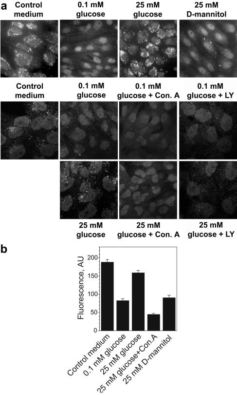
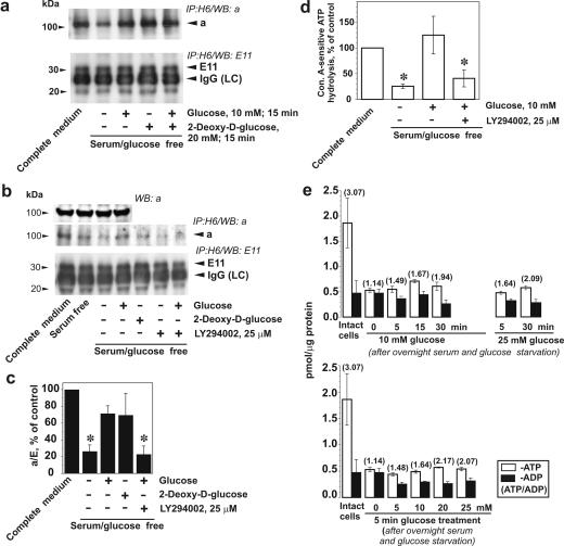
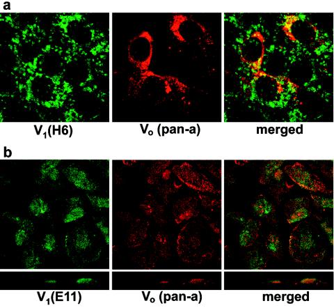
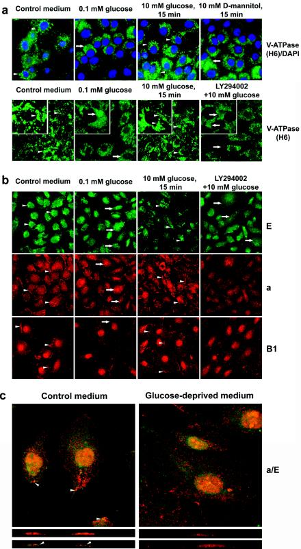
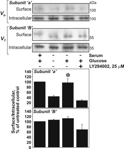
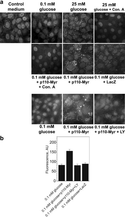
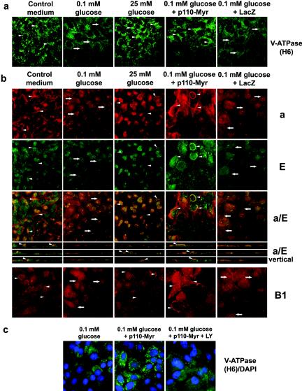
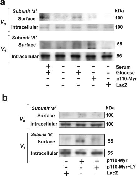
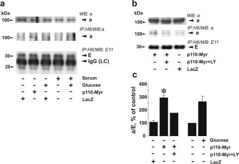
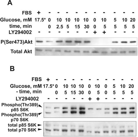
Vacuolar H+-ATPases (V-ATPases) are a family of ATP-driven proton pumps. They maintain pH gradients between intracellular compartments and are required for proton secretion out of the cytoplasm. Mechanisms of extrinsic control of V-ATPase are poorly understood. Previous studies showed that glucose is an important regulator of V-ATPase assembly in Saccharomyces cerevisiae. Human V-ATPase directly interacts with aldolase, providing a coupling mechanism for glucose metabolism and V-ATPase function. Here we show that glucose is a crucial regulator of V-ATPase in renal epithelial cells and that the effect of glucose is mediated by phosphatidylinositol 3-kinase (PI3K). Glucose stimulates V-ATPase-dependent acidification of the intracellular compartments in human proximal tubular cells HK-2 and porcine renal epithelial cells LLC-PK1. Glucose induces rapid ATP-independent assembly of the V1 and Vo domains of V-ATPase and extensive translocation of the V-ATPase V1 and Vo domains between different membrane pools and between membranes and the cytoplasm. In HK-2 cells, glucose stimulates polarized translocation of V-ATPase to the apical plasma membrane. The effects of glucose on V-ATPase trafficking and assembly can be abolished by pretreatment with the PI3K inhibitor LY294002 and can be reproduced in glucose-deprived cells by adenoviral expression of the constitutively active catalytic subunit p110alpha of PI3K. Taken together these data provide evidence that, in renal epithelial cells, glucose plays an important role in the control of V-ATPase-dependent acidification of intracellular compartments and V-ATPase assembly and trafficking and that the effects of glucose are mediated by PI3K-dependent signaling.


Figures

FIG. 1.
FIG. 1.
V-ATPase-dependent, LY294002 (LY)-sensitive acidification of intracellular compartments of renal epithelial cells in response to glucose deprivation/stimulation. HK-2 cells (a) or LLC-PK1 cells (a and b) were incubated in standard medium containing serum and glucose on coverslips. Sixteen to eighteen hours prior to the experiment cells were transferred to glucose-free medium containing dialyzed serum and 0.1 mM glucose. Concanamycin A (Con. A) (100 nM), LY294002 (25 μM), and vehicle were added 60 min before stimulation with glucose or d-mannitol (25 mM). Cells were labeled with DAMP, fixed, and stained with fluorescein-conjugated antidinitrophenol antibody. (a) Bright fluorescent structures are acidic compartments. (Upper panels) Acidification of intracellular compartments is induced by glucose but not by the same concentration of d-mannitol. (Lower panels) LY294002 and concanamycin A prevent the effect of glucose. Results shown are representative of five independent experiments. (b) Analysis of fluorescence intensity in LLC-PK1 cells labeled with DAMP. Image analysis of the average fluorescence intensities in cytoplasmic rectangular areas and fluorescence intensity profiles of cross-sections (see Fig. S3 in the supplemental material) were performed using NIH-Scion Image software. Average fluorescence (mean ± standard error of the mean, arbitrary units [AU]) for 18 to 24 cells from three or four randomly chosen fields is shown.
FIG. 2.
FIG. 2.
Glucose stimulates V-ATPase assembly and ATP hydrolytic activity. LLC-PK1 cells were cultured to 70 to 80% confluence in 199 medium containing 10% FBS and 5.5 mM glucose. Prior to the experiments, cells were incubated in glucose-free DMEM overnight and were then stimulated with 10 mM glucose and/or 20 mM 2-deoxyglucose for 15 min. If the effect of PI3K inhibition was studied, LY294002 (25 μM) and vehicle were added 30 min prior to glucose addition. (a) After cell stimulation, V-ATPase was immunoprecipitated from the lysates with H6.1 MAb, which binds the V1 domain on its E subunit. Coprecipitation of Vo and V1 domains of V-ATPase was probed by Western blotting using antibodies against subunits a (Vo domain) and E (MAb E11, V1 domain). The amount of a subunit present in the immunoprecipitate decreased after glucose and serum deprivation and was quickly restored by cell stimulation with glucose. Glucose-induced increase of subunit a coprecipitated by V1-specific antibody (upper panels) without changes in the amount of detected E subunit (lower panels) indicates an increase in V-ATPase assembly. (b) Effect of glucose on V-ATPase assembly is attenuated by PI3K inhibitor LY294002. (Top panels) Immunoblot detection of a subunit in the cell lysates after glucose deprivation and replacement before V-ATPase immunoprecipitation. No changes were observed. Glucose-stimulated increase of subunit a and E coprecipitation was prevented by preincubation of cells with 25 μM LY294002 (second and third panels from the top). (c) Densitometric analysis of the a/E ratio. Data are presented as percentages of the control (untreated cells). Shown are means ± standard errors of the means (n = 3 or 4); *, P < 0.05 (Student's t test) versus results for the glucose-treated group. (d) Concanamycin A (Con. A)-sensitive ATP hydrolysis by immunoprecipitated V-ATPase from LLC-PK1 cells after glucose deprivation/replacement. V-ATPase was immunoprecipitated from cell lysates with H6.1 antibody. After preincubation of aliquots of the immunoprecipitate in the presence or absence of a V-ATPase inhibitor (200 nM concanamycin A or, alternatively, 1 mM N-ethylmaleimide [NEM]), 3 mM ATP was added to start the reaction. After 30 min, the reaction was stopped and ATP hydrolysis was estimated by spectrophotometric measurement of inorganic phosphate. Ninety to ninety-five percent of the ATP hydrolytic activity in immunoprecipitates was concanamycin A/NEM sensitive (0.25 to 3.1 nmol of Pi/min/mg of cell protein in untreated cells). Data are presented as percentages of the control (untreated cells) as means ± standard errors of the means (n = 3); *, P < 0.05 (Student's t test) versus results for the glucose-treated group (e) Effect of glucose deprivation/stimulation on the levels of ATP and ADP and the ATP/ADP ratio. Cells were grown in complete medium. Prior to glucose treatments, cells were deprived overnight of serum and glucose. The cells were then incubated with 5 to 25 mM glucose for various periods of time up to 30 min, and cell extracts were prepared. ATP and ADP determination was performed as indicated in Materials and Methods. Data are presented as means ± standard errors of the means for three independent experiments performed in triplicate.
FIG. 3.
FIG. 3.
Localization of V1 and Vo domains of V-ATPase in renal tubular epithelial cells. LLC-PK1 (a) and HK-2 cells (b) were grown on coverslips in standard conditions and were then fixed and permeabilized followed by double staining with antibodies against V1 (H6.1 or E11) and Vo (pan-a) subunits as described in Materials and Methods. Confocal images (0.5-μm optical sections) are shown with overlays (merged) of two stainings, as well as lateral views (XZ vertical section) (b, lower panels).
FIG. 4.
FIG. 4.
Glucose-dependent, LY294002-sensitive translocation of V-ATPase in renal epithelial cells. (a) LLC-PK1 cells were grown on coverslips in standard conditions. Prior to the experiments, cells were incubated in glucose-free DMEM overnight and were then stimulated with 10 mM glucose or 10 mM d-mannitol (upper panels) for 15 min. If the effect of PI3K inhibition was studied, LY294002 (25 μM) and vehicle were added 30 min prior to glucose addition. Then cells were fixed, permeabilized, and stained with H6.1 V-ATPase antibody. Nuclei were (upper panels) or were not (lower panels) counterstained with DAPI. In the presence of glucose (before glucose deprivation and after glucose replacement) bright vesicular staining of V-ATPase is visible in the majority of cells (see arrowheads, for example). Glucose deprivation induced appearance of diffuse staining (see insets, arrows, for example). (b) HK-2 cells were grown on coverslips in standard conditions. Prior to the experiments, cells were incubated in glucose-free DMEM overnight and were then stimulated with glucose with or without LY294002, as described above. Then cells were fixed, permeabilized, and stained with antibodies to V1 subunits E and B1 and Vo subunit a. Vesicular staining of subunit E and peripheral perimembrane staining of B1 and a subunits in control conditions or after stimulation with glucose is seen (see arrowheads, for example). Glucose-deprived or LY294002-treated cells show more diffuse staining for the E subunit, disappearance of membrane staining for subunits a and B1, and translocation of subunit a to large vesicles (see arrow, for example). (c) Colocalization of a and E subunits in perimembrane and apical areas is observed only in the presence of glucose (arrowheads). Confocal images of optical 0.5-μM sections (upper panels) and vertical (XZ) sections (lower panels) are shown.
FIG. 5.
FIG. 5.
Glucose-induced increase in cell surface pool of V-ATPase in HK-2 cells. Subconfluent HK-2 cells were incubated in glucose-free DMEM overnight and were then stimulated with 10 mM glucose for 15 min. If the effect of PI3K inhibition was studied, LY294002 (25 μM) and vehicle were added 30 min prior to glucose addition. Then cell surface biotinylation was performed on ice, as described in Materials and Methods. Biotinylated proteins representing the cell surface pool were precipitated on streptavidin beads. Nonbiotinylated (intracellular) proteins remained in the supernatant. Both fractions were probed by immunoblotting with antibodies against a (Vo) and B1 (V1) subunits. Results shown are representative of four independent experiments. (Lower panel) Densitometric analysis of the surface V-ATPase/intracellular V-ATPase ratio. Data are presented as percentages of the control (untreated cells) as means ± standard errors of the means (n = 3 or 4); *, P < 0.05 (U-test) versus results for the glucose-deprived or LY294002-treated group.
FIG. 6.
FIG. 6.
V-ATPase-dependent acidification of intracellular compartments in renal epithelial cells expressing constitutively active PI3K. (a and b) LLC-PK1 cells were incubated in standard medium containing glucose and serum on coverslips. Cells were infected with Adeno/Myr-p110-Myc or Adeno/LacZ at MOI 10 for 48 h. Sixteen to eighteen hours prior to DAMP labeling cells were transferred to glucose-free medium containing dialyzed serum and 0.1 mM glucose. Concanamycin A (Con. A) (100 nM) and 10 mM NH4Cl were added 60 min before stimulation with glucose (25 mM). If the effect of PI3K inhibition was studied, LY294002 (LY) (25 μM) and vehicle were added 60 min prior to DAMP addition. Cells were labeled with DAMP, fixed, and stained with fluorescein-conjugated antidinitrophenol antibody. (b) Analysis of fluorescence intensity in LLC-PK1 cells labeled with DAMP. Image analysis of the average fluorescence intensity in a cytoplasmic rectangular area and fluorescence intensity profiles of cross sections (see Fig. S3 in the supplemental material) was performed using NIH-Scion Image software. Average fluorescence (mean ± standard error of the mean, arbitrary units [AU]) for 18 to 24 cells from three or four randomly chosen fields is shown.
FIG. 7.
FIG. 7.
Translocation of V-ATPase in renal epithelial cells overexpressing constitutively active PI3K. (a) LLC-PK1 cells were incubated in standard medium containing glucose and serum on coverslips. Cells were infected with Adeno/Myr-p110-Myc or Adeno/LacZ at an MOI of 10 for 48 h. Then cells were transferred to glucose-free medium containing dialyzed serum and 0.1 mM glucose for 16 to18 h followed by stimulation with 25 mM glucose for 15 min. Then cells were fixed, permeabilized, and stained with H6.1 V-ATPase antibody. Expression of constitutively active PI3K was sufficient to maintain vesicular staining of V-ATPase in glucose-deprived cells, similarly to cells incubated in standard medium or after stimulation with glucose (see arrowheads, for example). (b) HK-2 cells were grown on coverslips in standard conditions. Infection with Adeno/Myr-p110-Myc and Adeno/LacZ, serum deprivation, and stimulation were as described above. Cells were fixed, permeabilized, and double stained with antibodies for V1 subunit E and Vo subunit a or stained with antibody against V1 subunit B1. Confocal images of a and E double staining (0.5-μm optical sections) are shown together with overlays of two stainings (a/E). Overlaid images of vertical (XZ) sections for a/E double staining are also shown. (c) LLC-PK1 cells were incubated in standard medium containing glucose and serum on coverslips. Infection with Adeno/Myr-p110-Myc and Adeno/LacZ, serum deprivation, and stimulation were as described above. LY294002 (LY) (25 μM) and vehicle were added 60 min prior to cell fixation. Then the cells were fixed, permeabilized, and stained with H6.1 V-ATPase antibody. Nuclei were counterstained with DAPI.
FIG. 8.
FIG. 8.
Expression of constitutively active PI3K increases the cell surface pool of V-ATPase in HK-2 cells. Cells were grown in standard conditions. They were infected with Adeno/Myr-p110-Myc or Adeno/LacZ at an MOI of 10 for 48 h. Then cells were transferred to glucose- and serum-free medium for 16 to18 h followed by stimulation with 25 mM glucose for 15 min (a) or treatment with 25 μM LY294002 (LY) for 60 min (b). Then cell surface biotinylation was performed on ice. Cell surface (biotinylated) proteins were precipitated with streptavidin beads. Nonbiotinylated (intracellular) proteins remained in the supernatant. Both fractions were probed by immunoblotting with antibodies against a (Vo) and B1 (V1) subunits. Results shown are representative of two independent experiments.
FIG. 9.
FIG. 9.
Constitutively active PI3K stimulates assembly of V-ATPase in LLC-PK1 cells in the absence of glucose. LLC-PK1 cells were cultured in standard conditions and then were infected with Adeno/Myr-p110-Myc or Adeno/LacZ at an MOI of 10 for 48 h. Then cells were transferred to glucose- and serum-free medium for 16 to 18 h followed by stimulation with 25 mM glucose for 15 min (a) or treatment with 25 μM LY294002 (LY) for 60 min (b). After stimulation, V-ATPase was immunoprecipitated from the cell lysates with H6.1 MAb. Coprecipitation of Vo and V1 domains of V-ATPase was probed by Western blotting using antibodies against subunits a (Vo domain) and E (antibody E11, V1 domain). The upper panels in panels a and b show immunoblot detection of the a subunit in the cell lysates before V-ATPase immunoprecipitation. (c) Densitometric analysis of the ratio of the amount of coprecipitated a and E subunits represents changes in assembly of V1 and Vo domains. Data are presented as percentages of the control (glucose-deprived noninfected cells) as means ± standard errors of the means (n = 2 or 3); *, P < 0.01 (t test) versus LacZ-infected group.
FIG. 10.
FIG. 10.
Glucose induces rapid PI3K-dependent activation of Akt and p70 S6K in renal proximal tubular epithelial cells. HK-2 cells were incubated in glucose- and serum-free DMEM overnight and then were stimulated with 5 to 20 mM glucose for the indicated periods of time. If the effect of PI3K inhibition was studied, LY294002 (25 μM) and vehicle were added 30 min prior to glucose addition. The cell lysates were prepared as described in Materials and Methods, and 20-μg protein aliquots were resolved by SDS-PAGE and analyzed by immunoblotting with antibodies against phospho-Akt(Ser473) and total Akt (A) or phospho-p70 S6K(Thr389) and total p70 S6K (B). *, the source of glucose is serum-containing standard medium (about 5.5 and 17.5 mM for LLC-PK1 and HK-2 cells, respectively). The results shown are representative of three independent experiments.

References

    1. Anderson, R. G., J. R. Falck, J. L. Goldstein, and M. S. Brown. 1984. Visualization of acidic organelles in intact cells by electron microscopy. Proc. Natl. Acad. Sci. USA 81:4838-4842. - PMC - PubMed
    1. Bastani, B., H. Purcell, P. Hemken, D. Trigg, and S. Gluck. 1991. Expression and distribution of renal vacuolar proton-translocating adenosine triphosphatase in response to chronic acid and alkali loads in the rat. J. Clin. Invest. 88:126-136. - PMC - PubMed
    1. Belham, C., S. Wu, and J. Avruch. 1999. Intracellular signalling: PDK1-a kinase at the hub of things. Curr. Biol. 9:R93-R96. - PubMed
    1. Bollen, M., S. Keppens, and W. Stalmans. 1998. Specific features of glycogen metabolism in the liver. Biochem. J. 336(Part 1):19-31. - PMC - PubMed
    1. Bowman, E. J., A. Siebers, and K. Altendorf. 1988. Bafilomycins: a class of inhibitors of membrane ATPases from microorganisms, animal cells, and plant cells. Proc. Natl. Acad. Sci. USA 85:7972-7976. - PMC - PubMed

Publication types

MeSH terms