. 2005 Jan;25(2):612-20.
doi: 10.1128/MCB.25.2.612-620.2005.

The parafibromin tumor suppressor protein is part of a human Paf1 complex

Affiliations

The parafibromin tumor suppressor protein is part of a human Paf1 complex

Orit Rozenblatt-Rosen et al. Mol Cell Biol. 2005 Jan.

Abstract

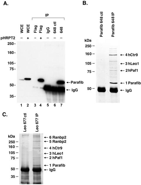
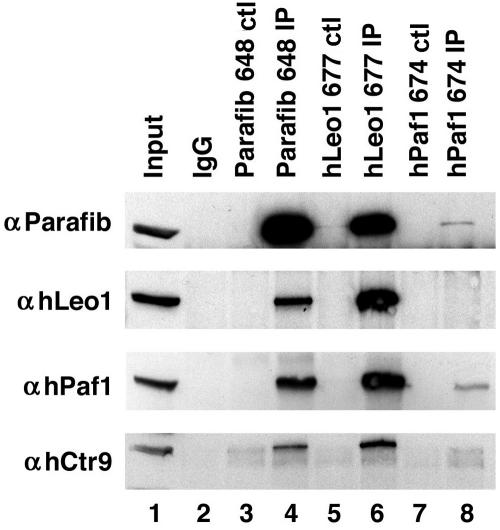
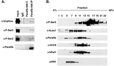
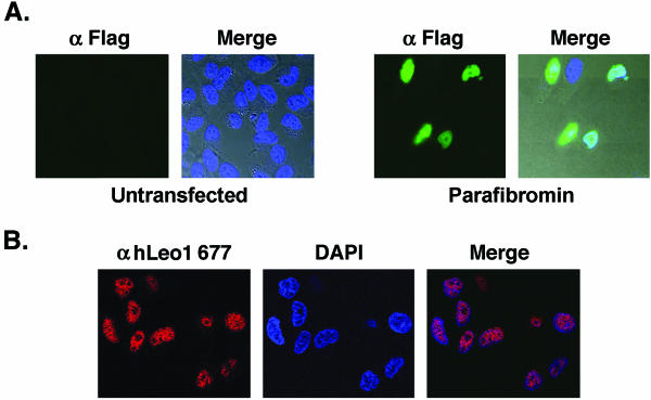
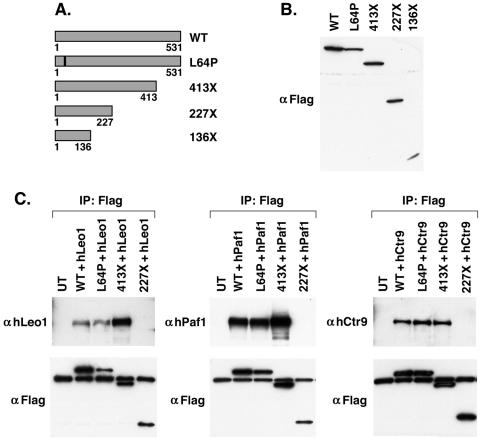
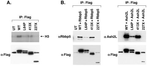
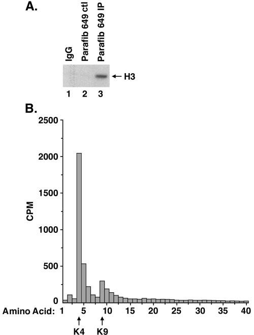
Parafibromin, the product of the HRPT2 (hyperparathyroidism-jaw tumor syndrome 2) tumor suppressor gene, is the human homologue of yeast Cdc73, part of the yeast RNA polymerase II/Paf1 complex known to be important for histone modification and connections to posttranscriptional events. By purifying cellular parafibromin and characterizing its associated proteins, we have identified a human counterpart to the yeast Paf1 complex including homologs of Leo1, Paf1, and Ctr9. Like the yeast complex, the parafibromin complex associates with the nonphosphorylated and Ser2 and Ser5 phosphorylated forms of the RNA polymerase II large subunit. Immunofluorescence experiments show that parafibromin is a nuclear protein. In addition, cotransfection data suggest that parafibromin can interact with a histone methyltransferase complex that methylates histone H3 on lysine 4. Some mutant forms of parafibromin lack association with hPaf1 complex members and with the histone methyltransferase complex, suggesting that disruption of these complexes may correlate with the oncogenic process.


Figures

FIG. 1.
FIG. 1.
Identification of parafibromin (Parafib) interacting proteins. (A) Characterization of antiparafibromin antibody Ab648 in transfected and untransfected cells. An immunoblot with Ab648 is shown. Whole-cell lysate (150 μg) of untransfected 293T cells (lane1) or 293T cells transfected with a plasmid encoding Flag-tagged parafibromin (pHRPT2; lane 2) is shown as a control. Proteins were immunoprecipitated (IP) from untransfected (lane 3) or transfected (lane 4) cell lysate with anti-Flag antibody (one-fifth of a 10-cm-diameter plate). Proteins were immunoprecipitated from lysates of untransfected cells (one 10-cm-diameter plate) with antiparafibromin antibody Ab648 (lane 7) and negative controls, normal rabbit IgG (lane 5) and antiparafibromin antibody Ab648 plus blocking peptide (lane 6). Proteins were resolved by SDS-8% PAGE and immunoblotted with antiparafibromin antibody Ab648. (B) Lysates of 293T cells were immunoprecipitated with antiparafibromin antibody Ab648 (right lane) or a peptide-blocked control (left lane), resolved by SDS-8% PAGE, and visualized by Coomassie blue staining. Bands 1 to 4 were subjected to mass spectrometric analysis. (C) Immunoprecipitation of protein from 293T cells with anti-hLeo1 antibody Ab677 (right lane) or a peptide-blocked control (left lane). Proteins were resolved by SDS-8% PAGE and visualized by silver staining. Numbered bands were identified by mass spectrometric analysis. WCE, whole-cell extract.
FIG. 2.
FIG. 2.
Parafibromin (Parafib) is part of a human Paf1 complex. Antiparafibromin antibody Ab648 (lane 4), anti-hLeo1 antibody Ab677 (lane 6), and anti-hPaf1 antibody Ab674 (lane 8) immunoprecipitates from 293T cell lysates were immunoblotted with antiparafibromin antibody Ab648 (top panel), anti-hLeo1 antibody Ab677 (second panel), anti-hPaf1 antibody Ab673 (third panel), and anti-hCtr9 antibody Ab679 (bottom panel). Cell lysate was loaded in lane 1. Negative control (ctl) immunoprecipitations (IP) were performed with normal rabbit IgG (lane 2) and peptide-blocked controls (lanes 3, 5, and 7).
FIG. 3.
FIG. 3.
Parafibromin (Parafib) interacts with the RNA polymerase large subunit. (A) 293T cell lysates were immunoprecipitated (IP) with antiparafibromin antibody Ab648, a peptide-blocked control (C), or a normal rabbit serum control and immunoblotted with antibodies to Rpb1 with unphosphorylated (Unphos.) CTD (top part), Ser5-phosphorylated CTD (second part), Ser2-phosphorylated CTD (third part), and parafibromin (bottom part). (B) 293T cell lysate was fractioned on a glycerol gradient. Forty-microliter aliquots of the fractions depicted were used for immunoblotting with anti-Rpb1 Ser5-phosphorylated CTD (top part), anti-hLeo1 antibody Ab677 (second part), antiparafibromin antibody Ab648 (third part), anti-hCtr9 antibody Ab679 (fourth part), anti-hPaf1 antibody Ab673 (fifth part), and anti-hRtf1 antibody (bottom part).
FIG. 4.
FIG. 4.
Parafibromin and hLeo1 are nuclear proteins. (A) Immunofluorescence assay with FITC-conjugated anti-Flag antibody on untransfected HeLa cells (Untransfected) or HeLa cells transfected with Flag-tagged WT parafibromin (Parafibromin). The left side of each pair of images shows the FITC signal, while the right side, marked “Merge,” is the integration of the FITC and DAPI signals overlaid upon the Nomarski-differential interference contrast images of the cells. (B) Immunofluorescence with anti-Leo1 antibody Ab677 (Cy3 red) was used to visualize endogenous Leo1 (left image), and DAPI staining (middle image) was used to mark nuclear DNA in HeLa cells. The right image, labeled “Merge,” shows the colocalization of the two signals.
FIG. 5.
FIG. 5.
Characterization of parafibromin truncation mutant constructs. (A) Schematic diagram of parafibromin truncation mutant constructs and the L64P mutant construct representing germ line alterations identified from HPT-JT patients. (B) 293T cell lysates from cells transfected with Flag-tagged WT parafibromin or Flag-tagged mutant forms of parafibromin were immunoblotted with anti-Flag antibodies. (C) Immunoprecipitations (IP) with anti-Flag antibodies from 293T cell lysates cotransfected with either hLeo1 (left), hPaf1 (middle), or hCtr9 (right) together with the indicated Flag-tagged WT parafibromin or Flag-tagged parafibromin mutant constructs. Transfectants were immunoblotted with anti-hLeo1, anti-hPaf1, or anti-hCtr9 antibodies (top) or with anti-Flag antibody (bottom) as shown. Untransfected (UT) 293T cells were used as a negative control.
FIG. 6.
FIG. 6.
Interaction between parafibromin and HMTase activity and complex members. (A) HMTase assays were performed on immunoprecipitates (IP) with anti-Flag antibodies from 293T cell lysates transfected with the indicated Flag-tagged WT parafibromin or Flag-tagged parafibromin mutant constructs (top). Transfectants were immunoblotted with anti-Flag antibody (bottom) as shown. Untransfected (UT) 293T cells were used as a negative control. (B) Immunoprecipitations with anti-Flag antibodies from 293T cell lysates cotransfected with either Rbbp5 (left) or Ash2L (right) together with the indicated Flag-tagged WT parafibromin or Flag-tagged parafibromin mutant constructs. Transfectants were immunoblotted with anti-Rbbp5 or anti-Ash2L antibodies (top) or with anti-Flag antibody (bottom) as shown. Untransfected 293T cells were used as a negative control.
FIG. 7.
FIG. 7.
Parafibromin (Parafib) interacts with an HMTase complex that methylates histone H3 on lysine 4. (A) Antiparafibromin antibody Ab649 (lane 3), normal rabbit IgG (lane 1), and peptide-blocked antibody Ab649 control (ctl; lane 2) immunoprecipitates were incubated with histone H3 and the methyl donor S-adenosyl-l-[methyl-3H]methionine, resolved by SDS-15% PAGE, amplified, dried, and fluorographed. (B) Scintillation counting of sequential Edman degradation products of histone H3, labeled by incubation with S-adenosyl-l-[methyl-3H]methionine and with immunoprecipitates of antiparafibromin antibody Ab649. The arrows at the bottom indicate lysines 4 and 9.

References

    1. Betz, J. L., M. Chang, T. M. Washburn, S. E. Porter, C. L. Mueller, and J. A. Jaehning. 2002. Phenotypic analysis of Paf1/RNA polymerase II complex mutations reveals connections to cell cycle regulation, protein synthesis, and lipid and nucleic acid metabolism. Mol. Genet. Genomics 268:272-285. - PubMed
    1. Carpten, J. D., C. M. Robbins, A. Villablanca, L. Forsberg, S. Presciuttini, J. Bailey-Wilson, W. F. Simonds, E. M. Gillanders, A. M. Kennedy, J. D. Chen, S. K. Agarwal, R. Sood, M. P. Jones, T. Y. Moses, C. Haven, D. Petillo, P. D. Leotlela, B. Harding, D. Cameron, A. A. Pannett, A. Hoog, H. Heath III, L. A. James-Newton, B. Robinson, R. J. Zarbo, B. M. Cavaco, W. Wassif, N. D. Perrier, I. B. Rosen, U. Kristoffersson, P. D. Turnpenny, L. O. Farnebo, G. M. Besser, C. E. Jackson, H. Morreau, J. M. Trent, R. V. Thakker, S. J. Marx, B. T. Teh, C. Larsson, and M. R. Hobbs. 2002. HRPT2, encoding parafibromin, is mutated in hyperparathyroidism-jaw tumor syndrome. Nat. Genet. 32:676-680. - PubMed
    1. Chang, M., D. French-Cornay, H. Y. Fan, H. Klein, C. L. Denis, and J. A. Jaehning. 1999. A complex containing RNA polymerase II, Paf1p, Cdc73p, Hpr1p, and Ccr4p plays a role in protein kinase C signaling. Mol. Cell. Biol. 19:1056-1067. - PMC - PubMed
    1. Gerber, M., and A. Shilatifard. 2003. Transcriptional elongation by RNA polymerase II and histone methylation. J. Biol. Chem. 278:26303-26306. - PubMed
    1. Goo, Y. H., Y. C. Sohn, D. H. Kim, S. W. Kim, M. J. Kang, D. J. Jung, E. Kwak, N. A. Barlev, S. L. Berger, V. T. Chow, R. G. Roeder, D. O. Azorsa, P. S. Meltzer, P. G. Suh, E. J. Song, K. J. Lee, Y. C. Lee, and J. W. Lee. 2003. Activating signal cointegrator 2 belongs to a novel steady-state complex that contains a subset of trithorax group proteins. Mol. Cell. Biol. 23:140-149. - PMC - PubMed

Publication types

MeSH terms