. 2005 Jan;25(2):699-705.
doi: 10.1128/MCB.25.2.699-705.2005.

Tenomodulin is necessary for tenocyte proliferation and tendon maturation

Affiliations

Tenomodulin is necessary for tenocyte proliferation and tendon maturation

Denitsa Docheva et al. Mol Cell Biol. 2005 Jan.

Abstract

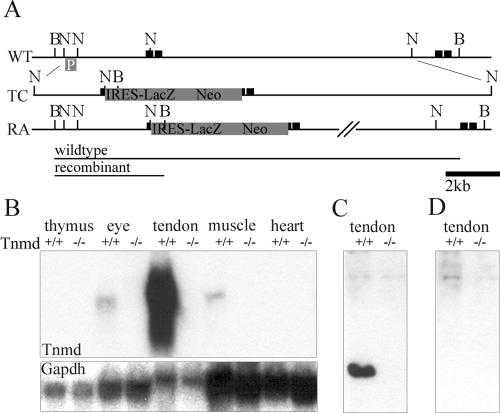
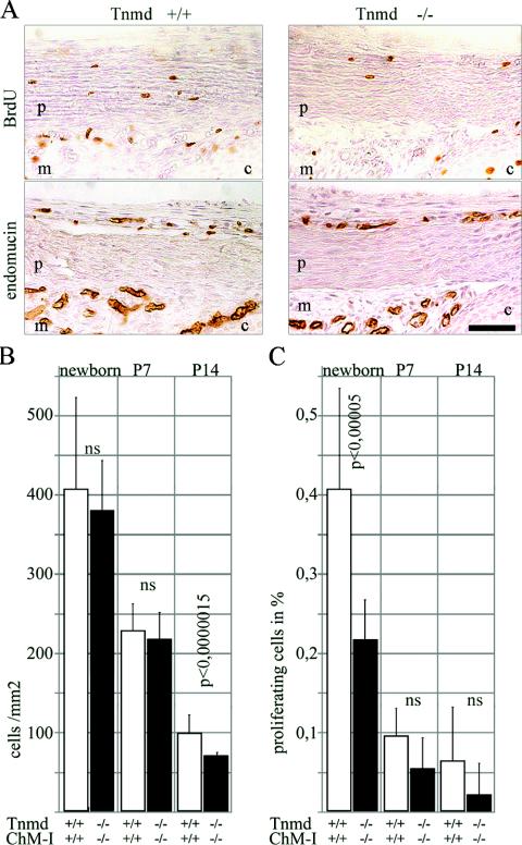
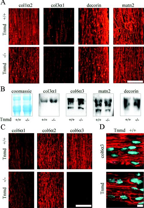
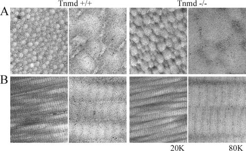
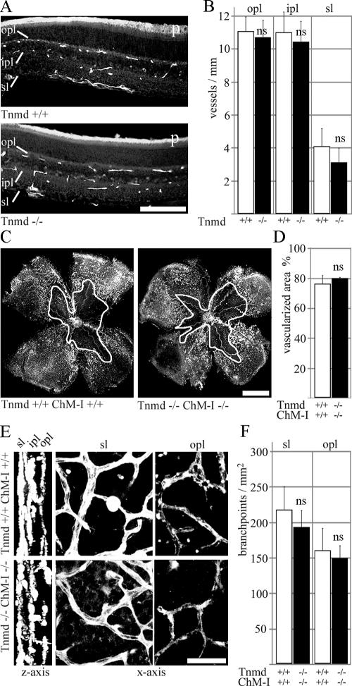
Tenomodulin (Tnmd) is a member of a new family of type II transmembrane glycoproteins. It is predominantly expressed in tendons, ligaments, and eyes, whereas the only other family member, chondromodulin I (ChM-I), is highly expressed in cartilage and at lower levels in the eye and thymus. The C-terminal extracellular domains of both proteins were shown to modulate endothelial-cell proliferation and tube formation in vitro and in vivo. We analyzed Tnmd function in vivo and provide evidence that Tnmd is processed in vivo and that the proteolytically cleaved C-terminal domain can be found in tendon extracts. Loss of Tnmd expression in gene targeted mice abated tenocyte proliferation and led to a reduced tenocyte density. The deposited amounts of extracellular matrix proteins, including collagen types I, II, III, and VI and decorin, lumican, aggrecan, and matrilin-2, were not affected, but the calibers of collagen fibrils varied significantly and exhibited increased maximal diameters. Tnmd-deficient mice did not have changes in tendon vessel density, and mice lacking both Tnmd and ChM-I had normal retinal vascularization and neovascularization after oxygen-induced retinopathy. These results suggest that Tnmd is a regulator of tenocyte proliferation and is involved in collagen fibril maturation but do not confirm an in vivo involvement of Tnmd in angiogenesis.


Figures

FIG. 1.
FIG. 1.
Generation of Tnmd-deficient mice. (A) An IRES-LacZ neomycin cassette was inserted into the translation start site in exon 1 of the Tnmd gene. (B) Hybridization of total RNA derived from newborn thymus, eye, tendon, muscle and heart tissues with the complete Tnmd cDNA showed the lack of any Tnmd transcript (1.4 kb) in the Tnmd gene-targeted mice. (C) A specific signal for Tnmd was detected at 16 kDa in urea tendon extracts of 1-month-old mice. (D) No signal was found in extracts from Tnmd-deficient mice or after incubation with the peptide, used for immunization. B, BamHI; N, NcoI; p, southern probe; black boxes, exons; WT, wild-type allele; TC, targeting construct; RA, recombinant allele.
FIG. 2.
FIG. 2.
Proliferation analysis in tendons. (A) BrdU staining of newborn patellaris tendon revealed a decrease of proliferating cells in Tnmd-deficient tendons. Staining with endomucin excluded endothelial origin of the proliferating cells. p, patellaris; m, meniscus; c, cartilage. Bar, 250 μm. (B) Significantly decreased cell densities were found at P14 but not in newborns or at P7. (C) Quantification revealed a significant proliferation deficit in newborns. At P7 and P14 the proliferation of Tnmd-null cells was still reduced compared to the wild type, but the differences were no longer significant (ns).
FIG. 3.
FIG. 3.
Analysis of ECM deposition in the tendon. (A) Similar signal intensities were observed by immunostaining for collagen types I (col1α2), matrilin-2 (matn2), and decorin in P14 Achilles tendon. Collagen type III (col3α1) signals were reduced in Tnmd-deficient tendon. Bar, 100 μm. (B) Coomassie blue staining of P14 tail tendon extracts showed similar intensities of the collagen α1(I) band. No differences in signal intensities were found by probing for collagen types III (col3α1, 130 kDa) and VI (col6α3, 180 to 200 kDa), matrilin-2 (matn2, 120 to 150 kDa), and decorin (90 to 120 kDa). (C) Immunostaining for the different α chains of collagen VI revealed similar signal intensities for the collagen VI α1 chain (col6α2) and the α2 chain (col6α2), whereas decreased signal intensity was obtained for the α3 chain (col6α3). Bar, 100 μm. (D) Tnmd and collagen VI α3 showed similar distribution patterns with a predominant pericellular localization. Bar, 10 μm.
FIG. 4.
FIG. 4.
Ultrastructural analysis of 6-month-old Tnmd-deficient tendons. (A) Cross-sections; (B) longitudinal sections. A shift of the fibril diameter distribution to large diameters was observed. In addition, Tnmd-null tendons showed an uneven and rough surface of the collagen fibrils. Magnifications: ×20,0001 and ×80,000.
FIG. 5.
FIG. 5.
Analysis of retinal neovascularization. (A) Endomucin staining of 1-month-old retinas. Superficial (sl), inner-plexiform (ipl), and outer plexiform (opl) vessel networks were completely formed in Tnmd-deficient retinas. (B) All layers had similar vessel numbers compared to wild-type eyes. p, photoreceptors. Bar, 100 μm. (C) Whole-mount lectin-staining failed to detect differences in the vascularized area of wild-type and double-null retinas after OIR at P7. The white outline indicates unvascularized area. Bar, 1 mm. (D) Quantification of the neovascular area/whole retina showed equal percentages for wild-type and double-null eyes. (E) After three-dimensional reconstruction, the three vascular layers were clearly distinguishable (z axis). Wild-type and double-null retinas displayed similar vascular morphology in the superficial (sl) and the outer plexiform vascular networks (opl). (F) Quantification of branchpoints in the superficial and outer plexiform layers did not reveal significant differences between wild-type and double-null eyes. ns, not significant. Bar, 20 μm.

References

    1. Alon, T., I. Hemo, A. Itin, J. Pe'er, J. Stone, and E. Keshet. 1995. Vascular endothelial growth factor acts as a survival factor for newly formed retinal vessels and has implications for retinopathy of prematurity. Nat. Med. 1:1024-1028. - PubMed
    1. Aszódi, A., J. F. Bateman, E. Hirsch, M. Baranyi, E. B. Hunzicker, N. Hauser, Z. Bösze, and R. Fässler. 1999. Normal skeletal development of mice lacking matrilin-1: redundant function of matrilins in cartilage? Mol. Cell. Biol. 19:7841-7845. - PMC - PubMed
    1. Azizan, A., N. Holaday, and P. J. Neame. 2001. Post-translational processing of bovine chondromodulin-I. J. Biol. Chem. 276:23632-23638. - PubMed
    1. Barr, P. J. 1991. Mammalian subtilisins: the long-sought dibasic processing endoproteases. Cell 66:1-3. - PubMed
    1. Benjamin, M., and J. R. Ralphs. 2000. The cell and developmental biology of tendons and ligaments. Int. Rev. Cytol. 196:85-130. - PubMed

Publication types

LinkOut - more resources